. 2016 Jan;31(1):52-64.
doi: 10.1002/jbmr.2592. Epub 2015 Aug 6.

NF-κB Has a Direct Role in Inhibiting Bmp- and Wnt-Induced Matrix Protein Expression

Affiliations

NF-κB Has a Direct Role in Inhibiting Bmp- and Wnt-Induced Matrix Protein Expression

Rohinton S Tarapore et al. J Bone Miner Res. 2016 Jan.

Abstract

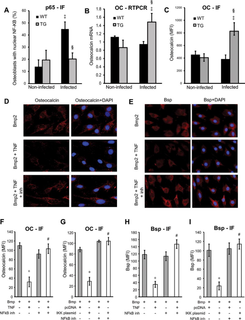
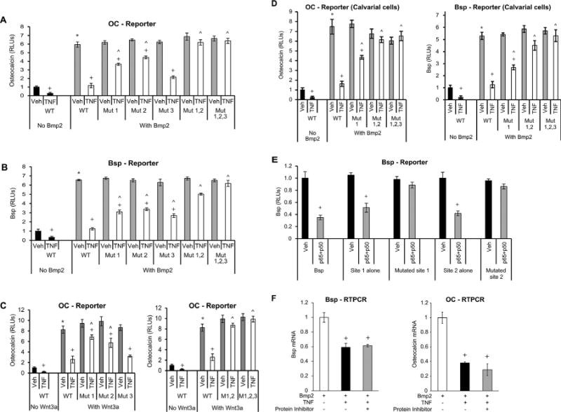
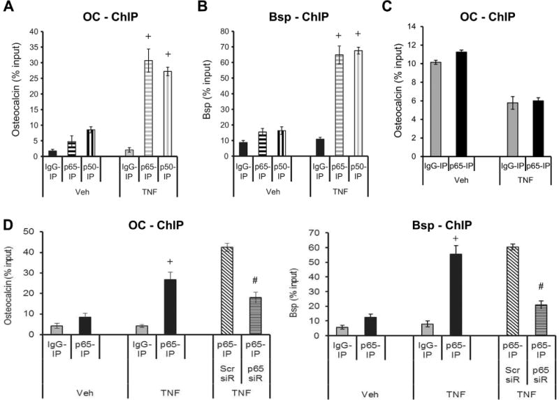
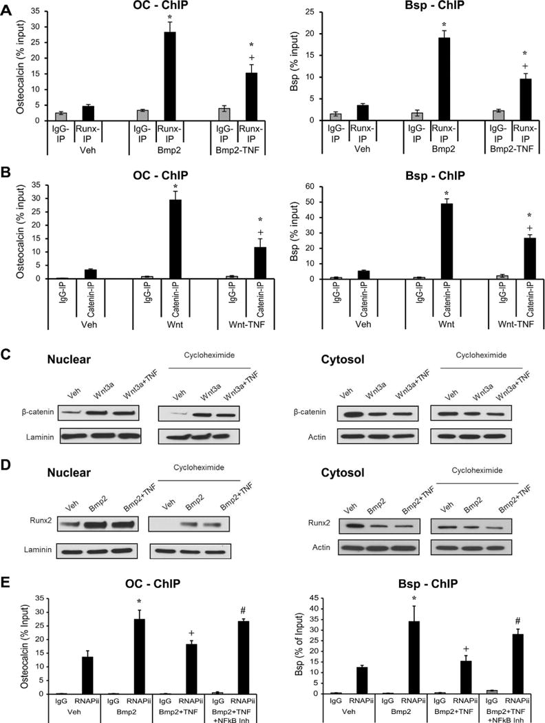
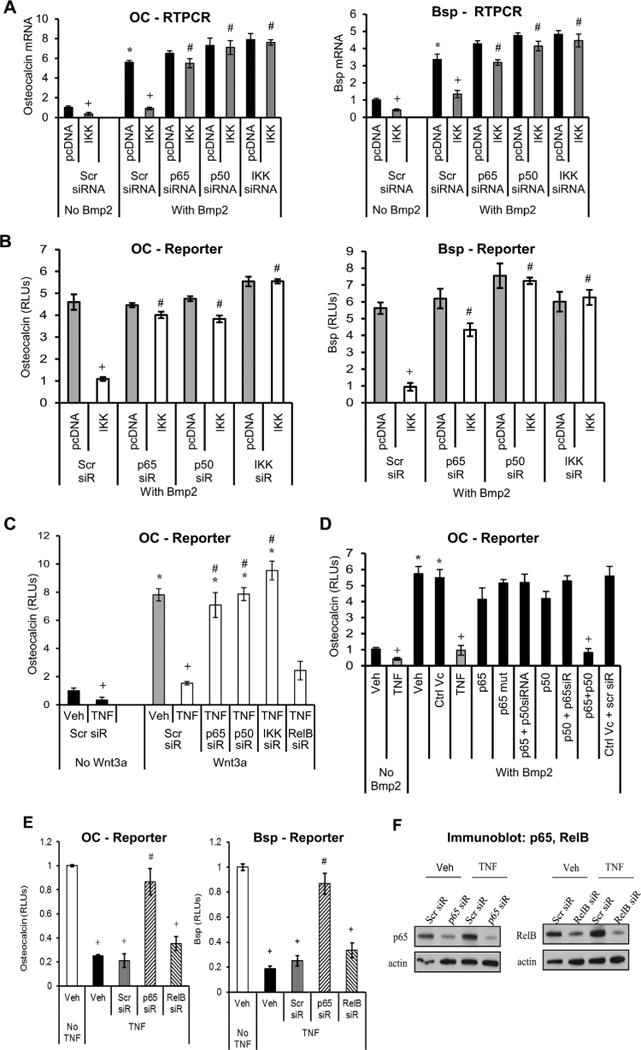
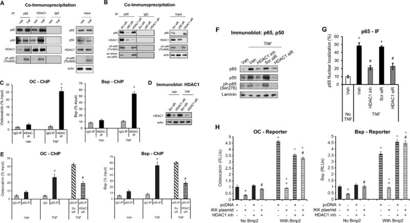
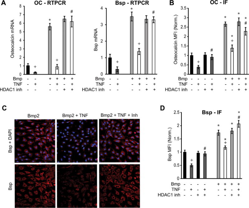
The host response to pathogens through nuclear factor κB (NF-κB) is an essential defense mechanism for eukaryotic organisms. NF-κB-mediated host responses inhibit bone and other connective tissue synthesis and are thought to affect the transcription of matrix proteins through multiple indirect pathways. We demonstrate that inhibiting NF-κB in osteoblasts increases osteocalcin expression in vivo in mice with periodontal disease. Mutating NF-κB binding sites on osteocalcin (OC) or bone sialoprotein (Bsp) promoters rescues the negative impact of NF-κB on their transcription and that NF-κB can inhibit Wnt- and Bmp-induced OC and Bsp transcription, even when protein synthesis is inhibited, indicating a direct effect of NF-κB. This inhibition depends on p65-p50 NF-κB heterodimer formation and deacetylation by HDAC1 but is not affected by the noncanonical NF-κB pathway. Moreover, NF-κB reduces Runx2 and β-catenin binding to OC/Bsp promoters independently of their nuclear localization. Thus, inflammatory signals stimulate the direct interaction of NF-κB with response elements to inhibit binding of β-catenin and Runx2 binding to nearby consensus sites and reduce expression of matrix proteins. This direct mechanism provides a new explanation for the rapid decrease in new bone formation after inflammation-related NF-κB activation.

Keywords: BMP; BONE FORMATION; INFLAMMATION; MATRIX PROTEINS; NF-κB; OSTEOBLASTS; TNFα; WNT.


Conflict of interest statement

Disclosures

All authors state that they have no conflicts of interests.

Figures

Fig. 1
Fig. 1
NF-κB-mediated response inhibits bone and matrix protein synthesis. Destructive periodontitis was initiated in IKK-DN transgenic mice (TG) or wild-type (WT) control mice by oral inoculation of the periodontal pathogens P. gingivalis plus F. nucleatum and compared with vehicle alone. Mice were euthanized 6 weeks after oral inoculation. Nuclear NF-κB was measured by immunofluorescence by colocalization of p65, a subunit of NF-κB and DAPI nuclear staining. (A) Number of NF-κB-positive cells was determined. Localization to bone-lining osteoblastic cells was determined in cells with a cuboidal appearance and with a nucleus that was not fusiform or parallel to the bone surface. (B) RNA was isolated from periodontal tissue, and osteocalcin mRNA levels were measured by real-time PCR. (C) OC expression was measured by immunofluorescence with antibody to OC and expressed as the mean fluorescence intensity. (DI) MC3T3 cells were incubated with TNFα or transfected with IKK-overexpression vector compared with pcDNA (empty vector) alone. NF-κB was inhibited with the specific inhibitor BAY117082. Cells were stimulated with Bmp2 for 48 hours. Cells were stained for OC or Bsp (red) and counterstained with DAPI (blue). Immunofluorescence was analyzed and expressed as mean expression above baseline (MFI). ‡Significantly different in infected compared with matched noninfected group; §significantly different in infected TG compared with infected WT. Data shown are representative of three independent experiments. +Significant inhibition with TNFα or IKK transfection; #significantly different between cells incubated with no inhibitor.
Fig. 2
Fig. 2
Mutation of NF-κB binding sites in osteocalcin or Bsp promoters rescues the negative effect of TNFα, indicating a direct effect of NF-κB. NF-κB binding sites were mutated as indicated (Mut1; Mut2; Mut3; Mut1,2; Mut1,2,3). Cells were transfected with wild-type and mutant constructs. After a 1-hour incubation with TNFα, cells were stimulated with Bmp2 or Wnt3a as indicated. (AC) OC or Bsp promoter activity was analyzed by luciferase reporter construct. Luciferase activity was normalized by renilla control and expressed as relative luciferase units (RLUs). (D) Primary mouse osteoblastic cells were transfected with osteocalcin or Bsp reporter construct with or without mutated NF-κB binding sites. Transcriptional activity was measured using a luciferase assay and expressed as relative luciferase units (RLUs). (E) Cells were transfected with wild-type or mutated Bsp luciferase constructs containing all the NF-κB putative sites. Using subcloning, Bsp mini genes were prepared from wild-type and mutated promoter regions as described in Materials and Methods. NF-κB was activated by cotransfecting cells with p65- and p50-overexpressing plasmids. (F) Cells were stimulated with Bmp2 for 48 hours and then incubated with TNFα for 6 hours without or with cycloheximide. Bsp or OC mRNA levels were analyzed by RT-PCR. *Significantly different from Bmp2-stimulated control; +significant inhibition with TNFα; ˄significantly different between cells transfected with WT (wild-type construct) versus matched control (p < 0.05).
Fig. 3
Fig. 3
NF-κB p65-p50 heterodimer binds to Bsp and osteocalcin promoters. Cells were stimulated with TNFα and compared with vehicle. After incubation, cells were fixed, DNA isolated, and subjected to chromatin immunoprecipitation using antibodies for p65 and p50 or matched IgG control. (A, B) ChIP was performed using antibodies for p65, p50 or control IgG. RT-PCR was performed using primers to Bsp or osteocalcin promoter that contained NF-κB response elements or (C) to a region further upstream of the promoter region. (D) Cells were incubated with TNFα after knockdown with p65 siRNA or scrambled siRNA and chromatin was immunoprecipitated with antibody to p65 or control IgG. RT-PCR was performed using primers to OC or Bsp promoter that contained NF-κB response elements. Data shown are representative of three independent experiments. +Significantly different with TNFα versus matched control; #significantly different between cells transfected with scrambled siRNA versus p65 siRNA (p < 0.05).
Fig. 4
Fig. 4
NF-κB activation decreases Bmp2-stimulated Runx2 and Wnt-stimulated β-catenin binding to osteocalcin or Bsp promoters. Cells were incubated with Bmp2 or Wnt3a for 4 hours with or without a 45-minute pre-incubation with TNFα. Some of the cells were incubated with NF-κB small molecule inhibitor (BAY-117082). Cells were fixed, DNA isolated, and subjected to ChIP using antibodies for Runx2, β-catenin, RNAP-II or control IgG. (A, B) RT-PCR was performed using primers to Bsp or OC promoter that contained NF-κB response elements. (C, D) Immunoblotting with antibody to beta-catenin, Runx2, actin or laminin of nuclear or cytosolic lysates of cells incubated with Wnt or Bmp for 4 hours, followed by a 3-hour cycloheximide incubation. Before stimulation, some of the cells were incubated with TNFα for 45 minutes. (E) ChIP assay was performed with antibody to RNAP-II or control IgG. RT-PCR was performed using primers to Bsp or OC promoter that contained binding regions of RNAP-II. *Significantly different from No-Bmp2/Wnt3a-stimulated control; +significant inhibition with TNFα; #significantly different between cells incubated with no inhibitor (p < 0.05).
Fig. 5
Fig. 5
The canonical NF-κB suppresses matrix protein transcription. Cells were stimulated with TNFα or transfected with IKK-, p65-, p50-overexpression vector compared with pcDNA (empty vector) alone. Some of the cells were also transfected with siRNA specific for IKK, the NF-κB subunits p65, p50, RelB, or scrambled siRNA (Scr). Cells were stimulated with Bmp2 or Wnt3a for 48 hours. (A) Osteocalcin or Bsp mRNA levels were measured by real-time PCR. (BE) Osteocalcin or Bsp promoter activity was analyzed by luciferase reporter construct in cells incubated with Bmp2 or Wnt. (F) Immunoblot analysis of whole-cell lysate probed for p65 and RelB. Data shown are representative of three independent experiments. +Significant inhibition with TNFα or IKK transfection versus matched control; #significantly different between cells transfected with scrambled siRNA versus targeted siRNA (p < 0.05).
Fig. 6
Fig. 6
HDAC1 interacts with NF-κB to regulate BSP and OC transcription activity. Cells were stimulated with TNFα alone with or without pre-incubation with HDAC1-specific inhibitor (parthenolide)(48) or transfection with siRNA specific for HDAC1 or scrambled (Scr). (A, B) Cell lysates were incubated with antibody to p65 or control IgG. Immunoprecipitated proteins or input cell lysate was then examined by immunoblot analysis with antibody to p65, p50, HDAC1, ph-p65 (Thr505), or acetylated lysine. (C, E) Cells were fixed, DNA isolated, and subjected to chromatin immunoprecipitation using antibodies for HDAC1, p65, or matched IgG control. RT-PCR was performed using primers to Bsp or OC promoter that contained NF-κB response elements. (D) Immunoblot analysis of whole-cell lysate probed for HDAC1. (F) Immunoblot analysis of nuclear lysate in cells incubated with HDAC1 inhibitor or transfected with HDAC1-siRNA were probed for p65, p50, or ph-p65. (G) Nuclear localization of NF-κB was measured by immunofluorescence with an antibody specific for p65 and counterstained with DAPI. (H) Cells were pre-incubated with the HDAC1 inhibitor (parthenolide), transfected with IKK or pcDNA (empty vector), and transfected with a Bsp or osteocalcin reporter vector with or without Bmp2 stimulation. Luciferase activity was normalized by renilla control and expressed as relative luciferase units (RLUs). Data shown are representative of three independent experiments. +Significantly different between TNFα versus matched control; #significantly different between cells transfected with scrambled siRNA versus HDAC1 siRNA or between cells incubated with no inhibitor; ˄significantly different between cells with Bmp2 stimulation (p < 0.05).
Fig. 7
Fig. 7
HDAC1 plays a role in decreased mRNA and protein expression of matrix proteins. Cells were stimulated with TNFα with or without pre-incubation with HDAC1-specific inhibitor (parthenolide). Some of the cells were stimulated with Bmp2. (A) OC or Bsp mRNA was measured by real-time PCR in cells incubated with TNFα and parthenolide, with or without Bmp2 stimulation. (BD) Protein was measured by immunofluorescence using specific antibody for osteocalcin or Bsp (red) and counterstained with DAPI. Protein expression was measured by immunofluorescence and expressed as mean fluorescence intensity (MFI). Values were normalized to unstimulated controls. Data are expressed as level above baseline. Data shown are representative of three independent experiments. *Significantly different from No-Bmp2-stimulated control; +significant inhibition with TNFα; #significantly different between cells with no inhibitor (p < 0.05).

Similar articles

Cited by

References

    1. Rahman MM, McFadden G. Modulation of tumor necrosis factor by microbial pathogens. PLoS Pathog. 2006;2(2):e4. - PMC - PubMed
    1. Vitiello M, Galdiero M, Finamore E, Galdiero S, Galdiero M. NF-kappaB as a potential therapeutic target in microbial diseases. Mol Biosyst. 2012;8(4):1108–20. - PubMed
    1. Marcu KB, Otero M, Olivotto E, Borzi RM, Goldring MB. NF-kappaB signaling: multiple angles to target OA. Curr Drug Targets. 2010;11(5):599–613. - PMC - PubMed
    1. Boyce BF, Yao Z, Xing L. Functions of nuclear factor kappaB in bone. Ann NY Acad Sci. 2010;1192:367–75. - PMC - PubMed
    1. Novack DV. Role of NF-kappaB in the skeleton. Cell Res. 2011;21(1):169–82. - PMC - PubMed

Publication types

MeSH terms