. 2015 Jun 18;161(7):1619-32.
doi: 10.1016/j.cell.2015.05.028.

A Single Kinase Generates the Majority of the Secreted Phosphoproteome

Affiliations

A Single Kinase Generates the Majority of the Secreted Phosphoproteome

Vincent S Tagliabracci et al. Cell. .

Abstract

The existence of extracellular phosphoproteins has been acknowledged for over a century. However, research in this area has been undeveloped largely because the kinases that phosphorylate secreted proteins have escaped identification. Fam20C is a kinase that phosphorylates S-x-E/pS motifs on proteins in milk and in the extracellular matrix of bones and teeth. Here, we show that Fam20C generates the majority of the extracellular phosphoproteome. Using CRISPR/Cas9 genome editing, mass spectrometry, and biochemistry, we identify more than 100 secreted phosphoproteins as genuine Fam20C substrates. Further, we show that Fam20C exhibits broader substrate specificity than previously appreciated. Functional annotations of Fam20C substrates suggest roles for the kinase beyond biomineralization, including lipid homeostasis, wound healing, and cell migration and adhesion. Our results establish Fam20C as the major secretory pathway protein kinase and serve as a foundation for new areas of investigation into the role of secreted protein phosphorylation in human biology and disease.


Figures

Figure 1
Figure 1. The Fam20C- and VLK-related secretory pathway kinases
(A, B) Phylogenetic trees built with MOLPHY using distances (-J option) calculated from an alignment of representative human proteins depicting (A) the Fam20C-related kinases and (B) the VLK-related kinases (Fjx1, Four-jointed box 1, DIA1; Deleted in Autism 1, DIA1R; DIA1-Related). See also Figure S1.
Figure 2
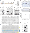
Figure 2. Fam20C activity is unique amongst the known secretory pathway kinases
(A) Schematic representation of human Fam20C depicting the signal peptide (SP; red), the putative pro-region (green), the kinase domain (tan) and the insertion domain (pink). The mature, C-terminally tagged Fam20C purified from conditioned medium of HEK293T cells is truncated by 92 amino acids as determined by Edman degradation. The amino acid sequence surrounding this region is shown. The N-linked glycosylation sites and the “DFG” metal binding residue, D478, are also depicted. (B) Protein immunoblotting of V5 and Flag immunoprecipitates from conditioned medium (CM) of U2OS cells co-expressing V5-tagged OPN (OPN:V5) with either WT Fam20C (20C:Flag), 91LQD93-91AAA93 Fam20C (20CLQD-AAA:Flag) or Fam20C lacking the signal peptide (ΔSP20C:Flag). Extracts were analyzed for Flag-Fam20C (or mutants) and GAPDH. (C) Protein immunoblotting of V5 and Flag immunoprecipitates from CM of U2OS cells co-expressing V5-tagged OPN (OPN:V5) with either WT Fam20C (20C:Flag), catalytically inactive D478A (20CD478A:Flag), or various N-linked glycosylation mutants. Extracts were analyzed for Flag-Fam20C (or mutants) and GAPDH. ** Denotes that 20C:Flag immunoprecipitates were treated with PNGase-F. (D) Protein immunoblotting and autoradiography of Flag-immunoprecipitates from CM of 32P-orthophosphate labeled U2OS cells expressing WT Fam20C (20C:Flag) or catalytically inactive D478A (20C D478A:Flag). (E) Protein immunoblotting of V5 and Flag immunoprecipitates from CM of U2OS cells co-expressing V5-tagged OPN (OPN:V5) with either WT Fam20C (20C:Flag) or potential S-x-E phosphosite Flag-tagged mutants (S41A, S106A and S372A). Extracts were analyzed for Flag-Fam20C (or mutants) and GAPDH. ** Denotes that 20C:Flag and OPN:V5 immunoprecipitates were treated with λ-phosphatase (λ-p’tase). (F) Characterization of a consensus Fam20C phosphorylation motif by phosphorylation of a positional scanning peptide library. The phosphorimage depicts incorporation of 32P into a mixture of biotinylated peptides bound to a streptavidin-coated membrane. The positions are numbered relative to the central phospho-acceptors. (G) Position-specific scoring matrix (PSSM) resulting from quantification of the in vitro phosphorylation of the positional scanning peptide library by Fam20C. (H) SDS-PAGE and Coomassie staining of MBP-tagged members of the Fam20C- and VLK -related kinases, and Sgk196, expressed and purified from CM of insect cells. (The tags for Fjx1 and Fam20B were cleaved with TEV). (I) The Fam20C- and VLK -related kinases, and Sgk196 were incubated with OPN in the presence of [γ-32P]ATP. The reaction products were analyzed by SDS-PAGE and autoradiography. (J) The Fam20C- and VLK -related kinases and Sgk196 were incubated with β28–40 in the presence of [γ-32P]ATP. Incorporated radioactivity was quantified by scintillation counting. Note: Fam198B was not included in these reactions because we could not produce the protein in mammalian or insect cells despite several attempts. However, when Flag-tagged Fam198B was coexpressed with V5-tagged Fetuin A (FetA) in mammalian cells metabolically labeled with 32P-orthophosphate, we did not observe 32P incorporation into V5-immunoprecipitates (not shown). FetA is an S-x-E containing phosphoprotein that is a Fam20C substrate (See Figure 4A and D). We therefore conclude that Fam20C activity is unique amongst the known secretory pathway kinases. (K) Protein immunoblotting of V5 and Flag immunoprecipitates from CM of U2OS cells co-expressing V5-tagged OPN (OPN:V5) with either WT (20C:Flag), catalytically inactive D478A (20C D478A:Flag), or Fam20CKDEL (20C:Flag-KDEL). Extracts were analyzed for Flag-Fam20C (or mutants) and GAPDH. See also Figure S2.
Figure 3
Figure 3. Fam20C generates the majority of the HepG2 extracellular phosphoproteome
(A) Schematic representation of the workflow used in these studies. (B) Volcano plot depicting the log2 of fold phosphorylation change (normalized to protein abundance) versus -log10(p-value) for phosphosites on secretory pathway proteins. The phosphopeptides marked in red displayed a statistically significant decrease in Fam20C KO HepG2 cells (log2 fold change ≤ −1; −log10(p-value) ≥ 1.3). (C) Zoomed in view of the volcano plot in (B) highlighting the phosphopeptides that significantly decreased in Fam20C KO cells. OPN; osteopontin, IGFBP1; insulin-like growth factor binding protein-1, FETUA; fetuin A, α-Feto; α-fetoprotein, ITIH2; Inter-alpha-trypsin inhibitor heavy chain H2, APOB; apolipoprotein B, FGA; fibrinogen gamma chain, QSOX1; quiescin Q6 sulfhydryl oxidase-1, Lam; laminin, SCG1; secretogranin-1, TF; transferrin, APOL1; apolipoprotein L1, BMP4; bone morphogenic protein-4, PCSK9; Proprotein convertase subtilisin/kexin type-9, APP; Aβ precursor protein, HRC; histidine rich calcium binding protein, C3; complement component C3, C4A; complement component. See also Figure S3.
Figure 4
Figure 4. Fam20C directly phosphorylates serum, plasma and CSF proteins
(A) Time dependent incorporation of 32P from [γ-32P]ATP into IGFBP3, PCSK9, Fetuin A (FetA), IGFBP5, fibronectin, and fibrinogen by Fam20C, or the D478A mutant. Reaction products were separated by SDS-PAGE, visualized by Coomassie staining, and radioactivity was detected by autoradiography. (B) Time dependent incorporation of 32P from [γ-32P]ATP into MBP-tagged calumenin (MBP-Calu), interleukin-6 (MBP-IL-6), stanniocalcin (MBP-STC2) and nucleobindin 1 (MBP-NucB1). MBP was also analyzed as a control for the indicated time point. Reaction products were analyzed as in (A). (C) Protein immunoblotting of V5-immunoprecipitates from conditioned medium (CM) of U2OS or HEK293T cells co-expressing V5-tagged IGFBP3, IGFBP4, IGFBP5, IGFBP7, proenkephalin (PENK), secretogranin 1 (SCG1), SCG2, secreted phosphoprotein 2 (Spp2), stanniocalcin 2 (STC2), secretogranin 3 (SCG3), cystatin C (CysC), ApoL1, SparcL1, trans Golgi network protein 2 (TGOLN2), NucB1 and IL-6 with WT (20C:Flag) or Fam20C-D478A (20C DA:Flag). V5-immunoprecipitates were treated with λ-phosphatase (λ-p’tase). (D) Protein immunoblotting and autoradiography of V5-immunoprecipitates from CM of 32P-orthophosphate labeled U2OS cells co-expressing V5-tagged apolipoprotein A1 (ApoA1), ApoE, FetA, quiescin Q6 sulfhydryl oxidase 1 (QSOX1), PCSK9, ApoA2, and Calu with 20C:Flag or 20C DA:Flag. (E) Protein immunoblotting and autoradiography of V5-immunoprecipitates from CM of 32P-orthophosphate labeled control and Fam20C KO U2OS cells expressing V5-tagged IGFBP3, IGFBP5, SCG1, histidine rich calcium binding protein (HRC), PCSK9, PENK, SCG2, and SCG3. Protein immunoblotting of V5-immunoprecipitates from CM (top) and autoradiography depicting 32P incorporation into the V5-tagged substrates (middle). Fam20C protein levels in CM (bottom). See also Table S1.
Figure 5
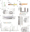
Figure 5. Fam20C exhibits broader substrate specificity than previously appreciated
(A) Weblogo depicting the phosphopeptides that displayed a significant reduction in Fam20C KO HepG2 cells. The number (n) of phosphopeptides analyzed to generate the Weblogo is shown in the insert. (B) Schematic representation of IGFBP1 depicting residues (red) whose phosphorylation decreased in Fam20C KO HepG2 cells (SP; signal peptide, TY, Thyroglobulin-1). (C) Control and Fam20C KO cells were metabolically labeled with 32P-orthophosphate and transfected with V5-tagged IGFBP1. Protein immunoblotting of V5 immunoprecipitates from conditioned medium (CM) (upper) and autoradiography depicting 32P incorporation into the V5-tagged IGFBP1 (middle). Fam20C protein levels in CM are also shown (lower). (D) Protein immunoblotting of V5 immunoprecipitates from CM of U2OS cells co-expressing V5-tagged IGFBP1 with either WT (20C:Flag) or catalytically inactive D478A (20C DA:Flag). V5-immunoprecipitates were treated with λ-phosphatase (λ-p’tase). (E) Time dependent incorporation of 32P from [γ-32P]ATP into IGFBP1 by recombinant Fam20C or the D478A mutant. Reaction products were separated by SDS-PAGE, visualized by Coomassie staining, and radioactivity was detected by autoradiography. (F) Representative MS/MS spectrum of a diphosphopeptide identifying phosphorylation of Thr193 and Ser194 on human IGFBP1 after Fam20C treatment. The precursor ion with m/z 669.74 eluted at 5.32 min. (G) Schematic representation of PRP1 depicting the two phosphoserines (site 8; green and site 22; red). (H) Recombinant Fam20C was incubated with peptides representing the PRP1 site 8 and 22 Ser residues in the presence of [γ-32P]ATP and incorporated radioactivity was quantified by means of scintillation counting. The β28–40 peptide was also analyzed. (I) A peptide representing the PRP1 site 22 Ser was synthesized and used to further assess the specificity determinants for Fam20C. Incorporation of phosphate was monitored by autoradiography. (J) Control and Fam20C KO cells were metabolically labeled with 32P-orthophosphate and transfected with V5-tagged PRP1. Protein immunoblotting of V5 immunoprecipitates from CM (upper) and autoradiography depicting 32P incorporation into PRP1 (middle). Fam20C protein levels in CM are also shown (lower). (K) Protein immunoblotting and autoradiography of V5-immunoprecipitates from CM of 32P-orthophosphate labeled U2OS cells co-expressing V5-tagged PRP1 or the site 8 and 22 Ala mutants with either WT (20C:Flag) or Fam20C-D478A (20C DA:Flag). See also Figure S6
Figure 6
Figure 6. GO term analysis of Fam20C substrates reveals an unexpected role for secreted protein phosphorylation in cell adhesion, migration and invasion
(A) DAVID GO term analysis using the HepG2 Fam20C-regulated gene set. The graph represents the statistically significant enriched gene clusters with the number of genes in each cluster indicated beside the bars. (B) Bar graph depicting cell detachment in a cell adhesion assay using WT and Fam20C KO MDA-MB-231 cells. Data report detached cells (% of total) after 10 min treatment with PBS-buffered EDTA and represent the mean (+/− SD) of 10 replicates in two independent experiments (*** p ≤ 0.0001 vs WT). (C) Bar graph depicting cell migration (area of wound covered) in a scratch wound-healing assay using WT and Fam20C KO MDA-MB-231 cells. Data was acquired 7 and 20 hours after the scratch and represents the mean (+/− SD) of three independent experiments (*** p ≤ 0.0001 vs WT). (D) Bar graph depicting cell migration/chemotaxis in a trans-well migration assay using WT and Fam20C KO MDA-MB-231 cells. Medium with 10% FBS was used as a chemoattractant. Data was acquired 24 hours after seeding in upper chamber of 8 μm pore size trans-wells and is expressed as average number of cells/field (+/− SD) from 3 independent experiments (* p<0.05 vs WT). (E) Bar graph depicting cell migration/invasion in a Matrigel trans-well migration assay using control and Fam20C KO MDA-MB-231 cells. Data was acquired 24 hours after seeding in upper chamber of 8 μm pore size trans-wells. Cells that invaded the Matrigel were quantified based on DNA content using CyQuant dye and normalized to WT. Data represent mean (+/− SD) from 3 independent experiments (*** p ≤ 0.0001 vs WT). (F) Bar graph depicting cell migration (area of wound covered) in a scratch wound-healing assay using WT and Fam20C KO MDA-MB-231 cells. Cells were incubated in SFM or conditioned medium (CM) from Fam20C WT or KO MDA-MB-231 cells as indicated during wound-healing. Data was acquired 6 hours after the scratch and depicts mean (+/− SD) from a representative experiment performed in triplicate (***p = 0.0002 vs WT, **p = 0.0025 vs KO). (G) Workflow used to screen candidate Fam20C substrates in the scratch-wound healing assay shown in Figure 6H. (H) Bar graph depicting cell migration (area of wound covered) in a scratch wound-healing assay using WT and Fam20C KO MDA-MB-231 cells. Cells were incubated in SFM or CM from U2OS cells that were transfected with either vector (Control), IGFBP7-V5 + WT Fam20C (pIGFBP7), or IGFBP7-V5 + D478A Fam20C (IGFBP7) as indicated during the wound-healing. Data was acquired 6 hours after the scratch and shows mean (+/− SD) of a representative experiment performed in triplicate (*p = 0.0146 vs IGFBP7, **p < 0.0001, ***p = 0.0098 vs KO). An immunoblot of V5-immunoprecipitates from the CM of U2OS cells used in the rescue experiments is shown in the insert. See also Figure S7

Comment in

Similar articles

Cited by

References

    1. Aziz A, Harrop SP, Bishop NE. Characterization of the deleted in autism 1 protein family: implications for studying cognitive disorders. PLoS One. 2011a;6:e14547. - PMC - PubMed
    1. Aziz A, Harrop SP, Bishop NE. DIA1R is an X-linked gene related to Deleted In Autism-1. PLoS One. 2011b;6:e14534. - PMC - PubMed
    1. Bahl JM, Jensen SS, Larsen MR, Heegaard NH. Characterization of the human cerebrospinal fluid phosphoproteome by titanium dioxide affinity chromatography and mass spectrometry. Anal Chem. 2008;80:6308–6316. - PubMed
    1. Bakrania P, Efthymiou M, Klein JC, Salt A, Bunyan DJ, Wyatt A, Ponting CP, Martin A, Williams S, Lindley V, et al. Mutations in BMP4 cause eye, brain, and digit developmental anomalies: overlap between the BMP4 and hedgehog signaling pathways. Am J Hum Genet. 2008;82:304–319. - PMC - PubMed
    1. Baxter RC. IGF binding proteins in cancer: mechanistic and clinical insights. Nat Rev Cancer. 2014;14:329–341. - PubMed

Publication types