. 2015 Jun 23;43(11):5465-75.
doi: 10.1093/nar/gkv434. Epub 2015 May 6.

CDK-mediated RNF4 phosphorylation regulates homologous recombination in S-phase

Affiliations

CDK-mediated RNF4 phosphorylation regulates homologous recombination in S-phase

Kuntian Luo et al. Nucleic Acids Res. .

Abstract

There are the two major pathways responsible for the repair of DNA double-strand breaks (DSBs): non-homologous end-joining (NHEJ) and homologous recombination (HR). NHEJ operates throughout the cell-cycle, while HR is primarily active in the S/G2 phases suggesting that there are cell cycle-specific mechanisms that regulate the balance between NHEJ and HR. Here we reported that CDK2 could phosphorylate RNF4 on T26 and T112 and enhance RNF4 E3 ligase activity, which is important for MDC1 degradation and proper HR repair during S phase. Mutation of the RNF4 phosphorylation sites results in MDC1 stabilization, which in turn compromised HR during S-phase. These results suggest that in addition to drive cell cycle progression, CDK also targets RNF4, which is involved in the regulatory network of DSBs repair.


Figures

Figure 1.
Figure 1.
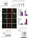
DSB induced degradation of MDC1 happens more promptly in S-phase. (A) HEK 293T cells were synchronized in G1 phase by double thymidine block or released into S phase. Then the cells were treated with or without irradiation (5 Gy) and harvested at indicated time points. Left panel: the level of endogenous MDC1 was examined with indicated antibodies by immunoblotting. Blots were quantified using ImageJ (National Institutes of Health). Right panel: the cell cycle profiles of the cells are analyzed by flow cytometry. (B) U2OS cells were synchronized in G1 phase by double thymidine block or released into S phase. Then the cells were treated with or without irradiation (2 Gy) and probed with indicated antibodies at the indicated time point. Representative images are shown in the left panels, the quantification of the percentage of cells displaying MDC1 (upper) or Rad51 (lower) foci are shown in right panels. Error bars represent SEM. from three independent experiments. **P < 0.01 two‐tailed Student's t‐test. >200 cells were counted per experiment. (C) HEK 293T cells transfected with HA-MDC1 and FLAG-SUMO1 were synchronized in G1 phase by double thymidine block or released into S phase. Then the cells were treated with or without irradiation (5 Gy) and harvested at indicated time points. MDC1 was then immunoprecipitated with anti-HA antibody and MDC1 sumoylation was examined using anti-FLAG antibody. Blots were quantified using ImageJ (National Institutes of Health). (D) HEK 293T cells transfected with Myc-RNF4 were synchronized by double thymidine block or released into S phase. The cells were irradiated (5 Gy) and then collected at indicated time points. RNF4 was then immunoprecipitated and subjected to in vitro ubiquitination reactions in the presence of HA-Ub, UbcH5a and UBE1. The ubiquitination of RNF4 were analyzed by immunoblot as indicated.
Figure 2.
Figure 2.
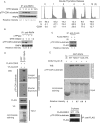
CDK2 phosphorylates RNF4 on T26 and T112 during S-phase. (A) HEK 293T cells were synchronized by double thymidine block and then released. Cells were collected at indicated time points. Left panel: RNF4 was then immunoprecipitated with anti-RNF4 antibody and the phosphorylation of RNF4 by CDKs were examined with indicated antibodies. Blots were quantified using ImageJ (National Institutes of Health). Right panel: the cell cycle profiles of the cells are analyzed by flow cytometry. (B) HEK 293T cells were synchronized by double thymidine block and then released. Cells were collected at indicated time points. RNF4 was then immunoprecipitated with anti-RNF4 antibody and treated with or without λ phosphatase. The phosphorylation of RNF4 by CDKs was examined with indicated antibodies. Blots were quantified using ImageJ (National Institutes of Health). (C) HEK 293T cells transfected with FLAG-RNF4 were synchronized by double thymidine block and then released into S phase. Then the cells were treated with or without irradiation (5 Gy). The cells were collected and the cell lysate were subjected to immunoprecipitated with anti-FLAG antibody. The phosphorylation of RNF4 by CDKs was examined with indicated antibodies. Blots were quantified using ImageJ (National Institutes of Health). (D) HEK 293T cells were transfected with FLAG-CDK2 and V5-CyclinE. 48 hr later, the cells were collected and the cell lysate were subjected to immunoprecipitated with anti-FLAG antibody. The protein conjugated to the beads were eluted with FLAG peptides and subjected to in vitro kinase assay. GST and GST-RNF4 expressed in bacterial were purified and subjected to kinase assay as substrates. The phosphorylation of RNF4 by CDK2 was examined with indicated antibodies. (E) GST and GST-RNF4 WT or mutants expressed in bacterial were purified and subjected to in vitro kinase assay with CDK2+Cyclin E (Millipore). The phosphorylation of RNF4 by CDK2 was examined using anti-phospho-CDKs substrate antibody. Blots were quantified using ImageJ (National Institutes of Health). (F) HEK 293T cells transfected with FLAG-RNF4 WT or 2TA mutant were synchronized by double thymidine block and then released into S phase. RNF4 was then immunoprecipitated with anti-FLAG antibody. The phosphorylation of RNF4 by CDKs was examined with indicated antibodies.
Figure 3.
Figure 3.
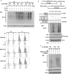
The phosphorylation on RNF4 affects its activity. (A) Upper panel: HEK 293T cells transfected with FLAG-RNF4 WT, 2TA or 2TE mutant were synchronized in G1 phase by double thymidine block or released into S phase. The cells were irradiated (5 Gy) and then collected at indicated time points. RNF4 was then immunoprecipitated and subjected to in vitro ubiquitination reactions in the presence of HA-Ub, UbcH5a and UBE1. The ubiquitination of RNF4 were analyzed by immunoblot as indicated. Lower panel: the cell cycle profiles of the cells were analyzed by flow cytometry. (B) Upper panel: schematic of the combined in vitro sumoylation and ubiquitination assay procedures. Lower panel: bacterially expressed GST‐MDC1 (aa 1818–2089) bound to GST‐sepharose were subjected to combined in vitro sumoylation and ubiquitination reactions as shown in the upper panel. The GST‐MDC1(aa 1818–2089) was subjected to in vitro sumoylation reactions in the presence of SUMO1 and then subjected to in vitro ubiquitination reactions in the presence or absence of HA‐Ub, RNF4‐WT, RNF4‐2TA, or RNF4-2TE mutant as indicated. The ubiquitination of GST‐sepharose‐bound protein were analyzed by immunoblot as indicated. (C) Cells depleted RNF4 with siRNA were cotransfected with HA-MDC1 and FLAG-RNF4-WT, 2TA or 2TE mutant. The cells were synchronized in G1 phase by double thymidine block or released into S phase and then irradiated (5 Gy). The ubiquitination of HA-MDC1 was then analyzed with indicated antibodies.
Figure 4.
Figure 4.
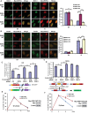
The phosphorylation of RNF4 by CDK2 regulates the HR pathway through targeting sumoylated MDC1. (A and B) U2OS cells transfected with Myc-RNF4 WT, 2TA or 2TE mutant were synchronized in G1 phase by double thymidine block or released into S phase. Then the cells were treated with irradiation (2 Gy) and probed with indicated antibodies 4 h later. Representative images are shown in the left panels, the quantification of the percentage of cells displaying MDC1 (A) or Rad51 (B) foci are shown in right panels. Error bars represent SEM. from three independent experiments. **P < 0.01 two‐tailed Student's t‐test. >200 cells were counted per experiment. (C and D) Cells depleted RNF4 with siRNA were reconstituted with FLAG-RNF4-WT, 2TA or 2TE mutant. Then the cells were subjected to HR (C) or NHEJ (D) assay as described in method. Error bars represent SEM. from three independent experiments. **P < 0.01 two‐tailed Student's t‐test. Lower panels: schematic of the reporters used in the assay. (E and F) U2OS cells transfected with Myc-RNF4 WT or 2TA mutant were synchronized in G1 phase by double thymidine block or released into S phase. Then the cells were treated with or without irradiation (2 Gy) and probed with anti-γH2AX, anti-Myc antibodies at indicated time point. The quantifications of the percentage of cells displaying γH2AX foci in G1 phase (E) or S phase (F) are shown. >200 cells were counted per experiment. Expression of Myc-RNF4 WT and 2TA mutant were examined using indicated antibodies (E and F, right panels).

Similar articles

Cited by

References

    1. Zhou B.B., Elledge S.J. The DNA damage response: putting checkpoints in perspective. Nature. 2000;408:433–439. - PubMed
    1. Lukas J., Lukas C., Bartek J. More than just a focus: The chromatin response to DNA damage and its role in genome integrity maintenance. Nat. Cell Biol. 2011;13:1161–1169. - PubMed
    1. Huen M.S., Chen J. Assembly of checkpoint and repair machineries at DNA damage sites. Trends Biochem. Sci. 2010;35:101–108. - PMC - PubMed
    1. Luo K., Zhang H., Wang L., Yuan J., Lou Z. Sumoylation of MDC1 is important for proper DNA damage response. EMBO J. 2012;31:3008–3019. - PMC - PubMed
    1. Warmerdam D.O., Kanaar R. Dealing with DNA damage: relationships between checkpoint and repair pathways. Mutat. Res. 2010;704:2–11. - PubMed

Publication types