. 2015 Apr 9;11(4):e1004822.
doi: 10.1371/journal.ppat.1004822. eCollection 2015 Apr.

The EBNA3 family of Epstein-Barr virus nuclear proteins associates with the USP46/USP12 deubiquitination complexes to regulate lymphoblastoid cell line growth

Affiliations

The EBNA3 family of Epstein-Barr virus nuclear proteins associates with the USP46/USP12 deubiquitination complexes to regulate lymphoblastoid cell line growth

Makoto Ohashi et al. PLoS Pathog. .

Abstract

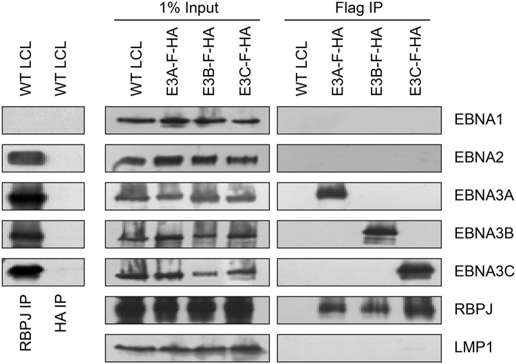
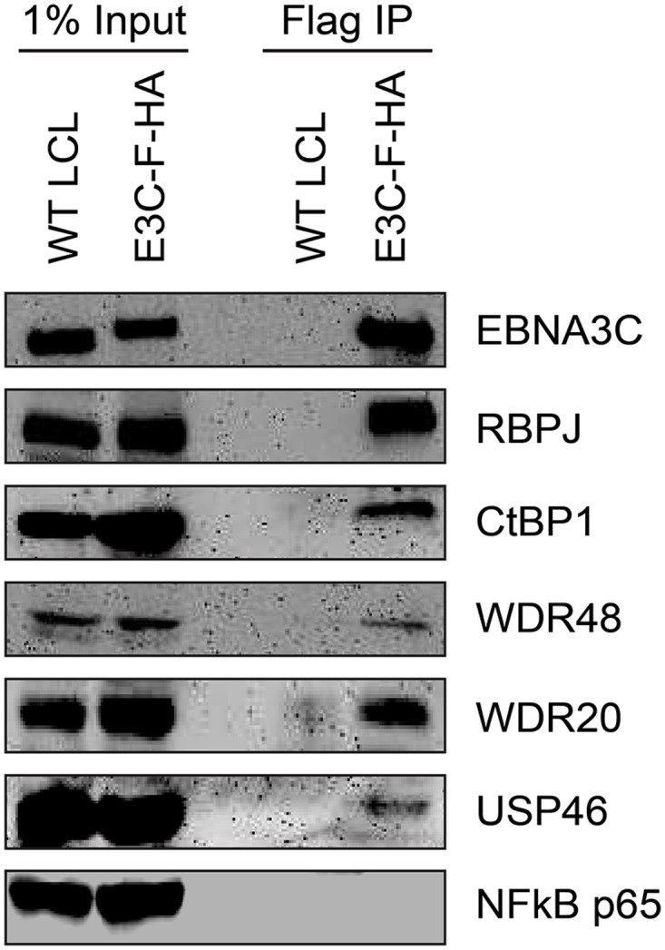
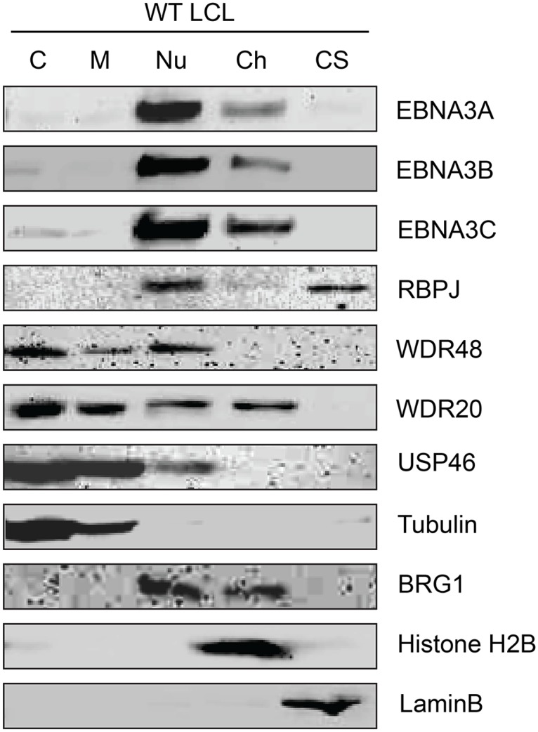
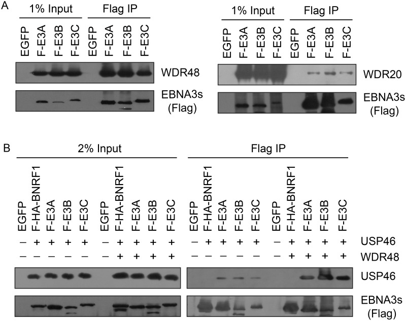
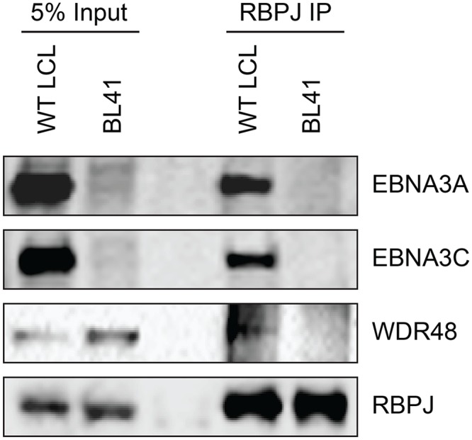
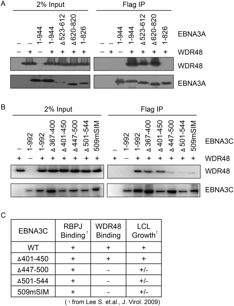
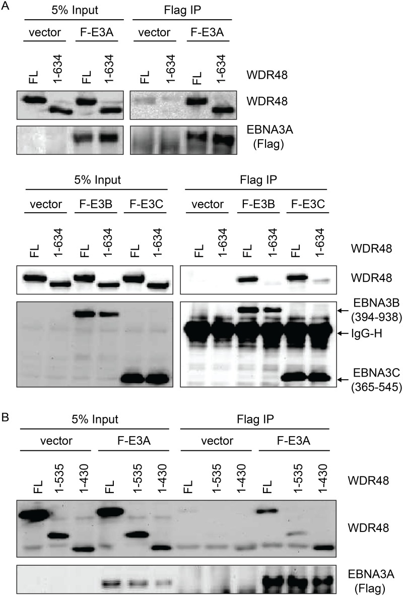
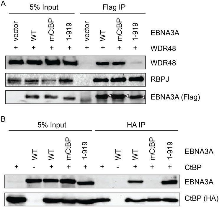
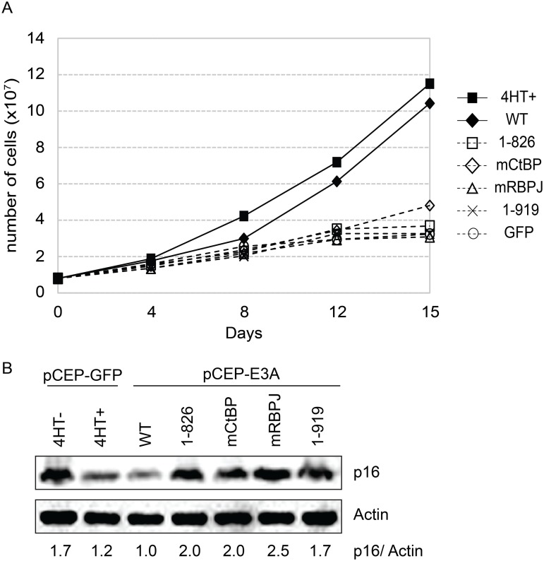
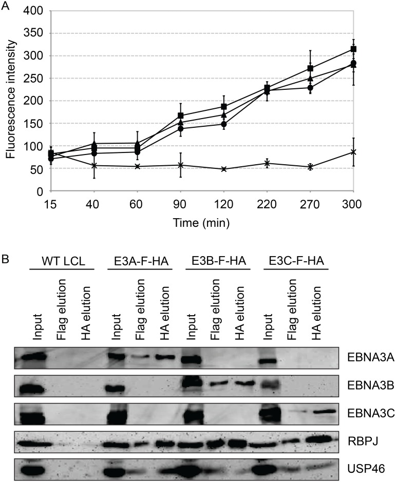
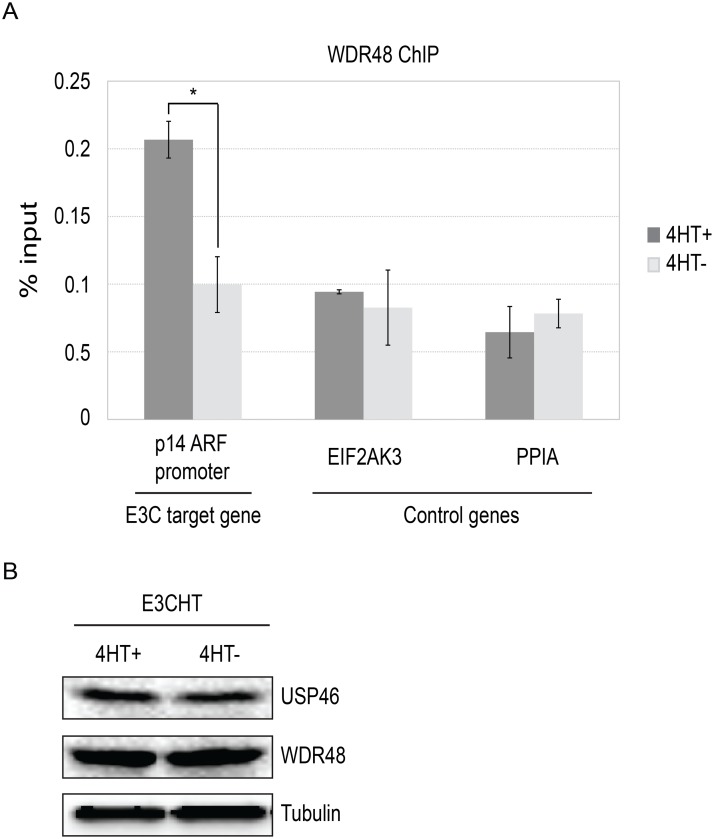
The Epstein-Barr virus (EBV) nuclear proteins EBNA3A, EBNA3B, and EBNA3C interact with the cell DNA binding protein RBPJ and regulate cell and viral genes. Repression of the CDKN2A tumor suppressor gene products p16(INK4A) and p14(ARF) by EBNA3A and EBNA3C is critical for EBV mediated transformation of resting B lymphocytes into immortalized lymphoblastoid cell lines (LCLs). To define the composition of endogenous EBNA3 protein complexes, we generated lymphoblastoid cell lines (LCLs) expressing flag-HA tagged EBNA3A, EBNA3B, or EBNA3C and used tandem affinity purification to isolate each EBNA3 complex. Our results demonstrated that each EBNA3 protein forms a distinct complex with RBPJ. Mass-spectrometry revealed that the EBNA3A and EBNA3B complexes also contained the deubquitylation complex consisting of WDR48, WDR20, and USP46 (or its paralog USP12) and that EBNA3C complexes contained WDR48. Immunoprecipitation confirmed that EBNA3A, EBNA3B, and EBNA3C association with the USP46 complex. Using chromatin immunoprecipitation, we demonstrate that WDR48 and USP46 are recruited to the p14(ARF) promoter in an EBNA3C dependent manner. Mapping studies were consistent with WDR48 being the primary mediator of EBNA3 association with the DUB complex. By ChIP assay, WDR48 was recruited to the p14(ARF) promoter in an EBNA3C dependent manner. Importantly, WDR48 associated with EBNA3A and EBNA3C domains that are critical for LCL growth, suggesting a role for USP46/USP12 in EBV induced growth transformation.


Conflict of interest statement

The authors have declared that no competing interests exist.

Figures

Fig 1
Fig 1. Characterization of EBNA3-F-HA LCLs, reveals that EBNA2, EBNA3A, EBNA3B, and EBNA3C form distinct RBPJ complexes.
Left panel: immunoprecipitation assay for RBPJ demonstrates it is in a complex with EBNA2, EBNA3A, EBNA3B, and EBNA3C. HA immunoprecipitation serves as negative control. Right panel: Selective immunoprecipitation of EBNA3A, EBNA3B, or EBNA3C complexes from EBNA3-F-HA LCLs or WT LCL using Flag agarose. Precipitated complexes were resolved by SDS-PAGE and probed for the indicated EBV latent proteins or RBPJ. For each LCL, one percent of the input lysate is shown for comparison.
Fig 2
Fig 2. EBNA3C associates with the WDR48/USP46 complex in EBNA3C-F-HA LCLs.
Immunoprecipitation assay using Flag agarose to retrieve protein complexes from EBNA3C-F-HA LCLs (E3C-F-HA) is compared to flag immunoprecipiates from untagged wildtype (WT) LCLs. One percent of total cell lysate (Input) or immunoprepicitated specimens using Flag agarose (Flag IP) were separated by SDS PAGE and probed using antibodies to EBNA3C, RBPJ, CtBP1, WDR48, WDR20, USP46, or NF-kB p65.
Fig 3
Fig 3. Subcellular localization of USP46 complexes in LCLs.
Wild type LCLs were extracted into five subcellular fractions [cytoplasm (C), membrane (M), soluble nuclear (Nu), chromatin (Ch), cytoskeleton (CS)], resolved by SDS-PAGE and probed for EBNA3 proteins, RBPJ, and components of the USP46 complex (USP46, WDR20, and WDR48). Fraction purity was assessed by probing for tubulin, BRG1, Histone H2B, and LaminB.
Fig 4
Fig 4. EBNA3 proteins preferentially bind the WDR48 subunit of the USP46 DUB complex.
(A) Immunoprecipitation assay in 293T cells demonstrating association of flag tagged EBNA3 proteins (F-E3A, F-E3B, and F-E3C) with WDR48 (left) and WDR20 (right). (B) Immunoprecipitation assay demonstrating WDR48 cotransfection enhanced USP46 association with EBNA3s (right panel, compare lanes 3–5 with 8–10). Epitope tagged BNRF1 (F-HA-BNRF1), an EBV protein of approximately the same size as the EBNA3 is included as an additional negative control. One percent (panel A) or two percent (panel B) of the input are shown for comparison.
Fig 5
Fig 5. WDR48 coimmunoprecipitates with RBPJ in EBV infected cells.
Co-immunoprecipitation assays comparing the association of RBPJ with WDR48 in LCLs with that observed in EBV negative BL41 cells. Cell lysates were immunoprecipitated with polyclonal RBPJ sera, separated by SDS PAGE, and probed for EBNA3A, EBNA3C, WDR48, and RBPJ (as indicated).
Fig 6
Fig 6. Identification of EBNA3A and EBNA3C domains that mediate WDR48 association.
Immunoprecipitation assays to map WDR48 binding regions within EBNA3A (A) and EBNA3C (B). 293T cells were co-transfected with Xpress tagged WDR48 and flag tagged full length EBNA3A, EBNA3C, or the indicated EBNA3A or EBNA3C deletion mutants. Cell lysates were immunoprecipitated with Flag agarose, separated by SDS PAGE, and probed for WDR48 (anti-Xpress) and EBNA3 proteins (anti-Flag). (C) Comparison of WDR48 binding results (from B) with previously published RBPJ binding results and LCL growth phenotype for each EBNA3C mutant [21].
Fig 7
Fig 7. WDR48 SLD2 mediates binding to EBNA3B and EBNA3C, but is not required for EBNA3A binding.
Immunoprecipitation assays were performed to assess effect of deleting the WDR48 SUMO-like domains (SLDs) on EBNA3 binding. (A) 293T cells were co-transfected with vector control, Xpress tagged full length WDR48 (FL) or a deletion mutant lacking the SLD2 domain (WDR48 1–634) and flag tagged EBNA3A 1–944 (F-E3A), EBNA3B 394–938 (F-E3B) or EBNA3C 365–545 (F-E3C). Cell lysates were immunoprecipitated with Flag agarose, separated by SDS PAGE, and probed with WDR48 or Flag antibody. (B) Immunoprecipitation assay to determine effect of deleting SLD1/2 on WDR48 binding to EBNA3A. Assays were performed as describe above with co-transfection of EBNA3A WT and either full length WDR48 (FL), WDR48 1–535, or WDR48 1–430 (which lacks both SLD1 and SLD2).
Fig 8
Fig 8. Deletion of EBNA3A residues 920–944 disrupts WDR48 binding without affecting CtBP1 association.
Co-immunoprecipitation assay to assess binding of EBNA3A mutants to WDR48 (A) and CtBP1 (B). For these assays, flag tagged full length EBNA3A (1–944), an EBNA3A CtBP1 binding mutant (mCtBP), an EBNA3A mutant lacking the C-terminal 25 residues (1–919), or vector control (pSG5) was co-transfected with Xpress-WDR48 or HA-CtBP1. Lysates were immunoprecipitated with Flag agarose (A) or HA agarose (B), separated by SDS PAGE, and probed with WDR48, RBPJ, flag, EBNA3A and HA antibodies.
Fig 9
Fig 9. EBNA3A1-919, which associates with CtBP1 but not WDR48, is impaired for LCL growth maintenance.
(A) Transcomplementation assay comparing growth of EBNA3A-HT cells transfected with EBNA3A WT (closed diamond), EBNA3A 1–826 (open square), EBNA3A mCtBP1 (open diamond), EBNA3A mRBPJ (open triangle), or EBNA3A 1–919 (X) in the absence of 4HT. EBNA3A-HT cells were also transfected with a control GFP expression plasmid, split, and maintained in either the presence (closed square) or absence (open circle) of 4HT. Cells were counted every 3 to 4 days, and diluted in fresh media to maintain a concentration of 200,000 cells/mL. Based on dilution factors, total cell number was calculated and is plotted on the Y-axis versus time. (B) Wild type EBNA3A, not mutant EBNA3A, suppresses p16 expression level in trans-complemented cells. After 15 days of EBNA3A WT or EBNA3A mutant (1–826, mCtBP1, mRBPJ, or 1–919) transfection, cells were harvested and protein expression was detected by immunoblotting with p16 antibody and actin as an internal control. As a control experiment, GFP expression plasmid was transfected into the cells and cultured with or without 4HT for 15 days. The ration of the p16 and actin bands was quantified and is indicated in the bottom panel.
Fig 10
Fig 10. Purified EBNA3 complexes exhibit DUB activity.
(A) Tandem affinity purification was performed on wild type or EBNA3-F-HA expressing LCLs. Purified complexes from wild type (X), E3A-F-HA (closed circle), E3B-F-HA (closed triangle), or E3C-F-HA (closed square) LCLs were assayed for DUB activity by fluorometric assay using Ub-AMC as a substrate. (B) Western blot of purified EBNA3 complexes used in (A) demonstrating selective precipitation of tagged EBNA3 protein complexes and co-purification of RBPJ and USP46 proteins. Lysates derived from the equivalent of approximately 1.5x10⁶ cells (input), 2.4x10⁶ cells (Flag elution), or 10x106 cells (HA elution) were loaded on each lane, separated by SDS PAGE, and probed with EBNA3s, Flag, EBNA3A, and HA antibodies.
Fig 11
Fig 11. Inability to derive USP46 null LCLs using CRISPR/ Cas9 mediated gene editing.
(A) Western blot for USP46 in 721 LCLs cells transfected with a plasmid expressing either of two guide RNAs targeting different USP46 exons (CRISPR 1 or CRISPR 2) and a hygromycin resistance gene. Prior to harvesting, cells were subjected to hygromycin selection for one month (resulting in 6 clones for CRISPR 1 and 8 clones CRISPR 2). Untransfected 721 cells are also shown (WT). As a loading control, lysates were probed for beta actin (bottom panel). (B) Western blots of 293T cells that were transfected same CRISPR plasmids as in panel A and also subjected to one month of hygromycin selection. Hygromycin resistance cells were harvested and blotted for USP46 (specific band is indicated by arrowhead). Untransfected 293T cells are also shown (WT).
Fig 12
Fig 12. ChIP assay for WDR48 at the p14ARF promoter.
Chromatin immunoprecipitation (ChIP) assays were performed using antibodies for WDR48 (A) from EBNA3C-HT LCLs that were grown in the presence of 4HT (dark gray) or after 14 days of growth in the absence of 4HT (light gray). Amount of genomic DNA was measured by real time PCR using primers specific to the EBNA3C binding site in the p14ARF promoters or sites near the EIF2AK3 and PPIA genes which bind cell transcription factors but not EBNA3C. The bar graph represents the amount of DNA precipitated relative to the amount of DNA in the corresponding input sample. The experiment shown is representative of four independent experiments and error bars indicating standard error of the mean within this experiment. Asterisk denotes that the difference in ChIP signal seen at the p14ARF promoter is statistically significant (p = 0.01). (B) Western blot for USP46, WDR48, and tubulin levels in whole cell lysates from EBNA3CHT LCLs grown in the presence of 4HT or after 14 days of growth in the absence of 4HT.

Similar articles

Cited by

References

    1. Rickinson AB, Kieff E (2007) Epstein-Barr Virus. Philadelphia: Lippincott Williams and Wilkins; 2655–2700 p.
    1. Henle W, Diehl V, Kohn G, Zur Hausen H, Henle G (1967) Herpes-type virus and chromosome marker in normal leukocytes after growth with irradiated Burkitt cells. Science 157: 1064–1065. - PubMed
    1. Pope JH, Horne MK, Scott W (1968) Transformation of foetal human keukocytes in vitro by filtrates of a human leukaemic cell line containing herpes-like virus. International journal of cancer 3: 857–866. - PubMed
    1. Rooney CM, Smith CA, Ng CY, Loftin SK, Sixbey JW, et al. (1998) Infusion of cytotoxic T cells for the prevention and treatment of Epstein-Barr virus-induced lymphoma in allogeneic transplant recipients. Blood 92: 1549–1555. - PubMed
    1. Thorley-Lawson DA, Hawkins JB, Tracy SI, Shapiro M (2013) The pathogenesis of Epstein-Barr virus persistent infection. Current opinion in virology 3: 227–232. 10.1016/j.coviro.2013.04.005 - DOI - PMC - PubMed

Publication types

MeSH terms