Review
. 2015 Apr;95(2):645-90.
doi: 10.1152/physrev.00026.2014.

Transient receptor potential channels in the vasculature

Affiliations
Review

Transient receptor potential channels in the vasculature

Scott Earley et al. Physiol Rev. 2015 Apr.

Abstract

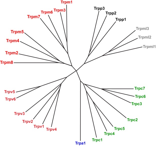
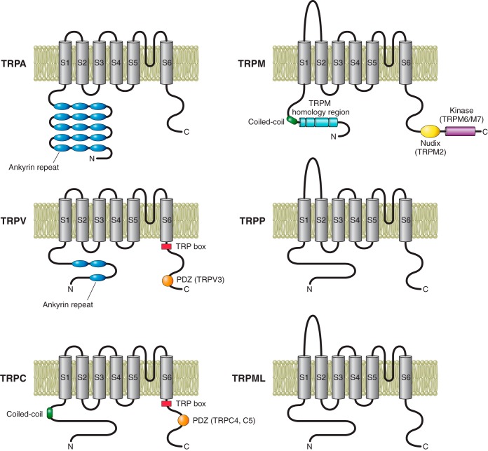
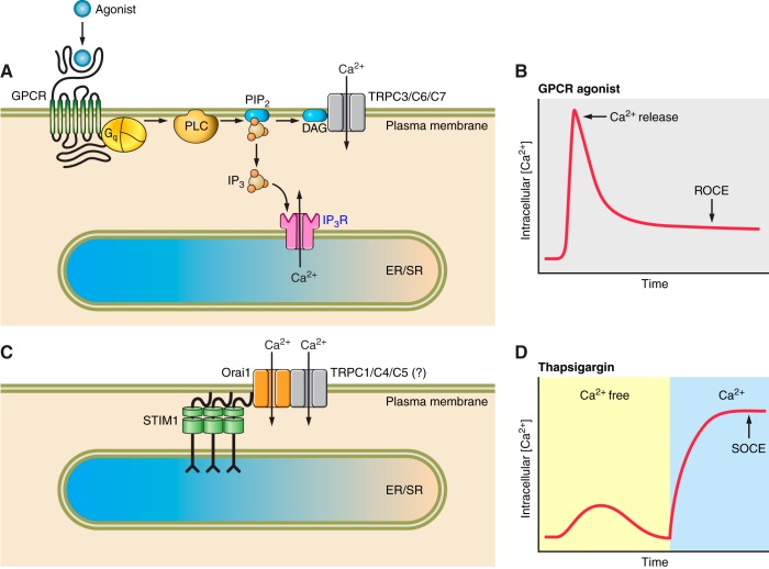
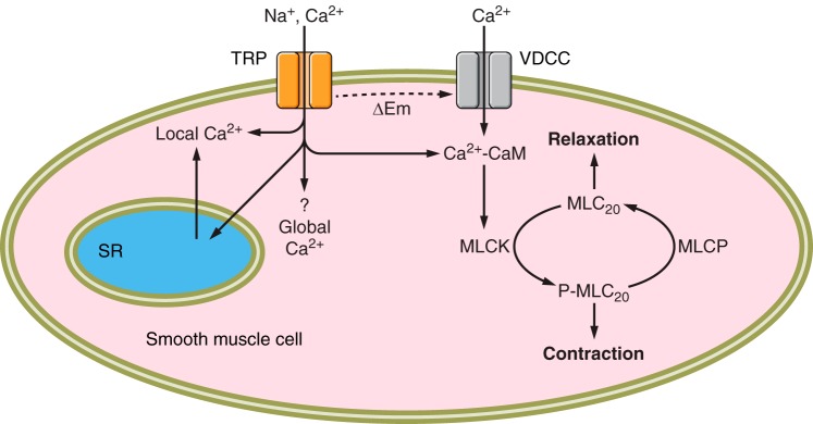
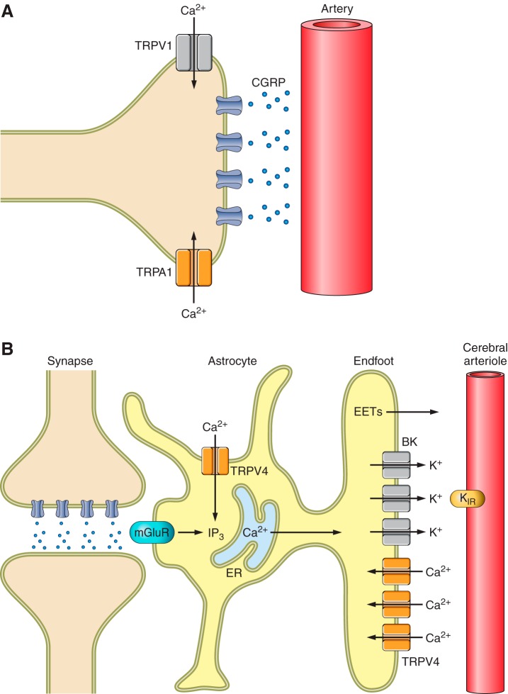
The mammalian genome encodes 28 distinct members of the transient receptor potential (TRP) superfamily of cation channels, which exhibit varying degrees of selectivity for different ionic species. Multiple TRP channels are present in all cells and are involved in diverse aspects of cellular function, including sensory perception and signal transduction. Notably, TRP channels are involved in regulating vascular function and pathophysiology, the focus of this review. TRP channels in vascular smooth muscle cells participate in regulating contractility and proliferation, whereas endothelial TRP channel activity is an important contributor to endothelium-dependent vasodilation, vascular wall permeability, and angiogenesis. TRP channels are also present in perivascular sensory neurons and astrocytic endfeet proximal to cerebral arterioles, where they participate in the regulation of vascular tone. Almost all of these functions are mediated by changes in global intracellular Ca(2+) levels or subcellular Ca(2+) signaling events. In addition to directly mediating Ca(2+) entry, TRP channels influence intracellular Ca(2+) dynamics through membrane depolarization associated with the influx of cations or through receptor- or store-operated mechanisms. Dysregulation of TRP channels is associated with vascular-related pathologies, including hypertension, neointimal injury, ischemia-reperfusion injury, pulmonary edema, and neurogenic inflammation. In this review, we briefly consider general aspects of TRP channel biology and provide an in-depth discussion of the functions of TRP channels in vascular smooth muscle cells, endothelial cells, and perivascular cells under normal and pathophysiological conditions.


Figures

Figure 1.
Figure 1.
Phylogenetic tree of the mouse TRP superfamily.
Figure 2.
Figure 2.
Membrane topology and functional domains of TRP subunits.
Figure 3.
Figure 3.
TRPV4 sparklets in endothelial cells and vascular smooth muscle cells. A: time-lapse image of a typical TRPV4 sparklet recorded from a primary cerebral artery endothelial cell using TIRF microscopy. The cell was stimulated with the selective TRPV4 agonist GSK1016790A (GSK; 100 nM). B: TRPV4 sparklets recorded from a tsA-201 cell transfected with TRPV4 (top) and from a native cerebral artery myocyte (bottom). The middle and right panels demonstrate the single channel-like behavior of these events. TRPV4 sparklets were stimulated with GSK. C: TRPV4 sparklets recorded from the intact endothelium of cerebral arteries from mice expressing the Ca2+-indicator protein GCaMP2 exclusively in the endothelium using high-speed, high-resolution confocal microscopy before and after stimulation with GSK. Experiments were performed in the presence of cyclopiazonic acid (CPA) to eliminate interference from ER Ca2+-release events. Representative traces indicating single channel-like events are shown below. [A from Sullivan and Earley (336). B from Mercado et al. (221). C from Sonkusare et al. (323), with permission from American Association for the Advancement of Science.]
Figure 4.
Figure 4.
Ca2+-influx mechanisms mediated by TRP channels. A: receptor-operated Ca2+ entry (ROCE). Agonist binding to GPCRs stimulates the activity of PLC, which cleaves the membrane phospholipid PIP2 into IP3 and DAG. IP3 binds to IP3Rs on the ER/SR to cause release of Ca2+ into the cytosol. DAG directly activates Ca2+ influx through TRPC3, TRPC6, or TRPC7 channels on the plasma membrane. B: example trace of changes in intracellular Ca2+ following stimulation of a GPCR, showing the phasic increase resulting from ER/SR Ca2+ release and sustained elevation resulting from ROCE. C: store-operated Ca2+ entry (SOCE). Depletion of Ca2+ in the ER/SR causes clustering of STIM1 in the ER/SR membrane proximal to the plasma membrane. STIM1 interacts with Oria1 at the plasma membrane to promote Ca2+ influx. TRPC1, TRPC4, and/or TRPC5 channels, either independently or in association with STIM1/Oria1, may participate in SOCE. D: example of a typical SOCE experiment. Under Ca2+-free conditions, cells are treated with the SERCA inhibitor thapsigargin, causing Ca2+ to leak from the ER/SR into the cytosol. Reintroduction of Ca2+ to the bathing solution after ER/SR stores are depleted causes SOCE.
Figure 5.
Figure 5.
TRP channels contribute to vascular smooth muscle cell contractility. Cation influx through TRP channels depolarizes the plasma membrane (ΔEm), initiating Ca2+ influx through VDCCs. Elevated Ca2+ levels increase binding of calmodulin-Ca2+ (Cd-Ca2+) complexes to MLCK, stimulating phosphorylation of the regulatory myosin light chain (MLC20). MLC20 is dephosphorylated by myosin light-chain phosphatase (MLCP). Some studies suggest that Ca2+ influx through TRP channels can directly initiate myocyte contraction.
Figure 6.
Figure 6.
A force-sensitive signaling network involving TRPC6 and TRPM4 channels in cerebral artery smooth muscle cells. A stretch-sensing pathway in cerebral artery myocytes that includes 1) activation of AT1R, Src tyrosine kinase, and PLC-γ1, leading to the generation of IP3; and 2) Ca2+ influx through TRPC6 channels onto IP3Rs, promoting CICR that results in activation of TRPM4 channels, which are responsible for pressure-induced depolarization of the plasma membrane. PM, plasma membrane; ΔVm, change in membrane potential. [From Gonzales et al. (118).]
Figure 7.
Figure 7.
Ca2+ influx through TRPV4 channels relaxes cerebral artery smooth muscle cells. Stimulation of Ca2+ influx through TRPV4 channels directly with 11,12-EET or through activation of Gq-coupled AT1Rs with ANG II, leading to generation of DAG by PLC and AKAP150-dependent PKC activity, causes an increase in the frequency of Ca2+ sparks mediated by ryanodine receptors (RyRs) on the SR through a CICR mechanism. Increased generation of Ca2+ sparks causes smooth muscle cell membrane hyperpolarization and relaxation by increasing K+ efflux through BK channels. [Modified from Earley et al. (79), with permission from Wolters Kluwer Health, and Mercado et al. (221).]
Figure 8.
Figure 8.
Mechanisms of endothelium-dependent vasodilation. In endothelial cells (EC), Ca2+ influx stimulates the activity of eNOS to generate NO, which diffuses through the IEL to cause relaxation of underlying vascular smooth muscle cells (SMC). COX activity generates PGI2, which also relaxes SMC. A variety of other substances, collectively known as EDHF, can cause smooth muscle cell hyperpolarization and relaxation. K+ efflux hyperpolarizes the endothelial cell plasma membrane (ΔEm). Electrotonic spread of this influence through myoendothelial gap junctions (MEGJs) promotes EDH, causing SMC relaxation.
Figure 9.
Figure 9.
Ca2+ pulsars in the endothelium. A: time course of a three-dimensional Ca2+ pulsar originating from within a hole in the IEL (white circle) shown in the leftmost image. Scale bar, 5 μm. B: Ca2+ pulsars recorded from a pressurized artery (80 mmHg) expressing the Ca2+-indicator protein GCaMP2. An endothelial cell and its nucleus are outlined (dotted lines), with the initiation sites indicated by red arrows. Scale bar, 10 μm. C: model of the functional effects of endothelial Ca2+ pulsars. IP3R-dense ER stores follow portions of the endothelial cell membrane that evaginate through holes in the IEL and interface with underlying SM cell membranes. Repetitive localized Ca2+ events (pulsars) originate from these deep Ca2+ stores, which are regionally delimited to the myoendothelial junction and the base of the endothelial cell. These ongoing dynamic Ca2+ signals are driven by constitutive IP3 production and are inherently dependent on the level of endothelial stimulation. The left detail depicts a single endothelial projection through the IEL. IK (KCa3.1) channels in the plasma membranes of these endothelial projections are in very close proximity to Ca2+ pulsars, eliciting persistent Ca2+-dependent hyperpolarization of the membrane potential at the myoendothelial junctions. The right detail illustrates the endothelial influence on the smooth muscle membrane potential at the myoendothelial interaction site where Ca2+ pulsars activate KCa3.1 channels and hyperpolarize the endothelial membrane. This hyperpolarization can be transmitted to the SM through gap junction channels or by activation of SM Kir channels by K+ ions released by endothelial KCa3.1 channels. Membrane hyperpolarization promotes relaxation of SM through a decrease in VDCC open probability. [From Ledoux et al. (180).]
Figure 10.
Figure 10.
TRPV4 Ca2+ sparklets in endothelial cells initiate vasodilation. TRPV4 Ca2+ influx from the extracellular space through as few as 3–4 TRPV4 channels activates Ca2+-activated K+ (KCa) channels, resulting in K+ efflux from the cell. This hyperpolarizes the endothelial cell (EC) membrane, which directly hyperpolarizes smooth muscle cells (SMCs) via gap junctions located within myoendothelial junctions. IEL, internal elastic lamina; Vm, membrane potential. [From Sullivan and Earley (336).]
Figure 11.
Figure 11.
TRP channels in perivascular cells. A: Ca2+ influx through TRPV1 or TRPA1 channels in perivascular nerves can stimulate the release of CGRP to cause arterial dilation. B: TRPV4 channels contribute to neurovascular coupling. Stimulation of metabotropic glutamate receptors (mGluR) on parenchymal astrocytes leads to the generation of IP3, which causes the release of Ca2+ from the endoplasmic reticulum (ER), stimulating K+ efflux through BK channels. The resulting increase in [K+] in the space between the astrocytic endfoot and cerebral arteriole activates K+ efflux through KIR channels present on vascular smooth muscle cells to cause dilation. Production of EETs or prostaglandins (e.g., PGE2) may also contribute. Ca2+ influx through TRPV4 channels contributes to the propagation of Ca2+ waves throughout the endfoot.

Similar articles

Cited by

References

    1. Aarts M, Iihara K, Wei WL, Xiong ZG, Arundine M, Cerwinski W, MacDonald JF, Tymianski M. A key role for TRPM7 channels in anoxic neuronal death. Cell 115: 863–877, 2003. - PubMed
    1. Abdullaev IF, Bisaillon JM, Potier M, Gonzalez JC, Motiani RK, Trebak M. Stim1 and Orai1 mediate CRAC currents and store-operated calcium entry important for endothelial cell proliferation. Circ Res 103: 1289–1299, 2008. - PMC - PubMed
    1. Adebiyi A, Narayanan D, Jaggar JH. Caveolin-1 assembles type 1 inositol 1,4,5-trisphosphate receptors and canonical transient receptor potential 3 channels into a functional signaling complex in arterial smooth muscle cells. J Biol Chem 286: 4341–4348, 2011. - PMC - PubMed
    1. Adebiyi A, Thomas-Gatewood CM, Leo MD, Kidd MW, Neeb ZP, Jaggar JH. An elevation in physical coupling of type 1 inositol 1,4,5-trisphosphate (IP3) receptors to transient receptor potential 3 (TRPC3) channels constricts mesenteric arteries in genetic hypertension. Hypertension 60: 1213–1219, 2012. - PMC - PubMed
    1. Adebiyi A, Zhao G, Narayanan D, Thomas-Gatewood CM, Bannister JP, Jaggar JH. Isoform-selective physical coupling of TRPC3 channels to IP3 receptors in smooth muscle cells regulates arterial contractility. Circ Res 106: 1603–1612, 2010. - PMC - PubMed

Publication types

MeSH terms

Substances