An Improved Reverse Genetics System to Overcome Cell-Type-Dependent Ebola Virus Genome Plasticity
- PMID: 25810440
- PMCID: PMC4564527
- DOI: 10.1093/infdis/jiu681
An Improved Reverse Genetics System to Overcome Cell-Type-Dependent Ebola Virus Genome Plasticity
Abstract
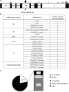
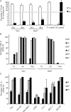
Reverse genetics systems represent a key technique for studying replication and pathogenesis of viruses, including Ebola virus (EBOV). During the rescue of recombinant EBOV from Vero cells, a high frequency of mutations was observed throughout the genomes of rescued viruses, including at the RNA editing site of the glycoprotein gene. The influence that such genomic instability could have on downstream uses of rescued virus may be detrimental, and we therefore sought to improve the rescue system. Here we report an improved EBOV rescue system with higher efficiency and genome stability, using a modified full-length EBOV clone in Huh7 cells. Moreover, by evaluating a variety of cells lines, we revealed that EBOV genome instability is cell-type dependent, a fact that has significant implications for the preparation of standard virus stocks. Thus, our improved rescue system will have an impact on both basic and translational research in the filovirus field.
Keywords: Ebola virus; RNA editing site; mutation; reverse genetics system.
Published by Oxford University Press on behalf of the Infectious Diseases Society of America 2015. This work is written by (a) US Government employee(s) and is in the public domain in the US.
Figures


Similar articles
-
Genomic RNA editing and its impact on Ebola virus adaptation during serial passages in cell culture and infection of guinea pigs.J Infect Dis. 2011 Nov;204 Suppl 3:S941-6. doi: 10.1093/infdis/jir321. J Infect Dis. 2011. PMID: 21987773
-
Generation of Recombinant Ebola Viruses Using Reverse Genetics.Methods Mol Biol. 2017;1628:177-188. doi: 10.1007/978-1-4939-7116-9_13. Methods Mol Biol. 2017. PMID: 28573619
-
Development of a New Reverse Genetics System for Ebola Virus.mSphere. 2021 May 5;6(3):e00235-21. doi: 10.1128/mSphere.00235-21. mSphere. 2021. PMID: 33952663 Free PMC article.
-
[Research progress on ebola virus glycoprotein].Bing Du Xue Bao. 2013 Mar;29(2):233-7. Bing Du Xue Bao. 2013. PMID: 23757858 Review. Chinese.
-
Ebola virus: unravelling pathogenesis to combat a deadly disease.Trends Mol Med. 2006 May;12(5):206-15. doi: 10.1016/j.molmed.2006.03.006. Epub 2006 Apr 17. Trends Mol Med. 2006. PMID: 16616875 Review.
Cited by
-
Species-Specific Evolution of Ebola Virus during Replication in Human and Bat Cells.Cell Rep. 2020 Aug 18;32(7):108028. doi: 10.1016/j.celrep.2020.108028. Cell Rep. 2020. PMID: 32814037 Free PMC article.
-
A review on the antagonist Ebola: A prophylactic approach.Biomed Pharmacother. 2017 Dec;96:1513-1526. doi: 10.1016/j.biopha.2017.11.103. Epub 2017 Dec 6. Biomed Pharmacother. 2017. PMID: 29208326 Free PMC article. Review.
-
In vivo characterization of the novel ebolavirus Bombali virus suggests a low pathogenic potential for humans.Emerg Microbes Infect. 2023 Dec;12(1):2164216. doi: 10.1080/22221751.2022.2164216. Epub 2023 Jan 18. Emerg Microbes Infect. 2023. PMID: 36580440 Free PMC article.
-
Growth-Adaptive Mutations in the Ebola Virus Makona Glycoprotein Alter Different Steps in the Virus Entry Pathway.J Virol. 2018 Sep 12;92(19):e00820-18. doi: 10.1128/JVI.00820-18. Print 2018 Oct 1. J Virol. 2018. PMID: 30021890 Free PMC article.
-
Recombinant Lloviu virus as a tool to study viral replication and host responses.PLoS Pathog. 2022 Feb 4;18(2):e1010268. doi: 10.1371/journal.ppat.1010268. eCollection 2022 Feb. PLoS Pathog. 2022. PMID: 35120176 Free PMC article.
References
-
- Feldmann H, Sanchez A, Geisbert T. Filoviridae: Marburg and Ebola viruses. In: Knipe D, Howley P, eds. Fields virology. 6th ed Philadelphia: Lippincott Williams & Wilkins, 2013:923–56.
-
- Pourrut X, Kumulungui B, Wittmann T, et al. The natural history of Ebola virus in Africa. Microbes Infect 2005; 7:1005–14. - PubMed
-
- Volchkov VE, Volchkova VA, Muhlberger E, et al. Recovery of infectious Ebola virus from complementary DNA: RNA editing of the GP gene and viral cytotoxicity. Science 2001; 291:1965–9. - PubMed
Publication types
MeSH terms
Substances
Grants and funding
LinkOut - more resources
Full Text Sources
Other Literature Sources
Medical

