. 2015 Mar 18;7(279):279ra39.
doi: 10.1126/scitranslmed.3010302.

Human skin is protected by four functionally and phenotypically discrete populations of resident and recirculating memory T cells

Affiliations

Human skin is protected by four functionally and phenotypically discrete populations of resident and recirculating memory T cells

Rei Watanabe et al. Sci Transl Med. .

Abstract

The skin of an adult human contains about 20 billion memory T cells. Epithelial barrier tissues are infiltrated by a combination of resident and recirculating T cells in mice, but the relative proportions and functional activities of resident versus recirculating T cells have not been evaluated in human skin. We discriminated resident from recirculating T cells in human-engrafted mice and lymphoma patients using alemtuzumab, a medication that depletes recirculating T cells from skin, and then analyzed these T cell populations in healthy human skin. All nonrecirculating resident memory T cells (TRM) expressed CD69, but most were CD4(+), CD103(-), and located in the dermis, in contrast to studies in mice. Both CD4(+) and CD8(+) CD103(+) TRM were enriched in the epidermis, had potent effector functions, and had a limited proliferative capacity compared to CD103(-) TRM. TRM of both types had more potent effector functions than recirculating T cells. We observed two distinct populations of recirculating T cells, CCR7(+)/L-selectin(+) central memory T cells (TCM) and CCR7(+)/L-selectin(-) T cells, which we term migratory memory T cells (TMM). Circulating skin-tropic TMM were intermediate in cytokine production between TCM and effector memory T cells. In patients with cutaneous T cell lymphoma, malignant TCM and TMM induced distinct inflammatory skin lesions, and TMM were depleted more slowly from skin after alemtuzumab, suggesting that TMM may recirculate more slowly. In summary, human skin is protected by four functionally distinct populations of T cells, two resident and two recirculating, with differing territories of migration and distinct functional activities.


Conflict of interest statement

Competing interests: The authors declare that they have no competing interests.

Figures

Figure 1
Figure 1. Skin T cells in a human engrafted mouse model recapitulate T cell populations in adult human skin
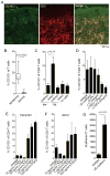
(A) A human engrafted mouse model was used to discriminate resident from recirculating T cells in skin. NSG mice were grafted with human foreskin and injected i.v. with allogeneic human PBMC, and alemtuzumab (αCD52) was used to deplete recirculating human T cells from grafted human skin. (B–E) human foreskins contained resident APC but few T cells. (B) Flow cytometry staining of collagenase digested foreskin demonstrated both CD11c+ DC and CD1a+ LC were present in foreskin. (C) Approximately four-fold more DC than T cells were present in foreskin, as assayed by the % viable cells of each cell type in collagenase digested foreskin. (D) T cells were 45-fold more frequent in healthy adult human skin than in foreskins; the percentage of CD3+ T cells in collagenase digested foreskin versus adult skin is shown. (E) The few T cells present in human foreskin lacked markers characteristic of TRM. The percent of T cells expressing each marker in collagenase treated foreskin and adult skin are shown. The mean and SEM of four foreskins (C–E) and seven adult skin donors (D,E) are shown (test: T-test). (F–H) allogeneic PBMC injected i.v. into grafted mice migrated specifically into the human skin graft and induced an inflammatory dermatitis. Images are representative of eight PBMC injected and eight saline injected mice. (F) H&E stained section demonstrating the junction between mouse and human skin in grafted mice. (G) H&E stain, 20X view of inflammatory dermatitis. (H) A cryosections of human grafted skin immunostained for CD3 (red) and keratin (green) are shown. T cells were evident in both the dermis and the epidermis. The dermoepidermal junction is indicated by a white line. (I) in the absence of infused allogeneic PBMC, no inflammatory infiltrate was observed and few or no T cells were visualized in skin. (J) CD69 up-regulation preceded CD103 up-regulation in T cells migrating into human skin grafts. Human skin grafts were harvested at the indicated time points, collagenase digested, and T cells were analyzed for CD69 and CD103 expression by flow cytometry. The mean and SEM of two grafted mice per time point are shown (test: T-test). (K,L) T cell populations in grafted skin recapitulate those observed in adult human skin. T cell expression of TCM (CCR7/L-selectin) and TRM (CD69/CD103) markers as assayed by flow cytometry are shown. T cells were isolated by collagenase digestion from healthy adult human skin or from skin grafts on PBMC infused mice. Representative histograms (K) and mean and SEM of expression of CD69 and CD103 are shown. The mean and SEM of three grafts and seven healthy skin donors are shown (test: T-test).
Figure 2
Figure 2. Alemtuzumab depletes recicrulating TCM from skin in human engrafted mice but spares a population of skin T cells
(A, B) Shown are human T cells isolated from peripheral blood (Blood) and collagenase digested foreskin grafts (Skin) stained for CD3 and analyzed by flow cytometry. Samples were obtained from human engrafted mice after treatment with either control IgG, alemtuzumab (αCD52) or with the anti-CD3-diphtheria immunotoxin A-dmDT(390)-bisFv(UCHT1) (αCD3-DT). Representative dot plots (A) and aggregate data (B) are shown from four control IgG (alemtuzumab control) injected mice, six alemtuzumab injected mice, two control IgG (αCD3-DT control) injected mice and two αCD3-DT treated mice. (C) CCR7+/L-selectin+ TCM were present in the blood and skin of human engrafted mice and were depleted in the skin by alemtuzumab. T cells isolated from the blood and skin of control IgG treated mice and the skin of alemtuzumab treated (αCD52) human engrafted mice are shown. (D) Aggregate data showing depletion of CCR7+/L-selectin+ TCM in skin grafts of human engrafted mice treated with control IgG (−) or alemtuzumab (+). Depletion of TCM among the CD3+, CD4+ and CD8+ T cell populations are shown. The mean and SEM of three grafted mice per group are shown. For comparison, the numbers of CCR7+/L-selectin+ TCM in the skin of five human CTCL patients (CTCL) before (−) and after (+) alemtuzumab therapy are shown.
Figure 3
Figure 3. Distinct populations of non-recirculating CD103+ and CD103 TRM exist in human skin engrafted mice and in human skin
(A) TRM cells remaining in the human skin grafts of mice after alemtuzumab treatment expressed CD69 and a subset coexpressed CD103. (B) The relative percentages of CD4+ and CD8+ T cells in human skin grafts did not change appreciably after alemtuzumab treatment. Shown are the percentage of total CD4+and CD8+ T cells in three control IgG (cont IgG) and four alemtuzumab (αCD52) treated mice (test: T-test). (C) Most CD4+ TRM cells were CD69+/CD103 whereas the majority of CD8+ T cells expressed both CD69 and CD103. (D) CD4+/CD69+/CD103 T cells were the most frequent TRM population in alemtuzumab treated human engrafted mice. The relative percentages of the indicated TRM subsets are shown. (E) CD103+ and CD103 TRM were found in both the epidermis and the dermis of human engrafted mice after alemtuzumab treatment. (F) Confirmatory studies in human patients demonstrated that CD103+ and CD103 TRM also exist in human skin following treatment with alemtuzumab. CD3+ T cells isolated from healthy skin (top row of histograms) and the skin of a CTCL patient after alemtuzumab treatment (bottom row of histograms) are shown. Similar to the findings in human engrafted mice, CD69 was expressed by virtually all TRM, CD4+ and CD8+ TRM of both subsets were observed, and CD4+ T cells were more frequently CD103 whereas CD8+ TRM were more frequently CD103+. Results are representative of data from three treated patients.
Figure 4
Figure 4. Two phenotypically and functionally distinct populations of TRM exist in healthy adult human skin
(A,B, C) Adult human skin was enzymatically separated into epidermis and dermis, T cells were then isolated and analyzed by flow cytometry for the expression of TRM markers CD69 and CD103. Representative histograms (A) and aggregate data from 5 donors (B, C) are shown. The percentages of CD69+/CD103+ (CD103+ TRM), CD69+/CD103 (CD103 TRM) and CD69/CD103 (Recirculating) T cells as a percentage of the separate CD4+ and CD8+ subsets are shown in (B) and proportions of each in CD3+ T cells as a whole (C) are shown. CD103+ TRM, both CD4+ and CD8+, were most frequent in the epidermis whereas CD103 TRM, in both CD4+ and CD8+ T cells, were more frequent in the dermis. Recirculating T cells were the minority among both CD4+ and CD8+ T cell populations in skin. (D) CD103+ TRM were superior to other T cell subsets in the production of effector cytokines. Shown are the relative production of the indicated effector cytokines by CD69+/CD103+ (CD103+ TRM), CD69+/CD103 (CD103 TRM) and CD69/CD103 (Recirculating) T cells isolated from healthy human skin. T cell subsets were isolated from either non-fractionated skin (whole skin), the epidermis alone and the dermis alone. The mean and SEM of analyses from three donors (whole skin), and five donors (epidermis and dermis) are shown (test:one way ANOVA).
Figure 5
Figure 5. CD103+ TRM have a lower proliferative capacity but increased effector function
Skin explants were injected with PBS, stimulatory anti-CD3and anti-CD28 antibodies (αCD3), or heat killed extracts of C. albicans and S. aureus. Two weeks later, T cells that had migrated out of the skin were collected and the proliferation of CD4+ TRM (A) and CD8+ TRM (C) were assayed by staining for Ki-67 and flow cytometry analysis. TNFα production was assayed by intracellular cytokine staining and flow cytometry analysis after stimulation with PMA and ionomycin. Results for CD4+ TRM (B) and CD8+ TRM (D) are shown. Results are shown for CD69+/CD103+ (CD103+ TRM), and CD69+/CD103 (CD103 TRM) The mean and SEM of three donors (PBS) and six donors (all other conditions) are shown (test:T-test).
Figure 6
Figure 6. T cell CD103 induction is enhanced by keratinocyte contact, depends on TGFβ and is independent of T cell keratinocyte adhesive interactions
(A,B) In human engrafted mice, T cells that migrating into the epidermis had a higher rate of CD103 up-regulation than dermal T cells. (A) Representative immunostains of human foreskin skin grafts 3 weeks after IV injection of CD103 PBMC are shown. CD103+ T cells were primarily localized to the epidermal compartment. The dermal-epidemal junction is highlighted by a white line. (B) The mean and SEM of 8 separate experiments are shown (test: T-test). (C) Keratinocyte contact induced up regulation of CD103 in CD4+ T cells from human blood stimulated with αCD3/αCD2/αCD28 beads. CD4+ T cells were isolated from human peripheral blood by magnetic bead separation and then co-cultured in direct contact (direct) with confluent monolayers of human keratinocytes (ker) or fibroblasts (fib) or in transwells separated from keratinocyte or fibroblasts monolayers (indirect) for one week in the presence of αCD3/αCD2/αCD28 beads. CD103 expression was then assessed by immunostaining and flow cytometry anlysis. The mean and SEM of four separate experiments are shown (test: ANOVA). (D). Blocking of adhesive interactions with keratinocytes had no effect on CD103 upregulation but neutralization of TGFβ did decrease CD103 induction. CD4+ T cells were cultured on confluent human keratinocyte monolayers for one week in the presence of αCD3/αCD2/αCD28 beads and the indicated control and function blocking antibodies. The mean and SEM of four experiments are shown (test: ANOVA). (E,F) TGFβ neutralizing antibodies decreased and exogenous TGFβ increased CD103 induction. T cells were cultured in transwells across from keratinocyte monolayers (E) or indirect contact with keratinocyte monolayers (F) for one week in the presence of αCD3/αCD2/αCD28 beads and the indicated neutralizing TGFβ antibodies (αTGFβ) or recombinant human TGFβ (hTGFβ). the mean and SEM of 2 separate experiments are shown (test: ANOVA). (G) Function blocking E-cadherin antibodies inhibited T cell adhesion to keratinocyte monolayers. CD4+ T cells were cultured on confluent human keratinocyte monolayers for one week in the presence of αCD3/αCD2/αCD28 beads in the presence of isotype control or E-cadherin function blocking antibodies. Nonadherent T cells were removed by rinsing and adherent T cells were then eluted from the wells by treatment with trypsin/EDTA. The mean and SEM of the number of adherent cells per well from three separate experiments are shown (test:T-test).
Figure 7
Figure 7. CCR7+/L-selectin T cells make up a second distinct population of recirculating T cells in human skin
(A) CCR7+/L-selectin T cells were present in healthy human skin and the skin of CTCL patients and were depleted by alemtuzumab therapy. (B) The proportion of TMM (CCR7+/L-selectin/CD69 T cells) in 10 healthy skin donors (NS) and ten CTCL patients before (untx) and after (αCD52) alemtuzumab therapy are shown (test: T-test). (C) TMM are the most frequent CLA+ memory T cell population in human blood. The relative percentages of CCR7+/L-selectin+ central memory (TCM), CCR7+/L-selectin migratory memory (TMM), and CCR7/L-selectin effector memory (TEM) T cells in the CD4+ and CD8+ T cells from human peripheral blood are shown. The mean and SEM of four donors are shown. Representative histograms are included in Fig. S4. (D) The effector functions of circulating TMM are intermediate between those of TCM and TEM. For panels, C and D, T cells were isolated from peripheral blood, stimulated with PMA and ionomycin in the presence of an L-selectin shedding inhibitor, stained for surface markers and intracellularly for cytokine production and then analyzed by CyTOF mass spectrometry. The mean and SEM of four donors are shown (test: ANOVA).
Figure 8
Figure 8. In human patients with CTCL, malignant T cells with a TMM phenotype were associated with expanding skin lesions with ill-defined borders and were depleted more slowly from skin then TCM after alemtuzumab therapy
(A) CTCL patients with malignant T cells of a TCM (CCR7+/L-selectin+) phenotype had diffuse erythema of the skin. In contrast, CTCL patients in whom the malignant T cells have a TMM phenotype (CCR7+/L-selectin) in lesional skin (B,C) or blood (D), presented with expanding discrete skin lesions with ill-defined borders. (E) A patient who had malignant T cells of both TCM and TMM phenotype had both diffuse erythema on presentation as well as discrete skin lesions with ill-defined borders. The patient was placed on alemtuzumab therapy and the skin was re-biopsied 18 days later. (F) 18 days after alemtuzumab initiation, the discrete erythema had receded, leaving the ill-defined skin lesions. Analysis of T cells from the skin demonstrated a relative depletion of TCM but persistence of TMM in skin.

Similar articles

Cited by

References

    1. Carbone FR, Mackay LK, Heath WR, Gebhardt T. Distinct resident and recirculating memory T cell subsets in non-lymphoid tissues. Current opinion in immunology. 2013 Jun;25:329. - PubMed
    1. Clark RA, et al. Skin effector memory T cells do not recirculate and provide immune protection in alemtuzumab-treated CTCL patients. Science Translational Medicine. 2012 Jan 18;4:117ra7. - PMC - PubMed
    1. Clark RA. Resident memory T cells in human health and disease. Science Translational Medicine. 2015 In press. - PMC - PubMed
    1. Siders WM, et al. Involvement of neutrophils and natural killer cells in the anti-tumor activity of alemtuzumab in xenograft tumor models. Leukemia & lymphoma. 2010 Jul;51:1293. - PubMed
    1. Jiang X, et al. Skin infection generates non-migratory memory CD8+ TRM cells providing global skin immunity. Nature. 2012 Mar 8;483:227. - PMC - PubMed

Publication types

MeSH terms