Review
. 2015 Aug 30:91:109-24.
doi: 10.1016/j.addr.2015.02.005. Epub 2015 Feb 19.

The neonatal Fc receptor, FcRn, as a target for drug delivery and therapy

Affiliations
Review

The neonatal Fc receptor, FcRn, as a target for drug delivery and therapy

Jonathan T Sockolosky et al. Adv Drug Deliv Rev. .

Abstract

Immunoglobulin G (IgG)-based drugs are arguably the most successful class of protein therapeutics due in part to their remarkably long blood circulation. This arises from IgG interaction with the neonatal Fc receptor, FcRn. FcRn is the central regulator of IgG and albumin homeostasis throughout life and is increasingly being recognized as an important player in autoimmune disease, mucosal immunity, and tumor immune surveillance. Various engineering approaches that hijack or disrupt the FcRn-mediated transport pathway have been devised to develop long-lasting and non-invasive protein therapeutics, protein subunit vaccines, and therapeutics for treatment of autoimmune and infectious disease. In this review, we highlight the diverse biological functions of FcRn, emerging therapeutic opportunities, as well as the associated challenges of targeting FcRn for drug delivery and disease therapy.

Keywords: Albumin; FcRn; Immunoglobulin G; Nanoparticle; Protein engineering.


Conflict of interest statement

J.S. and F.C.S. declare no financial conflicts of interest.

Figures

Figure 1
Figure 1. Structure of human FcRn in contact with hIgG1 and human serum albumin
Human IgG1 (green) contacts the α2 domain of human FcRn (red) and N-terminus of β2M (cyan) at the intersection of the CH2-CH3 domains within the Fc portion of IgG. Human serum albumin (purple) contacts a distinctly different binding site that spans the α1-α2 domains of FcRn and β2M. The HSA/hFcRn/hIgG1 model in this figure is based upon the HSA/hFcRn/Fc-YTE structure (PDB code 4N0U) and the full length human IgG1 structure (PDB code 1HZH). As there is no crystal structure of the full length hIgG1/hFcRn complex, full length hIgG1 was aligned to human FcRn based upon the human Fc-YTE/FcRn structure using Chimera v1.6.1 (UCSF, San Francisco, CA).
Figure 2
Figure 2. Amino acid residues on albumin and the IgG1 Fc-domain involved in binding to human FcRn and that have been mutated to alter albumin and IgG half-life
(a) The human FcRn heavy chain (HC) and β2M light chain (LC) residues involved in the IgG (red, HC; salmon, LC) and albumin (blue, HC; light blue, LC) interaction. Residues involved in the IgG-FcRn interaction are restricted to the α-2 domain of the HC and Ilu1 on the β2M LC, whereas the residues involved in the albumin-FcRn interaction span the α-1 and α-2 HC domains and include a number of residues on the β2M LC,,,. (b) Human serum albumin residues involved in pH-dependent binding to human FcRn. Residues that interact with the hFcRn HC are colored blue, β2M LC are colored light blue, and those that interact with both the HC and LC are colored green. The residues are mostly located within domain III (DIII) with a smaller contribution from domain I (DI). The critical HH-loop residues on albumin that result in pH-dependent binding to the WW-loop on hFcRn are located in DIII. Amino acid residues that have been mutated to increase the albumin-FcRn affinity at pH 6 are labeled red and cluster in DIII,. Residues that contribute to the endogenous albumin-FcRn interaction and also have been mutated to increase affinity are colored purple. (c) Human IgG1 Fc-domain residues involved in pH-dependent binding to human FcRn are colored blue and purple. The residues predominately cluster at the CH2-CH3 domain interface and include the key protonatable histidine residues H310 and H435. The residues in the Fc-domain of hIgG1 that have been mutated to alter the binding affinity to human FcRn are colored red. Again the residues cluster at the CH2-CH3 domain interface and most are distinct from the FcRn contact residues shown in blue. The residues colored purple are both involved in the endogenous IgG-FcRn interaction and have been mutated to alter IgG affinity for FcRn.
Figure 3
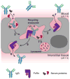
Figure 3. The FcRn-mediated recycling and transcytosis model
FcRn-mediated recycling initiates upon non-specific fluid-phase pinocytosis of serum IgG into FcRn expressing cells (1). As IgG is trafficked along the endosomal pathway (2) the pH decreases to 6 resulting in association with endosomal FcRn. The IgG-FcRn complex is recycled back to the plasma membrane (3) where IgG is released into blood due to its weak affinity for FcRn at blood pH (4). FcRn-mediated transcytosis of IgG across polarized epithelial cells, such as in the gut or lung, follows a similar cellular trafficking mechanism that directs the FcRn-IgG complex to the opposing cell membrane (5) where IgG can be released into the interstitial tissue space (6) due to the elevated pH. Serum proteins that do not bind a salvage receptor are trafficked to the lysosome and catabolized (7).
Figure 4
Figure 4. Inhibitors of the FcRn-IgG interaction
From left to right: 1) High dose IVIg can saturate FcRn and accelerate the clearance of endogenous IgG, 2) anti-FcRn heavy chain antibodies and 3) anti-β2m light chain antibodies bind FcRn epitopes that overlap with the IgG-Fc domain binding site, thereby inhibiting FcRn function, 4) Fc-engineered IgGs that that have increased, pH-independent affinity for FcRn (Abdegs), and 5) peptides and 6) small molecules that compete with IgG for binding to FcRn.
Figure 5
Figure 5. Proposed mechanisms of non-invasive protein delivery, mucosal vaccination, and mucosal viral neutralization
(a) Fc-fusion proteins or Fc functionalized nanoparticles (Fc-NP) delivered to the lung airspace or intestinal luman transcytosis the polarized epithelial barrier mediated by FcRn. Once delivered to the tissue interstitial space, Fc-fusions or Fc-NPs may access the systemic circulation directly by crossing the capillary endothelial cell barrier or indirectly by first entering the lymphatic system prior to accessing the blood,. Alternatively, Fc-fusions or Fc-NPs may be phagocytosed by resident dendritic cells or macrophages expressing Fcγ receptors. Fc-fusions or Fc-NPs may be recycled by FcRn-expressing antigen presenting cells or be diverted to the lysosome where the proteolytic peptides can be loaded onto MHC molecules. APCs then migrate to the lymph node and stimulate a T-cell response resulting in immune induction. (b) Broadly neutralizing antibodies (bnAbs) extravasate from the blood after systemic administration, accumulate in the tissue interstitial space, and are subsequently transported by FcRn across the epithelium, during which IgG may intercept and neutralize intracellular virus, or be released into the mucosa to neutralize invading pathogens, such as HIV or influenza,. IgG-opsonized virus may be degraded in the lysosome or be cleared from the mucosa via alternative mechanisms. Alternatively, IgG-opsonized virus may be reverse transcytosed by FcRn across the epithelial barrier and delivered to the tissue interstitial and initiate infection. The latter has not been proven in vivo.
Figure 6
Figure 6. Schematic of various fusion protein domains that can hijack FcRn
(a) Cartoon depiction of full length human IgG1 and (b) albumin are shown on the top. (c) The Fc-domain of hIgG1 used to construct Fc-fusion proteins. Typical Fc-fusions are constructed by fusing the C-terminus of a protein of interest to the N-terminus of the Fc hinge with either two drugs attached to a homodimeric Fc (left) or one drug attached to a heterodimeric Fc (right). (d) Monomeric Fc-domain fusion. Fc-domain residues are mutated to generate monoFc-fusions or residues are mutated to incorporate N-linked glycans resulting in a monomeric Fc. (e) Monomeric CH2 and CH3 derived from IgG1-Fc. (f) Engineered FcRn-binding affibody (cartoon derived from PDB 2M5A; a representative albumin binding affibody structure). (g) FcBP fusion (cartoon derived from PDB code 3M17).
Figure 7
Figure 7. Potential IgG engineering strategies to alter Fc interactions with FcRn or hijack FcRn transport
(a) Bi-specific IgG-Fc derived from various combination of wild-type or Fc-engineered IgGs may be constructed by various mechanism (e.g. Knobs n’ hole). Given the bivalent nature of the IgG-FcRn interaction, such bispecific IgG-Fc molecules may have interesting FcRn affinity relationships that may alter their recycling or transport properties. (b) The selection of IgGs (or alternative protein scaffolds) that bind an exosite on FcRn, which does not compete with endogenous IgG or albumin binding, may be a useful reagent to piggy-back on the FcRn an alter the biodistribtuion and/or clearance of fusion proteins and drug carriers.
Figure 8
Figure 8. Alternative IgG salvage theories that act in combination with FcRn
(a) In this “bait n’ switch” type mechanism an unknown cell-membrane anchored receptor, Factor X, binds IgG in serum (1) triggering receptor-mediated endocytosis (2). As IgG is trafficked along the endosomal pathway (3) the pH decreases to 6 resulting in dissociation of IgG from Factor X (4) and subsequent transfer to FcRn (5). FcRn then recycles IgG back to the plasma membrane (6) where IgG is released into blood due to its weak affinity for FcRn at blood pH (7). FcBP fusion proteins would not be readily endocytosed because they cannot bind Factor X. (b) In the PArtner Inhibited Recycling mechanism IgG is endocytosed into FcRn expressing cells by non-specific pinocytosis (1) and is trafficked to the endosome. The IgG binding site on FcRn is blocked by Factor X at steady state; however, IgG can effectively compete with the Factor X (2) resulting in dissociation of Factor X from FcRn and the formation of the FcRn-IgG complex (3). FcRn then recycles IgG back to the plasma membrane (4) where IgG is released into blood due to its weak affinity for FcRn at blood pH (5). FcBP fusion proteins cannot compete with the Factor X-FcRn interaction and are not salvaged by FcRn. (c) Alternatively, in a PArtner Initiated Recycling process an additional, unknown cell-membrane anchored FcRn binding partner increases the affinity of FcRn for IgG at physiological pH (1) resulting in FcRn-mediated endocytosis of IgG (2). As the complex is trafficked along the endosomal pathway (3) the pH decreases to 6 resulting in dissociation of Factor X from the FcRn-IgG complex (4). IgG stays bound to FcRn due to its high affinity at pH 6 and is trafficked back to the plasma membrane (5) where IgG is released into blood due to its weak affinity for FcRn at blood pH in the absence of Factor X (6). In all cases, IgG, FcBP fusion proteins, or additional serum components that do not bind FcRn or cannot compete with Factor X for binding FcRn are trafficked to the lysosome and degraded.

Similar articles

Cited by

References

    1. Brambell FW, Hemmings WA, Morris IG. A theoretical model of gamma-globulin catabolism. Nature. 1964;203:1352–4. - PubMed
    1. Rodewald R. pH-dependent binding of immunoglobulins to intestinal cells of the neonatal rat. J Cell Biol. 1976;71:666–9. - PMC - PubMed
    1. Jones EA, Waldmann TA. The mechanism of intestinal uptake and transcellular transport of IgG in the neonatal rat. J Clin Invest. 1972;51:2916–27. - PMC - PubMed
    1. Rodewald R, Kraehenbuhl JP. Receptor-mediated transport of IgG. J Cell Biol. 1984;99:159s–164s. - PMC - PubMed
    1. Simister NE, Mostov KE. An Fc receptor structurally related to MHC class I antigens. Nature. 1989;337:184–7. - PubMed

Publication types

MeSH terms