. 2015 Feb 17;6(1):e01670-14.
doi: 10.1128/mBio.01670-14.

Gammaherpesvirus small noncoding RNAs are bifunctional elements that regulate infection and contribute to virulence in vivo

Affiliations

Gammaherpesvirus small noncoding RNAs are bifunctional elements that regulate infection and contribute to virulence in vivo

Kevin W Diebel et al. mBio. .

Abstract

Many viruses express noncoding RNAs (ncRNAs). The gammaherpesviruses (γHVs), including Epstein-Barr virus, Kaposi's sarcoma-associated herpesvirus, and murine γHV68, each contain multiple ncRNA genes, including microRNAs (miRNAs). While these ncRNAs can regulate multiple host and viral processes in vitro, the genetic contribution of these RNAs to infection and pathogenesis remains largely unknown. To study the functional contribution of these RNAs to γHV infection, we have used γHV68, a small-animal model of γHV pathogenesis. γHV68 encodes eight small hybrid ncRNAs that contain both tRNA-like elements and functional miRNAs. These genes are transcribed by RNA polymerase III and are referred to as the γHV68 TMERs (tRNA-miRNA-encoded RNAs). To determine the total concerted genetic contribution of these ncRNAs to γHV acute infection and pathogenesis, we generated and characterized a recombinant γHV68 strain devoid of all eight TMERs. TMER-deficient γHV68 has wild-type levels of lytic replication in vitro and normal establishment of latency in B cells early following acute infection in vivo. In contrast, during acute infection of immunodeficient mice, TMER-deficient γHV68 has reduced virulence in a model of viral pneumonia, despite having an enhanced frequency of virus-infected cells. Strikingly, expression of a single viral tRNA-like molecule, in the absence of all other virus-encoded TMERs and miRNAs, reverses both attenuation in virulence and enhanced frequency of infected cells. These data show that γHV ncRNAs play critical roles in acute infection and virulence in immunocompromised hosts and identify these RNAs as a new potential target to modulate γHV-induced infection and pathogenesis.

Importance: The gammaherpesviruses (γHVs) are a subfamily of viruses associated with chronic inflammatory diseases and cancer, particularly in immunocompromised individuals. These viruses uniformly encode multiple types of noncoding RNAs (ncRNAs) that are not translated into proteins. It remains unclear how virus-expressed ncRNAs influence the course and outcome of infection in vivo. Here, we generated a mouse γHV that lacks the expression of multiple ncRNAs. Notably, this mutant virus is critically impaired in the ability to cause disease in immunocompromised hosts yet shows a paradoxical increase in infected cells early during infection in these hosts. While the original mouse virus encodes multiple ncRNAs, the expression of a single domain of one ncRNA can partially reverse the defects of the mutant virus. These studies demonstrate that γHV ncRNAs can directly contribute to virus-induced disease in vivo and that these RNAs may be multifunctional, allowing the opportunity to specifically interfere with different functional domains of these RNAs.


Figures

FIG 1
FIG 1
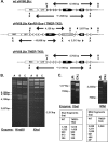
Genomic organization of the γHV68 TMER genes and TMER mutagenesis strategy. (A) Genetic details of the left end of the γHV68 genome. The TMER genes are represented as colored triangles numbered 1 through 8. The ORFs for M1 and M2 are shown as black arrows. Locations of the γHV68 miRNA genes are shown as dashed lines and numbered miR-M1-1 through -15. *miR-M1-11 is the only γHV68 miRNA not directly embedded within a TMER primary transcript (10, 11). miRNA genes are named according to the miRBase nomenclature (49). (B) Schematic of a single TMER (TMER1) identifying the vtRNA- and miRNA-containing stem-loops. (C) Schematics of the γHV68 genomic inserts in the pLE-WT and pLE-TKO plasmids (23). The grayed-out triangles in the pLE-TKO insert represent TMER genes for which the RNA Pol III promoter sequences have been deleted. Shown below the pLE-TKO schematic is a generic representation of the RNA Pol III promoter region removed from each γHV68 TMER gene.
FIG 2
FIG 2
γHV68 TMER-TKO mutant construction. (A) Schematic showing the genomic organization of the γHV68 TMER locus in the BAC DNAs for parental WT γHV68.βla, intermediate viral recombinant γHV68.βla Kanr/I-Sce-I TMER-TKO (for antibiotic selection in bacteria), and γHV68.βla TMER-TKO (in which the Kanr-encoding gene is removed). TMERs are indicated by gray triangles in WT γHV68 and as white triangles indicating the deletion of RNA Pol III promoter elements in the TMER-TKO recombinants. Restriction enzyme sites (H for HindIII, X for XhoI) and distances between these sites are indicated. (B) Agarose gel electrophoresis of restriction digests of BAC DNA for WT γHV68.βla (lane A), γHV68.βla Kanr/I-Sce-I TMER-TKO (lane B), and γHV68.βla TMER-TKO (lane C) following digestion with HindIII (left) or XhoI (right). (C) Agarose gel electrophoresis of restriction digests of PCR amplicons from the WT γHV68.βla and γHV68.βla TMER-TKO following digestion with XbaI (left) or MfeI (right). Predicted sizes of restriction digest fragments are indicated. The 25-bp MfeI fragment of the WT γHV68.βla PCR amplicon cannot be visualized on this gel. Molecular size markers are shown beside each gel.
FIG 3
FIG 3
Validation of the γHV68 TMER-TKO RNA analysis of WT and TMER-TKO virus-infected samples demonstrates that the TMER-TKO virus completely and specifically ablates transcription of the TMERs with no effect on neighboring genes. (A) RLM-RT-PCR analysis of mature miRNAs from total RNA isolated from WT or TMER-TKO (TKO) virus- or mock-infected 3T12 cells (MOI of 5) at 24 or 48 h p.i. miRNAs are identified to the left of the gels, and the associated TMERs are identified on the right. TMER-TKO virus-infected samples uniformly failed to express viral miRNAs. mmu-miR-21 is a host miRNA to control for miRNA detection in all the samples. (B) Northern blot analysis for TMER1, TMER5, and TMER7 from total RNA isolated from WT or TKO virus-infected or mock-infected 3T12 cells (MOI of 5) at 24 h p.i. Ethidium bromide-stained 5S rRNA (shown below the blots) served as a loading control. TMER1 and TMER5 have an alternative termination site after the first hairpin that results in two different sizes of pri-miRNA products, where TMER7 does not and results in only one band for the pri-miRNA. TKO virus-infected samples had undetectable expression of TMER1, -5, and -7. (C) RT-PCR analysis for the γHV68 M1, M2, and Rta/ORF50 transcripts with total RNA isolated from WT or TKO virus- or mock-infected 3T12 cells (MOI of 5) at 24 h p.i. A no-template control (NTC) was included. Amplification occurred only in the presence of reverse transcriptase (+RT), demonstrating that all amplifications reflect detection of RNA and not genomic DNA. (D) Quantitative RT-PCR analysis of the γHV68 M1, M2, and M3 transcripts from total RNA isolated from WT and TKO virus-infected 3T12 cell samples at 24 h p.i. All quantification are standardized relative to 18S rRNA levels.
FIG 4
FIG 4
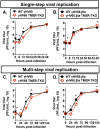
The γHV68 TMERs are dispensable for in vitro replication in fibroblasts. Analysis of single (A and B) and multiple (C and D) rounds of viral replication in 3T12 fibroblasts, comparing infection with the WT γHV68 and γHV68 TMER-TKO viruses (A and C) and infection with the WT γHV68.βla and γHV68.βla TMER-TKO viruses that contain the ORF73-βla reporter gene (B and D). (A and B) Single-step replication analysis with WT (black filled symbols) or TMER-TKO (open red symbols) with cells infected at an MOI of 5. Cells and supernatants were collectively harvested at the postinfection times indicated. (C and D) Multistep replication analysis was done comparably to single-step analysis, with cells infected at an MOI of 0.05. Viral titers were assessed by plaque assay, and the data depict the mean ± the standard error of the mean of three independent experiments, with one to three replicates per experiment. All plots include error bars; in cases where error bars are not shown, the standard error of the mean is very low. Statistically significant differences were calculated by unpaired t test comparing WT and TKO values at each individual time, and statistically significant differences are indicated (*, P < 0.05).
FIG 5
FIG 5
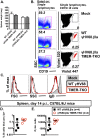
The γHV68 TMERs are dispensable for the establishment of latent infection of B cells in B6 mice. B6 mice were infected with either WT or TMER-TKO virus as indicated and harvested at day 14 p.i. (A) Spleen weights of mice infected with the viruses indicated. Shown is the mean value ± the standard error of the mean of five mice per group. (B) Representative flow cytometric analysis of cells from the spleens of B6 mice at day 14 p.i. comparing mock-infected samples and samples infected with the WT γHV68.βla or γHV68.βla TMER-TKO virus. Data depict the frequency of CD19+ B cells among lymphocytes that are single cells (left column) and the frequency of CD19+ B cells that are virus infected (βla-expressing cells), with βla+ cells identified in the top right quadrant and identified as red dots within the identified polygon (right column). (C) Analysis of cell size (FSC), granularity (SSC), and IgD expression comparing B cells from mock-infected spleen cells (gray) to WT γHV68.βla (black) or γHV68.βla TMER-TKO (red) virus-infected CD19+ βla+ B cells. (D and E) Quantitation of the frequency of virus-infected βla+ cells among CD19+ B cells (D) and the total number of virus-infected βla+ cells from WT γHV68.βla (black) or γHV68.βla TMER-TKO (red) virus-infected splenocytes (E). Each symbol indicates an individual mouse value, where horizontal black lines indicate the mean value ± the standard error of the mean. The data in panels B to E are from two independent experiments with two mice per group per experiment.
FIG 6
FIG 6
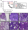
The γHV68 TMERs are required for virulence in a model of acute lethal pneumonia in BALB.IFN-γ−/− mice. (A) Survival of BALB.IFN-γ−/− mice following infection with a series of viral recombinants. The survival of mock-infected mice (gray diamonds) is compared with that of mice infected with WT (closed black symbols) or TMER-TKO (open red symbols) γHV68 with or without the βla reporter. γHV68Δ9473 (gray circles) is a γHV68 variant that lacks the entire TMER locus and the M1, M2, M3, and M4 genes (31). The number of mice in each group is indicated in the box. Hematoxylin-and-eosin-stained lung tissue from mock-infected (B and E) and WT γHV68.βla (C and F) and γHV68.βla TMER-TKO (D and G) virus-infected mice at either low (×40, B to D) or high (×200, E to G [circled areas in panels B to D]) magnification. Lungs were harvested at 8 days p.i. from virus-infected mice and 14 days p.i. from mock-infected mice. In this model, 8 to 11 days p.i. is the peak of disease signs, where there is notable infiltration and severe pneumonia. Statistical analysis of survival curves was done by log-rank (Mantel-Cox) test performing pairwise comparisons of virus mutants relative to the appropriate WT control (WT γHV68 or WT γHV68.βla). Statistically significant differences from the WT are indicated.
FIG 7
FIG 7
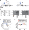
The γHV68 TMER1-only and vtRNA1-only mutant viruses partially reverse the deficit of the γHV68 TMER-TKO recombinant. (A) Schematic showing the genomic organization of the γHV68 TMER locus in the γHV68.βla TMER1-only and vtRNA1-only viruses. (B) Diagram of TMER-derived RNAs (left) and northern blot analysis of TMER1-derived products from WT, TMER-TKO (TKO), TMER1-only, or vtRNA1-only virus-infected or mock-infected 3T12 cells at 24 h p.i. (MOI of 5). Ethidium bromide-stained 5S rRNA (bottom) served as a loading control. Molecular size markers are indicated at the left; a nonspecific band is indicated by an asterisk. (C) Survival of BALB.IFN-γ−/− mice infected with γHV68.βla TMER1-only (brown squares) and vtRNA1-only (blue triangles) recombinants, relative to that of mock-infected and WT γHV68 and TMER-TKO virus-infected mice, indicated in gray (these data sets are the same data shown in Fig. 6A). ns, not significant. (D) Analysis of viral titers in the lungs of infected BALB.IFN-γ−/− mice at either day 5 (left) or day 8 (right) p.i. comparing WT and TMER mutant viruses, where each individual symbol represents a value from an individual mouse. The horizontal black lines indicate the mean titer of each group ± the standard error of the mean. The horizontal dashed line at 2 PFU/ml indicates the limit of detection of the plaque assay. The number of mice per group is indicated. Statistical analysis of survival curves was done by log-rank (Mantel-Cox) test performing pairwise comparisons of mutant viruses and WT γHV68.βla. Statistically significant differences from the WT are indicated. There were no statistically significant differences in virus titer as assessed by one-way ANOVA and Dunnett’s multiple-comparison test, with all comparisons done relative to WT γHV68.βla.
FIG 8
FIG 8
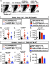
The γHV68 TMERs limit the frequency of virus-infected cells in BALB.IFN-γ−/− mice. (A) Representative flow cytometric analysis of virus-infected cells from the lungs of infected BALB.IFN-γ−/− mice at day 8 p.i., as measured by the detection of βla-expressing cells, with βla+ cells identified in the top right quadrant as red dots within the identified polygon. Plots were gated by using a large gate based on cell size and granularity, followed by doublet exclusion. Quantitation of the frequency (B) and number (C) of βla+ cells in the lungs of mice at 8 days p.i. with WT γHV68.βla, γHV68.βla TMER-TKO, and γHV68.βla vtRNA1-only viruses, with each symbol indicating an individual mouse value and horizontal black lines indicating the mean value ± the standard error of the mean. (D) Total cellularity in lung tissue samples in panels B and C. Quantitation of the frequency (E) and number (F) of βla+ cells in the spleens of mice at 8 days p.i. with WT γHV68.βla, γHV68.βla TMER-TKO, and γHV68.βla vtRNA1-only viruses. (G) Total cellularity in spleen tissue from samples in panels E and F. Data are from two or three independent experiments, with 7 to 10 mice per group. Statistical significance was assessed by one-way ANOVA and Dunnett’s multiple-comparison test with adjusted P values as indicated; all comparisons were done relative to WT γHV68.βla.

Similar articles

Cited by

References

    1. Speck SH, Virgin HW. 1999. Host and viral genetics of chronic infection: a mouse model of gamma-herpesvirus pathogenesis. Curr Opin Microbiol 2:403–409. doi:10.1016/S1369-5274(99)80071-X. - DOI - PubMed
    1. Cesarman E. 2011. Gammaherpesvirus and lymphoproliferative disorders in immunocompromised patients. Cancer Lett 305:163–174. doi:10.1016/j.canlet.2011.03.003. - DOI - PMC - PubMed
    1. Conrad NK, Fok V, Cazalla D, Borah S, Steitz JA. 2006. The challenge of viral snRNPs. Cold Spring Harb Symp Quant Biol 71:377–384. doi:10.1101/sqb.2006.71.057. - DOI - PubMed
    1. Takada K. 2001. Role of Epstein-Barr virus in Burkitt’s lymphoma. Curr Top Microbiol Immunol 258:141–151. doi:10.1007/978-3-642-56515-1_9. - DOI - PubMed
    1. Sun R, Lin SF, Gradoville L, Miller G. 1996. Polyadenylylated nuclear RNA encoded by Kaposi sarcoma-associated herpesvirus. Proc Natl Acad Sci U S A 93:11883–11888. doi:10.1073/pnas.93.21.11883. - DOI - PMC - PubMed

Publication types

LinkOut - more resources