Comparative Study
. 2014 Nov 10:5:5407.
doi: 10.1038/ncomms6407.

Progressive contraction of the latent HIV reservoir around a core of less-differentiated CD4⁺ memory T Cells

Affiliations
Comparative Study

Progressive contraction of the latent HIV reservoir around a core of less-differentiated CD4⁺ memory T Cells

S Jaafoura et al. Nat Commun. .

Abstract

In patients who are receiving prolonged antiretroviral treatment (ART), HIV can persist within a small pool of long-lived resting memory CD4(+) T cells infected with integrated latent virus. This latent reservoir involves distinct memory subsets. Here we provide results that suggest a progressive reduction of the size of the blood latent reservoir around a core of less-differentiated memory subsets (central memory and stem cell-like memory (TSCM) CD4(+) T cells). This process appears to be driven by the differences in initial sizes and decay rates between latently infected memory subsets. Our results also suggest an extreme stability of the TSCM sub-reservoir, the size of which is directly related to cumulative plasma virus exposure before the onset of ART, stressing the importance of early initiation of effective ART. The presence of these intrinsic dynamics within the latent reservoir may have implications for the design of optimal HIV therapeutic purging strategies.


Figures

Figure 1
Figure 1. During HIV infection the relative size of the TSCM subset within memory CD4 T cells remains stable.
(a) The gating strategy used to sort the memory cell subsets: CD4-enriched PBMC were stained with a cocktail of antibodies (see Methods) and doublets were excluded on the basis of both forward scatter (FSC) and side scatter (SSC). Resting CD4+ T cells were gated after exclusion of CD19+, CD14+, CD8+, HLADR+, CD69+, CD25+ cells. Resting TSCM were sorted on the basis of the following phenotype: CD45RA+ CD45RO CCR7+ CD62L+ CD27+ CD95+. CXCR3 and CD122 expression by TSCM is also shown. (b) The absolute number of cells in each memory subset (TSCM, TCM, TEM and TIM) was determined in HIV-infected patients with undetectable plasma viral load on ART and with CD4 cell counts above 500 per mm3, who were tested for integrated virus (UND, n=38), as well as in age- and sex-matched viremic patients (VIR, n=18) and HIV-seronegative healthy donors (HD, n=20). The Kruskal–Wallis and Dunn tests were used for statistical analysis (*P<0.05, **P<0.01, ****P<0.0001). Boxes represent the median and the 25th and 75th percentiles; whiskers represent the 10th and 90th percentiles. (c) Shows the percentage of each memory subset within the compartment formed by the four memory subsets analysed. The χ2 test was used to analyse differences in the distribution of the four memory subsets (****P<0.0001).
Figure 2
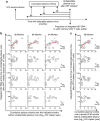
Figure 2. Slow reduction of the latent HIV reservoir around a core formed mainly by TSCM and TCM.
(a) Integrated HIV DNA was quantified in TSCM, TCM, TEM and TIM isolated from 38 strictly selected patients in whom viral load was undetectable for 24–189 months on ART. Recovery of TEM and TSCM was suboptimal in three patients. TIM could not be isolated from nine patients. The results are expressed as the integrated HIV DNA copy number per 105 cells. Taking inter-patient variability into account, the data shown in a were analysed with the Monte Carlo algorithm with 106 computer simulations with random samplings for each latently infected memory subset (see Methods). For each simulation, the number of integrated HIV DNA copies per 105 cells at the time of undetectable plasma virus following ART initiation (y0) and the slope of decay (λ) were fitted. The distributions of y0 and λ obtained from the 106 simulations are shown in Supplementary Fig. 8. The mean values of y0 and λ were used to plot the decay curves shown in a. The mean half-life of decay (−ln2/λ) is indicated for each latently infected memory subset. Statistical significance is indicated as * (for statistical analysis, see the Monte Carlo section of Methods). (b) Extends the data shown in a to the entire blood compartment. To analyse the decay in absolute numbers of latently infected memory subsets in the whole blood compartment, we used the 106 simulation values of y0 and slopes of decay obtained for each memory subset following Monte Carlo analysis. The predictions shown in b correspond to the average values of y0 and slopes. For the other parameters required to calculate the absolute numbers of latently infected cells (body weight, CD4 T-cell count and the percentages of the memory subsets among total CD4 T cells), we used the median values from our cohort of 38 patients. Total blood volume was estimated to represent 7% of total body weight. Note that the predictions in b remained consistent when random sampling (Monte Carlo approach) was used for body weight, the CD4+ T-cell count and the percentages of memory subsets among total CD4+ T cells, based on the median values and variance for the patients analysed. We considered 50 time points from 0 to 200 years (a total of 50 × 106 values for each memory subset). Comparison between the memory subsets was performed as in a. (See in Methods the Monte Carlo section). Statistical significance is indicated *, #. (c) The immunological dynamics of the CD4+ T-cell latent reservoir, corresponding to changes, at chosen time-points, in the relative size of each memory subset within the compartment formed by the four latently infected memory subsets in the entire blood compartment (see above). The surface areas are proportional to the numbers of integrated HIV DNA copies in each memory CD4 subset in the entire blood volume (predictions shown in b).
Figure 3
Figure 3. The frequency of latent infection of TSCM is related to cumulative plasma virus exposure.
(a,b) Cumulative viremia, expressed as log10 virus copy-years during periods of 36, 60 and 84 months before plasma virus became undetectable, was determined in patients (n=9–13) for whom virological follow-up was available for a period of 84 months. Associations were sought between cumulative viremia and the frequency of latent infection of each memory subset at the time when plasma virus became undetectable after ART initiation. For each patient, the frequency of latent infection of each memory subset at the time when plasma virus became undetectable after ART initiation was calculated from the slopes of the decay curves shown in Fig. 2a. The number of patients for whom virological follow-up was available over a period longer than 84 months before undetectable plasma virus, was insufficient for statistical analysis. In c, the same analysis was performed for all patients (n=16–22) for whom virological follow-up was available over a period of 36 months before plasma virus became undetectable. The Spearman rank test was used for statistical analysis (*P<0.05, **P<0.01).
Figure 4
Figure 4. PD-1 is less strongly expressed on TSCM.
Surface PD-1 expression on blood memory CD4+ T-cell subsets was analysed in 15 patients with CD4+ T-cell counts >500 per mm3 and undetectable plasma virus for at least 24 months on ART (a) and in 15 viremic patients (median CD4 T-cell count=477 per mm3, median viral load=3.3 log10, b). Data are expressed as the difference in PD-1 mean fluorescence intensity (ΔMFI) relative to naive CD4+ T cells. Boxes represent the median and the 25th and 75th percentiles; whiskers represent the 10th and 90th percentiles. Representative PD-1 staining is shown. The data were analysed with the Friedman test for paired data and with Dunn’s multiple comparison test (*P<0.05, **P<0.01, ***P<0.001, ****P<0.0001).

Similar articles

Cited by

References

    1. Chun T. W. et al. Quantification of latent tissue reservoirs and total body viral load in HIV-1 infection. Nature 387, 183–188 (1997). - PubMed
    1. Chun T. W. et al. Presence of an inducible HIV-1 latent reservoir during highly active antiretroviral therapy. Proc. Natl Acad. Sci. USA 94, 13193–13197 (1997). - PMC - PubMed
    1. Finzi D. et al. Latent infection of CD4+ T cells provides a mechanism for lifelong persistence of HIV-1, even in patients on effective combination therapy. Nat. Med. 5, 512–517 (1999). - PubMed
    1. Finzi D. et al. Identification of a reservoir for HIV-1 in patients on highly active antiretroviral therapy. Science (New York, NY) 278, 1295–1300 (1997). - PubMed
    1. Chomont N. et al. HIV reservoir size and persistence are driven by T cell survival and homeostatic proliferation. Nat. Med. 15, 893–900 (2009). - PMC - PubMed

Publication types

Substances