. 2014 Nov 6;10(11):e1004503.
doi: 10.1371/journal.ppat.1004503. eCollection 2014 Nov.

IFI16 restricts HSV-1 replication by accumulating on the hsv-1 genome, repressing HSV-1 gene expression, and directly or indirectly modulating histone modifications

Affiliations

IFI16 restricts HSV-1 replication by accumulating on the hsv-1 genome, repressing HSV-1 gene expression, and directly or indirectly modulating histone modifications

Karen E Johnson et al. PLoS Pathog. .

Erratum in

Abstract

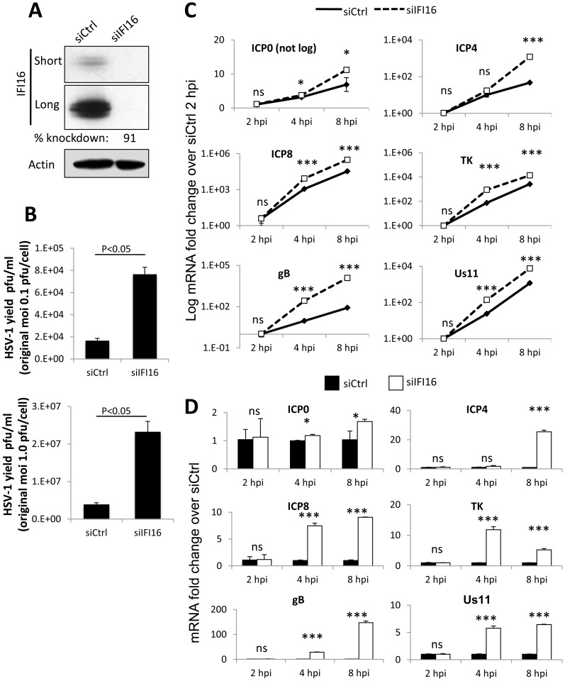
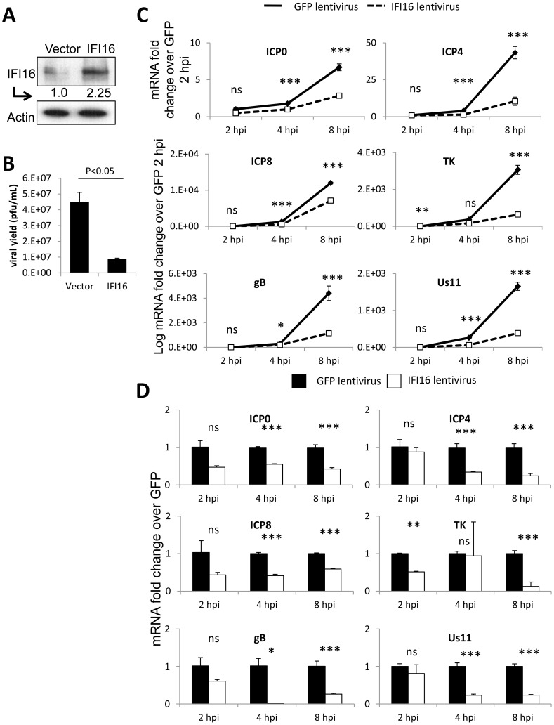
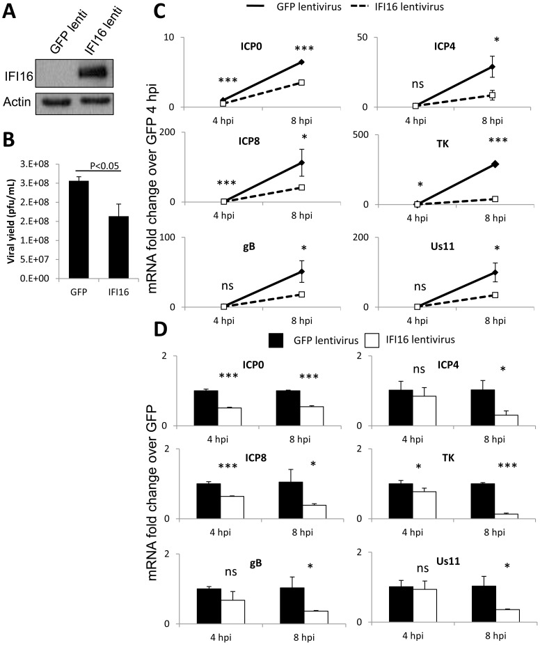
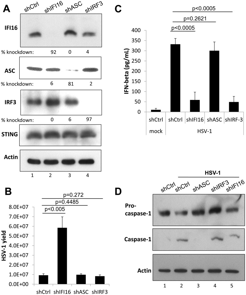
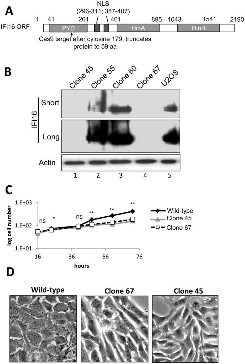
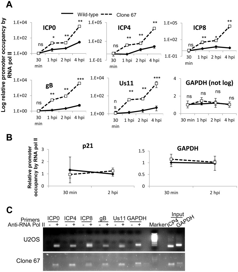
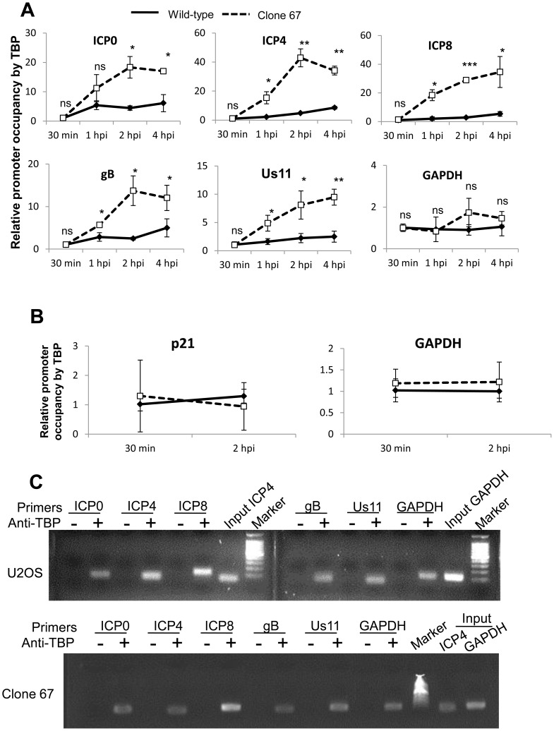
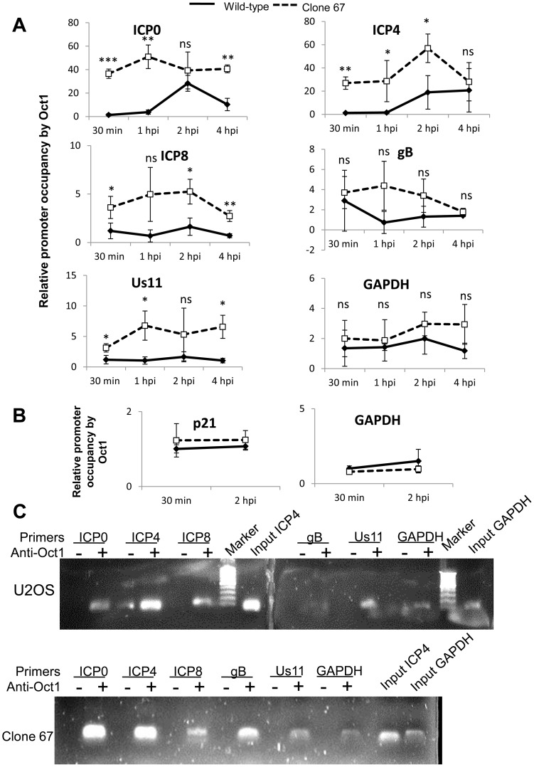
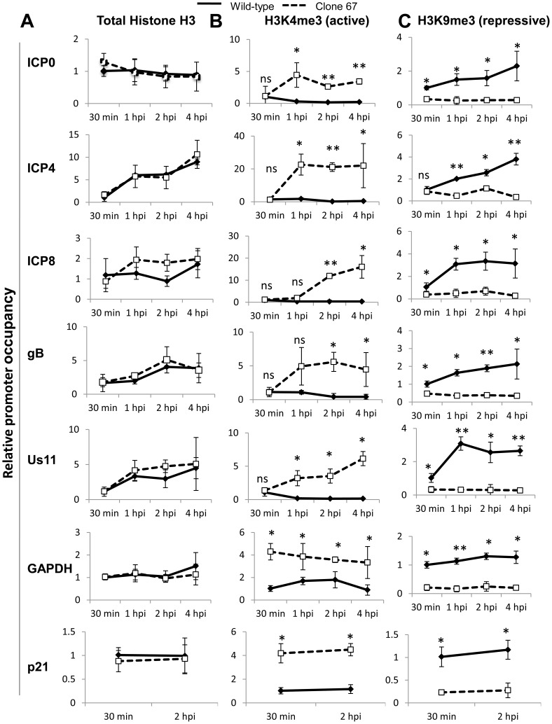
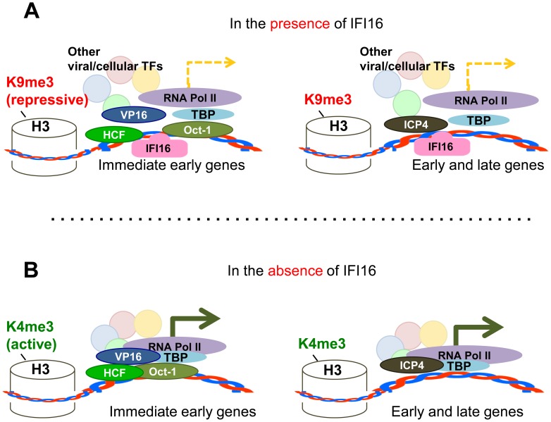
Interferon-γ inducible factor 16 (IFI16) is a multifunctional nuclear protein involved in transcriptional regulation, induction of interferon-β (IFN-β), and activation of the inflammasome response. It interacts with the sugar-phosphate backbone of dsDNA and modulates viral and cellular transcription through largely undetermined mechanisms. IFI16 is a restriction factor for human cytomegalovirus (HCMV) and herpes simplex virus (HSV-1), though the mechanisms of HSV-1 restriction are not yet understood. Here, we show that IFI16 has a profound effect on HSV-1 replication in human foreskin fibroblasts, osteosarcoma cells, and breast epithelial cancer cells. IFI16 knockdown increased HSV-1 yield 6-fold and IFI16 overexpression reduced viral yield by over 5-fold. Importantly, HSV-1 gene expression, including the immediate early proteins, ICP0 and ICP4, the early proteins, ICP8 and TK, and the late proteins gB and Us11, was reduced in the presence of IFI16. Depletion of the inflammasome adaptor protein, ASC, or the IFN-inducing transcription factor, IRF-3, did not affect viral yield. ChIP studies demonstrated the presence of IFI16 bound to HSV-1 promoters in osteosarcoma (U2OS) cells and fibroblasts. Using CRISPR gene editing technology, we generated U2OS cells with permanent deletion of IFI16 protein expression. ChIP analysis of these cells and wild-type (wt) U2OS demonstrated increased association of RNA polymerase II, TATA binding protein (TBP) and Oct1 transcription factors with viral promoters in the absence of IFI16 at different times post infection. Although IFI16 did not alter the total histone occupancy at viral or cellular promoters, its absence promoted markers of active chromatin and decreased those of repressive chromatin with viral and cellular gene promoters. Collectively, these studies for the first time demonstrate that IFI16 prevents association of important transcriptional activators with wt HSV-1 promoters and suggest potential mechanisms of IFI16 restriction of wt HSV-1 replication and a direct or indirect role for IFI16 in histone modification.


Conflict of interest statement

The authors have declared that no competing interests exist.

Figures

Figure 1
Figure 1. Effect of IFI16 depletion on HSV-1 gene expression, replication and viral yield.
HFF cells were microporated with siCtrl or siIFI16 RNA. (A) Western blot analysis of IFI16 knockdown at 48 h post microporation. Shown are short and long exposures of the same blot. Actin is used as a loading control. (B) Viral yield at 24 h p.i. from cells originally infected at an moi of 0.1 pfu/cell (top) or 1 pfu/cell (bottom). (C and D) Representative HSV-1 gene expression from cells infected at an moi of 1 pfu/cell, normalized to GAPDH cDNA and levels at 2 h p.i. Shown are mRNA fold-change over siCtrl at 2 h p.i. (C) or concurrent siCtrl samples (D), ± standard deviation. Statistics were done using Student's T test (ns: not significant, *p<0.05, **p<0.005, ***p<0.0005).
Figure 2
Figure 2. Effect of IFI16 overexpression on HSV-1 gene expression, replication and viral yield in U2OS cells.
Lentivirus with the coding region of IFI16 or GFP was transduced into U2OS cells. (A) Western blot analysis of IFI16 expression, 48 h post transduction. Actin is a loading control. (B) Viral yield at 24 h p.i. from cells originally infected at an moi of 0.1 pfu/cell. (C) Representative HSV-1 gene expression from cells infected at an moi of 1 pfu/cell. Shown are mRNA fold-change over siCtrl at 2 h p.i. (C) or concurrent siCtrl samples (D) normalized to GAPDH, ± standard deviation. Statistics were done using Student's T test (ns: not significant, *p<0.05, **p<0.005, ***p<0.0005).
Figure 3
Figure 3. Effect of IFI16 overexpression on HSV-1 gene expression, replication and viral yield in MCF7 cells.
Lentivirus encoding IFI16 or GFP was transduced into IFI16-negative MCF7 cells. (A) Western blot analysis of IFI16 expression at 48 h post transduction. Actin is a loading control. (B) Viral yield at 24 h p.i. from cells originally infected at an moi of 0.1 pfu/cell. (C) Representative HSV-1 gene expression from cells infected at an moi of 1 pfu/cell. Shown are mRNA fold-change over siCtrl at 2 h p.i. (C) or concurrent siCtrl samples (D), normalized to GAPDH, ± standard deviation. Statistics were done using Student's T test (ns: not significant, *p<0.05, **p<0.005, ***p<0.0005).
Figure 4
Figure 4. Effect of IFI16, ASC, and IRF3 knockdown on HSV-1 replication.
HFF cells were transduced with lentiviruses encoding scrambled shRNA (shCtrl), and shRNA targeting IFI16, ASC, or IRF3. (A) Western blot showing IFI16, ASC, IRF3, and STING, 48 h post transduction, using actin as a loading control. (B) Viral yield at 24 h p.i. from transduced HFF cells originally infected at an moi of 1 pfu/cell, 48 h post transduction. (C) Cell culture supernatant IFNβ levels as determined by ELISA from transduced HFF cells infected at an moi of 1 pfu/cell at 6 h p.i. 48 h post transduction. Statistics were done using Student's T test (ns: not specific). (D) Western blot showing procaspase-1 and caspase 1, from transduced HFF cells infected at an moi of 1 pfu/cell at 6 h p.i., 48 h post transduction, using actin as a loading control.
Figure 5
Figure 5. Cas9-mediated IFI16 gene editing and permanent depletion from U2OS cells.
IFI16 was depleted from U2OS cells using Cas9 and guided RNA targeting the PYD of IFI16. (A) Schematic representation of Cas9 target site within the 2190 bp IFI16 ORF. NLS: Nuclear localization signal; PYD: Pyrin domain; HinA and HinB: DNA binding domains. (B) Western blot of IFI16 in wt U2OS and a selection of clonal U2OS cells. Short and long exposure of the same IFI16 blot are shown. Actin was used as a loading control. (C) wt-U2OS, clone 45, and clone 67 cell growth. (D) Phase micrograph of wt-U2OS, clone 67, and clone 45 cells at 40×. Statistics were done using Student's T test (ns: not significant, *p<0.05, **p<0.005).
Figure 6
Figure 6. Effect of Cas9-mediated U2OS cell IFI16 gene edition on HSV-1 gene expression and replication, and comparison with wt U2OS cells.
(A) HSV-1 yield at 24 h p.i. from wt U2OS cells and clone 67 U2OS cells transduced with empty vector or IFI16 expression vector originally infected at an moi of 0.1 pfu/cell. (B) Western blot of IFI16 and actin in uninfected wt U2OS cells and clone 67 U2OS cells transduced with empty vector (V) or IFI16 expression vector (IFI16), or cells infected with HSV-1 for 24 h at an moi of 1 pfu/cell. (C–D) Representative HSV-1 gene expression from cells infected at an moi of 1 pfu/cell. Shown are mRNA fold-change over GFP at 2 h p.i. (C) or concurrent GFP samples (D), ± standard deviation. Statistics were done using Student's T test (ns: not significant, *p<0.05, **p<0.005, ***p<0.0005). (E) Western blot of IFI16 and ICP0 in U2OS and clone 67 U2OS cells infected with HSV-1 at an moi of 1 pfu/cell for the times indicated (upper panel). Actin is used as a loading control. Densitometric analysis of ICP0 band intensities relative to actin from western blots (lower panel).
Figure 7
Figure 7. Analysis of HSV-1 genome entry into the nucleus and association with IFI16.
U2OS cells were mock infected or infected with EdU-labeled HSV-1 at an moi of 1 pfu/cell for 30 or 60 min. (A) Immunofluorescence analysis of IFI16 (red) and EdU detection of HSV-1 DNA (green). Boxed areas are enlarged on the right. Yellow arrows indicate colocalized IFI16 and HSV-1 DNA. White arrows indicate cytoplasmic IFI16. (B) EdU detection of HSV-1 DNA (red) and PLA for IFI16/IFI16 (green), including intensity plots for red and green channels (lower panels). Boxed areas are enlarged on the right. Yellow arrows indicate colocalized IFI16 and HSV-1 DNA. White arrows indicate cytoplasmic IFI16.
Figure 8
Figure 8. ChIP analysis of IFI16 binding to the HSV-1 genome at the transcriptional start site of viral genes.
(A) HFF, U2OS, and clone 67 cells were infected with BrdU-labeled HSV-1 at an moi of 1 pfu/cell for 15, 30, or 90 min before fixation. Cells were immunostained for BrdU. The percent of cells with nuclear BrdU was quantified by scoring BrdU localization in 4 fields of cells per sample (total 150–200 cells/sample). (B–G) U2OS and clone 67 cells were mock infected or infected with wt HSV-1 at an moi of 1 pfu/cell. (B) The relative quantity of nuclear HSV-1 DNA at various times post infection was quantified by qPCR of HSV-1-infected nuclear extracts from U2OS and clone 67 U2OS cells using primers for the ICP4 start site. (C) ChIP analysis of IFI16 in HFF and U2OS cells. IFI16 was immunoprecipitated from cells infected with HSV-1 (1 pfu/cell) for 30 min, 1, 2, or 4 hours. Bound DNA was analyzed by real-time PCR with primers to regions flanking the transcriptional start sites of the genes indicated. Values were normalized to IFI16-bound GAPDH and input ICP4. (D) ChIP analysis of IFI16 binding the cellular promoters for GAPDH and p21 in U2OS cells, performed as above. (E) ChIP analysis of IFI16 binding cellular promoters in uninfected U2OS cells or cells infected with HSV-1 at an moi of 1 pfu/cell for 1 hour, performed as above. (F) Agarose gels showing HSV-1 and cellular promoter DNA precipitated with antibody to IFI16 or control IgG in U2OS (U) or clone 67 (67) cells at 1 h p.i., and an moi of 1 pfu/cell. (G) Agarose gels showing the amplification of the indicated HSV-1 and cellular promoter regions after IFI16 ChIP at 4 h p.i.
Figure 9
Figure 9. IFI16 prevents RNA Pol II accumulation at HSV-1 promoters.
U2OS and clone 67 cells were infected with HSV-1 at an moi of 1 pfu/cell for 30 min, 1, 2, or 4 h. ChIP analysis was done with RNA pol II antibody and primers to the indicated HSV-1 genes, normalized to input ICP4, and cellular promoters normalized to GAPDH and are shown as relative to the association of RNA pol II and the indicated promoter at 30 min p.i. (A and B). Statistics were done using Student's T test (ns: not significant, *p<0.05, **p<0.005, ***p<0.0005). (C) Representative amplification products of the indicated promoters precipitated with beads alone or antibody to RNA polymerase II at 4 h p.i.
Figure 10
Figure 10. IFI16 prevents TBP accumulation at HSV-1 promoters.
Wt U2OS and clone 67 U2OS cells were infected with HSV-1 at an moi of 1 pfu/cell for 30 min, 1, 2, or 4 h. ChIP analysis was done with TBP antibody and real-time PCR primers to the indicated HSV-1 genes, normalized to input ICP4 for viral promoters and to GAPDH for cellular promoters, and are shown as relative to the association of TBP and the indicated promoter at 30 min p.i. (A and B). Statistics were done using Student's T test (ns: not significant, *p<0.05, **p<0.005, ***p<0.0005). (C) Representative amplification products of the indicated promoters precipitated with beads alone or antibody to TBP at 4 h p.i.
Figure 11
Figure 11. IFI16 prevents Oct1 accumulation at HSV-1 promoters.
U2OS and clone 67 cells were infected with HSV-1 at an moi of 1 pfu/cell. ChIP analysis was done with Oct1 antibody and real-time PCR with primers to the indicated HSV-1 genes, normalized to input ICP4, and cellular promoters normalized to GAPDH and are shown relative to the association of Oct1 and the indicated promoter in U2OS cells. Statistics were done using Student's T test. (ns: not significant, *p<0.05, **p<0.005, ***p<0.0005) (A and B). (C) Representative amplification products of the indicated promoters precipitated with beads alone or antibody to Oct1 at 4 h p.i.
Figure 12
Figure 12. IFI16 induces changes in histone modifications at HSV-1 promoters.
Wt U2OS and clone 67 U2OS cells were infected with HSV-1 at an moi of 1 pfu/cell for 30 min, 1, 2, or 4 h. ChIP analysis was done with antibodies to total histone H3 (A), H3K4me3 (B), or H3K9me3 (C) and primers to the indicated genes, normalized to input ICP4 for HSV-1 genes and to input GAPDH for cellular genes and are shown relative to the association of total histone or modified histone at the indicated promoter at 30 min p.i. in each cell type. Statistics were done using Student's T test (ns: not significant, *p<0.05, **p<0.005, ***p<0.0005).
Figure 13
Figure 13. Schematic model: IFI16 inhibits HSV-1 gene expression by modulating histone modifications and binding to HSV-1 promoters, specifically preventing or decreasing the association of transcription factors at viral promoters.
In the presence of IFI16 (A) markers for heterochromatin are loaded onto HSV-1 gene transcription start sites and transcription factors including TBP, RNA pol II, and (in the case of IE genes) Oct1 associate with HSV-1 promoters to stimulate gene expression at low levels. In the absence of IFI16 (B), IFI16 and markers for euchromatin are loaded onto HSV-1 gene transcription start sites, and association of transcription factors with viral gene promoters is increased, thereby significantly increasing viral gene expression.

Similar articles

Cited by

References

    1. Knipe DM, Howley PM, editors (2006) Fields' Virology. Philadelphia: Lippincott Williams & Wilkins. 3177 p.
    1. Streilein JW, Dana MR, Ksander BR (1997) Immunity causing blindness: five different paths to herpes stromal keratitis. Immunol Today 18: 443–449. - PubMed
    1. Whitley RJ, Gnann JW (2002) Viral encephalitis: familiar infections and emerging pathogens. Lancet 359: 507–513. - PubMed
    1. Whitley RJ, Soong SJ, Linneman C Jr, Liu C, Pazin G, et al. (1982) Herpes simplex encephalitis. Clinical Assessment. JAMA 247: 317–320. - PubMed
    1. Honess RW, Roizman B (1974) Regulation of herpesvirus macromolecular synthesis. I. Cascade regulation of the synthesis of three groups of viral proteins. J Virol 14: 8–19. - PMC - PubMed

Publication types