. 2015 Jan;89(1):415-27.
doi: 10.1128/JVI.02591-14. Epub 2014 Oct 15.

Kaposi's sarcoma-associated herpesvirus-encoded replication and transcription activator impairs innate immunity via ubiquitin-mediated degradation of myeloid differentiation factor 88

Affiliations

Kaposi's sarcoma-associated herpesvirus-encoded replication and transcription activator impairs innate immunity via ubiquitin-mediated degradation of myeloid differentiation factor 88

Qinglan Zhao et al. J Virol. 2015 Jan.

Abstract

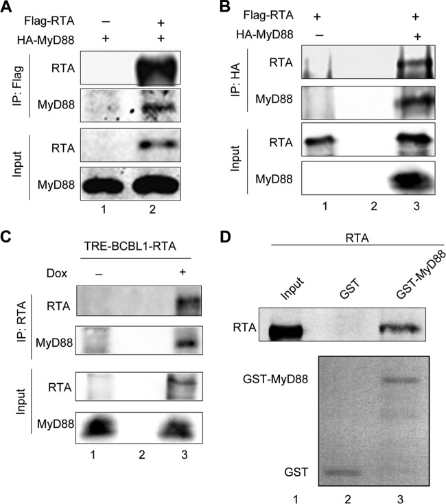
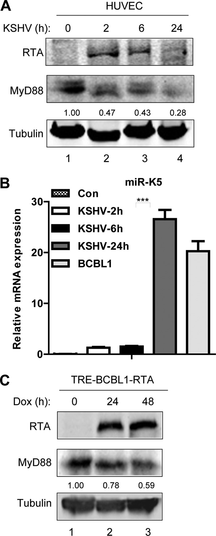
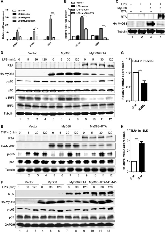
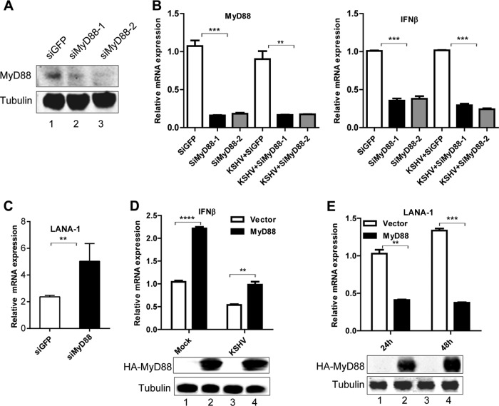
Kaposi's sarcoma-associated herpesvirus (KSHV) is a human gammaherpesvirus with latent and lytic reactivation cycles. The mechanism by which KSHV evades the innate immune system to establish latency has not yet been precisely elucidated. Toll-like receptors (TLRs) are the first line of defense against viral infections. Myeloid differentiation factor 88 (MyD88) is a key adaptor that interacts with all TLRs except TLR3 to produce inflammatory factors and type I interferons (IFNs), which are central components of innate immunity against microbial infection. Here, we found that KSHV replication and transcription activator (RTA), which is an immediate-early master switch protein of viral cycles, downregulates MyD88 expression at the protein level by degrading MyD88 through the ubiquitin (Ub)-proteasome pathway. We identified the interaction between RTA and MyD88 in vitro and in vivo and demonstrated that RTA functions as an E3 ligase to ubiquitinate MyD88. MyD88 also was repressed at the early stage of de novo infection as well as in lytic reactivation. We also found that RTA inhibited lipopolysaccharide (LPS)-triggered activation of the TLR4 pathway by reducing IFN production and NF-κB activity. Finally, we showed that MyD88 promoted the production of IFNs and inhibited KSHV LANA-1 gene transcription. Taken together, our results suggest that KSHV RTA facilitates the virus to evade innate immunity through the degradation of MyD88, which might be critical for viral latency control.

Importance: MyD88 is an adaptor for all TLRs other than TLR3, and it mediates inflammatory factors and IFN production. Our study demonstrated that the KSHV RTA protein functions as an E3 ligase to degrade MyD88 through the ubiquitin-proteasome pathway and block the transmission of TLRs signals. Moreover, we found that KSHV inhibited MyD88 expression during the early stage of de novo infection as well as in lytic reactivation. These results provide a potential mechanism for the virus to evade innate immunity.


Figures

FIG 1
FIG 1
RTA reduces MyD88 expression. (A) One μg pCDH-RTA or vector control was transfected into HeLa cells in 12-well plates. After 36 h, protein levels of MyD88, TRIF, MAVS, IRAK1, IRF3, and IRF7 were determined by Western blotting. GAPDH was the loading control. Values represent percentages of the target proteins normalized against GAPDH and compared to the vector control. (B) HeLa cells were transfected with increasing doses of pCDH-RTA (0 μg, 0.5 μg, 1 μg, and 2 μg). After 36 h, endogenous MyD88, TRIF, and IRF7 were analyzed by Western blotting. Values represent percentages of the target proteins normalized against tubulin and compared to the signal of a 0-μg dose. (C) iSLK cells were treated with 0.002 μg/μl DOX for the indicated times, and endogenous MyD88 was analyzed by Western blotting. Values represent percentages of MyD88 proteins normalized against tubulin and compared to the signal at 0 h. (D) HeLa cells were transfected with 1 μg pCDH-RTA or vector control. After 36 h, RNA was collected for reverse transcription and used to quantify MyD88 and actin by qRT-PCR.
FIG 2
FIG 2
RTA alters the stability of MyD88 protein and RTA degrades MyD88 through the proteasome pathway. (A) 293T cells were transfected with HA-tagged MyD88 with or without flag-RTA and treated with cycloheximide (CHX) as indicated. Anti-flag and anti-HA antibodies were used for Western blotting. (B) Values are calculated from the experiments shown in panel A. Signal intensities first were normalized to the corresponding tubulin and then normalized to the signal at 0 h of each group. (C) 293T cells were transfected with HA-tagged MyD88 with or without flag-RTA and treated with 10 μM MG132 as indicated. Anti-flag and anti-HA antibodies were used for Western blotting. (D) Values are calculated from the experiments shown in panel C. Signal intensities first were normalized to the corresponding tubulin value and then normalized to the signal of each group at 21 h.
FIG 3
FIG 3
RTA promotes polyubiquitination of MyD88. (A) 293T cells were cotransfected with His-tagged ubiquitin, HA-tagged MyD88, and flag-tagged RTA, and after 24 h cells were treated with MG132 for 16 h. Whole-cell extracts were precipitated with Ni-NTA agarose beads for 6 h, followed by Western blotting with anti-HA. (B) 293T cells were cotransfected with strep-tagged MyD88, His-tagged ubiquitin, and pCR3.1-RTA, and after 24 h, cells were treated with MG132 for 16 h. Whole-cell extracts were precipitated with Strep-Tactin agarose beads for 6 h, followed by Western blotting with anti-His.
FIG 4
FIG 4
RTA interacts with MyD88 directly. (A) 293T cells were transfected with HA-tagged MyD88 alone or cotransfected with constructs with flag-tagged RTA. Twelve h later, cells were treated with MG132 for 24 h. Extracts then were immunoprecipitated with M2 beads and analyzed by Western blotting with anti-HA antibody. (B) 293T cells were transfected with flag-tagged RTA alone or cotransfected with HA-tagged MyD88. Twelve h later, cells were treated with MG132 for 24 h. Cells then were extracted and immunoprecipitated with anti-HA and analyzed by Western blotting with anti-RTA. (C) TRE-BCBL1-RTA cells were mock induced or DOX induced for 36 h, MG132 was added for the final 12 h, and then cells were extracted and immunoprecipitated with anti-RTA antibody and analyzed by Western blotting with anti-MyD88 antibody. (D) In vitro GST affinity binding assay. Bacterially expressed GST alone and GST-MyD88 attached to beads were incubated with in vitro-translated RTA (lane 1). RTA binding to beads was detected by Western blotting.
FIG 5
FIG 5
RTA functions as E3 Ub-ligase for MyD88. (A) 293T cells were cotransfected with HA-tagged MyD88, WT RTA, and RTA mutants (C131S, C141S, and H145L). Thirty-six h later, proteins were collected for Western blotting. (B) 293T cells were cotransfected with HA-tagged MyD88, WT RTA, and RTA mutant RTA141-145. Thirty-six h later, proteins were collected for Western blotting. (C) In vitro polyUb conjugation of MyD88 by RTA. Cell-free in vitro ubiquitination reactions were carried out by incubation with the indicated purified GST-MyD88 proteins. RTA or RTA141-145 bound to M2 beads was used at catalytic levels, and GST-MyD88 was used at substrate levels (1 μg). (Bottom) Western blotting detected polyUb-conjugated and unconjugated 60-kDa GST-MyD88 using anti-MyD88.
FIG 6
FIG 6
MyD88 expression is impaired at the early stage of KSHV de novo infection and during lytic reactivation. (A) MyD88 expression was downregulated in response to KSHV de novo infection. KSHV (MOI, 20) was used to infect confluent primary HUVECs. Western blotting was used to detect MyD88 protein. Values represent percentages of MyD88 proteins normalized against tubulin and compared to the signal at 0 h. (B) KSHV (MOI, 20) was used to infect confluent primary HUVECs. RNA was collected for testing the KSHV miR-K5 level by qRT-PCR. RNA from BCBL1 cells was a positive control. (C) MyD88 expression was downregulated during reactivation. TRE-BCBL1-RTA cells were mock induced or DOX induced for 24 h and 48 h. Western blotting was used to detect MyD88 expression. Values represent percentages of MyD88 proteins normalized against tubulin and compared to the signal at 0 h.
FIG 7
FIG 7
RTA inhibits MyD88-mediated TLR4 signaling. (A) After transfection of HeLa cells for 24 h, cells were stimulated with 800 ng/ml LPS for 12 h, and mRNA levels were determined for IFN-α1, IFN-α2, and IFN-β. Relative quantitative values from RT-PCR assays are shown as fold changes in IFN mRNA over basal levels obtained with control vector DNA without LPS stimulation. (B) Effect of RTA on MyD88-induced expression from reporter luciferase genes driven by NF-κB and IFN-β promoters. HeLa cells were transfected with NF-κB-luciferase and IFN-β-luciferase plasmids and the indicated plasmids for 24 h, followed by stimulation with 800 ng/ml LPS for 12 h before luciferase assays. Luciferase activity is shown as the fold increase over basal levels obtained with control vector DNA without LPS stimulation. (C) Lysates from luciferase assays with IFN-β promoters were subjected to Western blotting. (D) HeLa cells were transfected as indicated. After 36 h, cells were stimulated with 800 ng/ml LPS by time course as indicated, and lysates were used to detect p65 and IRF3 phosphorylation by Western blotting. (E) HeLa cells were transfected as indicated. After 36 h, cells were stimulated with 10 ng/ml TNF-α by time course as indicated, and lysates were used to detect p65 phosphorylation by Western blotting. (F) HeLa cells were transfected as indicated. After 36 h, cells were stimulated with 800 ng/ml LPS by time course as indicated, and lysates were used to detect p65 phosphorylation by Western blotting. (G) Primary HUVECs were infected by KSHV (MOI, 4) for 24 h, and RNA was collected for testing the TLR4 mRNA level by qRT-PCR. (H) iSLK cells were induced by DOX for RTA expression for 24 h. RNA was collected for testing the TLR4 mRNA level by qRT-PCR.
FIG 8
FIG 8
MyD88 promotes production of IFNs and inhibits KSHV latent LANA-1 gene transcription during KSHV de novo infection. (A) Effect of MyD88 on IFN production during KSHV infection by knockdown of MyD88. Primary HUVECs were transfected with MyD88 siRNA and GFP siRNA as a control. After 24 h, cells were collected for detecting MyD88 by Western blotting. (B) Primary HUVECs were transfected with MyD88 siRNA and GFP siRNA (siGFP) as a control. After 36 h, cells were infected with KSHV (MOI, 4). RNA was collected to analyze MyD88 and IFN mRNA levels. Relative quantitative values of results from RT-PCR are shown as fold changes in IFN mRNA over basal levels obtained with siGFP-treated samples. Data are presented as means ± standard errors of the means, n = 3. *, P < 0.05; **P < 0.01; ***, P < 0.001. (C) Effect of MyD88 on LANA-1 transcription after knockdown of MyD88. Primary HUVECs were transfected with the first MyD88 siRNA and with GFP siRNA as a control. After 24 h, HUVECs were infected with KSHV (MOI, 4) for 72 h. RNA was collected to analyze KSHV LANA-1 mRNA. Relative quantitative values of RT-PCR assays are shown as fold changes in LANA-1 mRNA over basal levels obtained with siGFP-treated samples. (D) Effect of MyD88 on IFN-β production during KSHV infection with overexpression of MyD88. HeLa cells were transfected with MyD88 or control vector DNA. After 24 h, cells were infected with KSHV (MOI, 0.6) for 24 h. RNA was collected to analyze IFN-β mRNA. Relative quantitative values of RT-PCR assays are shown as fold changes in IFN-β mRNA over basal levels with vector alone without KSHV infection. Proteins were analyzed by Western blotting to show MyD88 overexpression with anti-HA antibody. (E) Effect of MyD88 on LANA-1 transcription with overexpression of MyD88. HeLa cells were transfected with MyD88 or control vector DNA. After 24 h, cells were infected with KSHV (MOI, 0.6). RNA was collected over a time course to analyze LANA-1 transcription. Relative quantitative values of qRT-PCR assays are shown as fold changes in LANA-1 mRNA over basal levels with vector alone with KSHV infection. Proteins were analyzed by Western blotting to show MyD88 overexpression with anti-HA antibody.

Similar articles

Cited by

References

    1. Kawai T, Akira S. 2009. The roles of TLRs, RLRs and NLRs in pathogen recognition. Int Immunol 21:317–337. doi:10.1093/intimm/dxp017. - DOI - PMC - PubMed
    1. Hornung V, Ablasser A, Charrel-Dennis M, Bauernfeind F, Horvath G, Caffrey DR, Latz E, Fitzgerald KA. 2009. AIM2 recognizes cytosolic dsDNA and forms a caspase-1-activating inflammasome with ASC. Nature 458:514–518. doi:10.1038/nature07725. - DOI - PMC - PubMed
    1. Yanai H, Savitsky D, Tamura T, Taniguchi T. 2009. Regulation of the cytosolic DNA-sensing system in innate immunity: a current view. Curr Opin Immunol 21:17–22. doi:10.1016/j.coi.2009.01.005. - DOI - PubMed
    1. Janeway CA., Jr 1992. The immune system evolved to discriminate infectious nonself from noninfectious self. Immunol Today 13:11–16. doi:10.1016/0167-5699(92)90198-G. - DOI - PubMed
    1. Dolganiuc A, Oak S, Kodys K, Golenbock DT, Finberg RW, Kurt-Jones E, Szabo G. 2004. Hepatitis C core and nonstructural 3 proteins trigger toll-like receptor 2-mediated pathways and inflammatory activation. Gastroenterology 127:1513–1524. doi:10.1053/j.gastro.2004.08.067. - DOI - PubMed

Publication types

LinkOut - more resources