

Importance of exosome depletion protocols to eliminate functional and RNA-containing extracellular vesicles from fetal bovine serum
- PMID: 25317276
- PMCID: PMC4185091
- DOI: 10.3402/jev.v3.24783
Importance of exosome depletion protocols to eliminate functional and RNA-containing extracellular vesicles from fetal bovine serum
Abstract
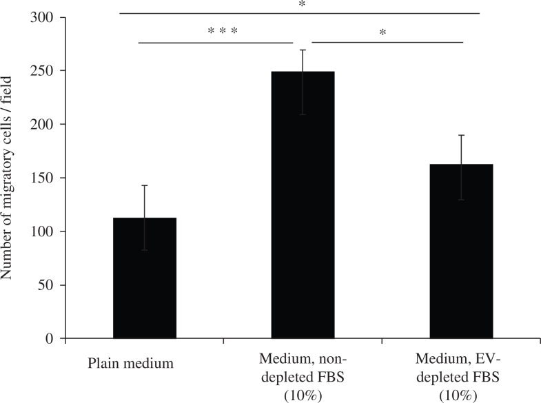
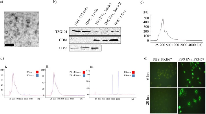
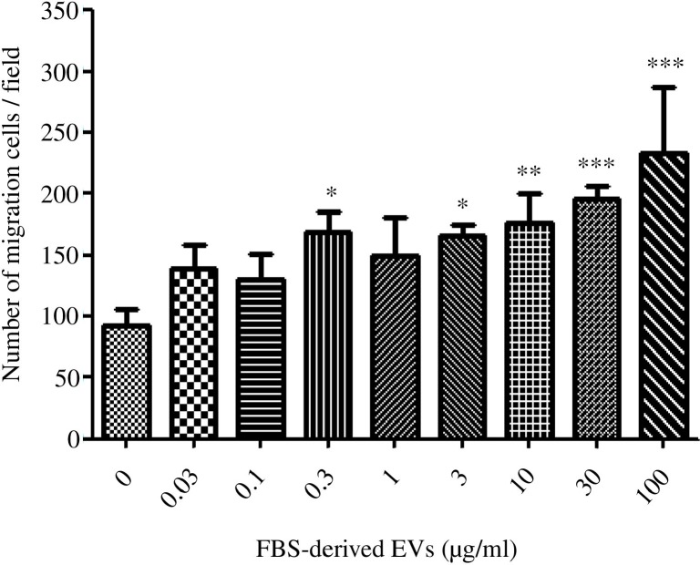
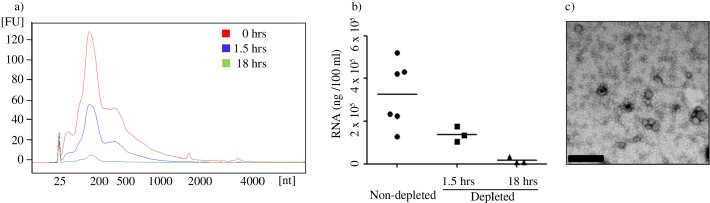
Extracellular vesicles (EVs), including the nano-sized exosomes, have the capacity to transfer multiple functional molecules between cells. In cell culture experiments, fetal bovine serum (FBS) is often used to supplement cell culture medium as a nutrient, but it is important to know that the FBS also contain significant quantities of EVs. The aim of the current study was to determine whether the FBS EVs can influence cultured cell phenotype, and secondly to determine the efficiency of FBS-EV elimination protocols. Firstly, FBS that had not been depleted of EVs induced a migratory phenotype in a lung cancer epithelial cell line (A549 cells), an effect that could be mimicked by isolated FBS EVs alone. FBS-derived EVs also contained RNA, which was protected from consecutive proteinase K and RNase A treatment. Comparison of common isolation protocols suggested that an 18-hour centrifugation period eliminates approximately 95% of RNA-containing FBS EVs, whereas a 1.5-hour protocol is insufficient. In conclusion, this study shows that FBS EVs substantially influence cultured cell behaviour, but also that they can be virtually removed by an 18-hour ultracentrifugation protocol.
Keywords: Depletion protocol; Exosomes; Extracellular vesicles; Fetal bovine serum; RNA.
Figures
Similar articles
-
Fetal Bovine Serum-Derived Extracellular Vesicles Persist within Vesicle-Depleted Culture Media.Int J Mol Sci. 2018 Nov 9;19(11):3538. doi: 10.3390/ijms19113538. Int J Mol Sci. 2018. PMID: 30423996 Free PMC article.
-
Efficient ultrafiltration-based protocol to deplete extracellular vesicles from fetal bovine serum.J Extracell Vesicles. 2018 Jan 21;7(1):1422674. doi: 10.1080/20013078.2017.1422674. eCollection 2018. J Extracell Vesicles. 2018. PMID: 29410778 Free PMC article.
-
Heat inactivation of foetal bovine serum performed after EV-depletion influences the proteome of cell-derived extracellular vesicles.J Extracell Vesicles. 2024 Jan;13(1):e12408. doi: 10.1002/jev2.12408. J Extracell Vesicles. 2024. PMID: 38263378 Free PMC article.
-
The dark side of foetal bovine serum in extracellular vesicle studies.J Extracell Vesicles. 2022 Oct;11(10):e12271. doi: 10.1002/jev2.12271. J Extracell Vesicles. 2022. PMID: 36214482 Free PMC article. Review.
-
Ribonucleic artefacts: are some extracellular RNA discoveries driven by cell culture medium components?J Extracell Vesicles. 2017 Jan 12;6(1):1272832. doi: 10.1080/20013078.2016.1272832. eCollection 2017. J Extracell Vesicles. 2017. PMID: 28326168 Free PMC article. Review.
Cited by
-
Transformed extracellular vesicles with high angiogenic ability as therapeutics of distal ischemic tissues.Front Cell Dev Biol. 2022 Aug 31;10:869850. doi: 10.3389/fcell.2022.869850. eCollection 2022. Front Cell Dev Biol. 2022. PMID: 36120585 Free PMC article.
-
Techniques used for the isolation and characterization of extracellular vesicles: results of a worldwide survey.J Extracell Vesicles. 2016 Oct 31;5:32945. doi: 10.3402/jev.v5.32945. eCollection 2016. J Extracell Vesicles. 2016. PMID: 27802845 Free PMC article.
-
In Vitro and In Vivo Analysis of Extracellular Vesicle-Mediated Metastasis Using a Bright, Red-Shifted Bioluminescent Reporter Protein.Adv Genet (Hoboken). 2022 Jan 19;3(1):2100055. doi: 10.1002/ggn2.202100055. eCollection 2022 Mar. Adv Genet (Hoboken). 2022. PMID: 36619349 Free PMC article.
-
Exosomes and pancreatic diseases: status, challenges, and hopes.Int J Biol Sci. 2019 Jul 20;15(9):1846-1860. doi: 10.7150/ijbs.35823. eCollection 2019. Int J Biol Sci. 2019. PMID: 31523187 Free PMC article. Review.
-
Stem Cell-Derived Exosomes as Nanotherapeutics for Autoimmune and Neurodegenerative Disorders.ACS Nano. 2019 Jun 25;13(6):6670-6688. doi: 10.1021/acsnano.9b01004. Epub 2019 May 29. ACS Nano. 2019. PMID: 31117376 Free PMC article.
References
-
- Denzer K, van Eijk M, Kleijmeer MJ, Jakobson E, de Groot C, Geuze HJ. Follicular dendritic cells carry MHC class II-expressing microvesicles at their surface. J Immunol. 2000;165:1259–65. - PubMed
-
- Valadi H, Ekström K, Bossios A, Sjöstrand M, Lee JJ, Lötvall JO. Exosome-mediated transfer of mRNAs and microRNAs is a novel mechanism of genetic exchange between cells. Nat Cell Biol. 2007;9:654–9. - PubMed
LinkOut - more resources
Full Text Sources
Other Literature Sources
