. 2014 Sep 25;10(9):e1004412.
doi: 10.1371/journal.ppat.1004412. eCollection 2014 Sep.

Exploitation of the complement system by oncogenic Kaposi's sarcoma-associated herpesvirus for cell survival and persistent infection

Affiliations

Exploitation of the complement system by oncogenic Kaposi's sarcoma-associated herpesvirus for cell survival and persistent infection

Myung-Shin Lee et al. PLoS Pathog. .

Abstract

During evolution, herpesviruses have developed numerous, and often very ingenious, strategies to counteract efficient host immunity. Specifically, Kaposi's sarcoma-associated herpesvirus (KSHV) eludes host immunity by undergoing a dormant stage, called latency wherein it expresses a minimal number of viral proteins to evade host immune activation. Here, we show that during latency, KSHV hijacks the complement pathway to promote cell survival. We detected strong deposition of complement membrane attack complex C5b-9 and the complement component C3 activated product C3b on Kaposi's sarcoma spindle tumor cells, and on human endothelial cells latently infected by KSHV, TIME-KSHV and TIVE-LTC, but not on their respective uninfected control cells, TIME and TIVE. We further showed that complement activation in latently KSHV-infected cells was mediated by the alternative complement pathway through down-regulation of cell surface complement regulatory proteins CD55 and CD59. Interestingly, complement activation caused minimal cell death but promoted the survival of latently KSHV-infected cells grown in medium depleted of growth factors. We found that complement activation increased STAT3 tyrosine phosphorylation (Y705) of KSHV-infected cells, which was required for the enhanced cell survival. Furthermore, overexpression of either CD55 or CD59 in latently KSHV-infected cells was sufficient to inhibit complement activation, prevent STAT3 Y705 phosphorylation and abolish the enhanced survival of cells cultured in growth factor-depleted condition. Together, these results demonstrate a novel mechanism by which an oncogenic virus subverts and exploits the host innate immune system to promote viral persistent infection.


Conflict of interest statement

The authors have declared that no competing interests exist.

Figures

Figure 1
Figure 1. Detection of C5b-9 and C3d depositions on KS spindle tumor cells.
(A–C) Representative illustration of immunohistochemical detection of C5b-9 deposition (A), C3d deposition (B) and KSHV latent protein LANA (C) in human KS tumors. Typical KS tumor spindle cells are indicated by red arrows. Black arrows show C3d-negative infiltrated immune cells (B). (D–E) Negative staining in KS tumors using matched non-immune sera for C5b-9 (D) and C3d (E) antibodies, respectively. (F–H) Negative control staining for C5b-9 deposition (F), C3d deposition (G) and LANA (H) in the uninvolved adjacent tissues using the respective antibodies. The scale bar is 10 µm.
Figure 2
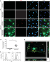
Figure 2. Detection and quantification of C5b-9 and C3b depositions on latently KSHV-infected endothelial cells.
(A) Detection of C5b-9 deposition on TIME-KSHV but not TIME cells. Cells were incubated with 10% normal or heat-inactivated human serum for 30 min and stained for C5b-9 deposition by immunofluorescence staining. The scale bar is 50 µm. (B) Quantification of C5b-9 deposition on TIME and TIME KSHV cells. Ten images from each treatment shown in Figure S2 were analyzed for C5b-9-positive areas using the ImageJ software. The average C5b-9 staining area per cell was calculated by dividing the total pixels of C5b-9 staining in a microscopic field by the cell number. Each dot represents the analyzed value from one field. The average values of ten analyses were indicated as horizontal black bars. *** P<0.001. (C) Flow cytometry analysis of C3b staining on TIME and TIME-KSHV cells. White areas are C3b-positive cells. Grey areas are cells stained with an isotype-matched antibody control. (D) Cell surface localization of C5b-9 deposition on TIME-KSHV cells. TIME-KSHV cells were incubated with 10% normal human serum for 30 min, and stained for C5b-9 (red), integrin αVβ3 (captured in the far-red channel but pseudo-colored in green to facilitate visualization) to label plasma membrane and DAPI (blue) to localize the nucleus. Z-stack images were acquired with confocal laser-scanning microscopy. Three-dimensional software was used to generate z-projection images from at least 70 confocal images of 0.1 µm sections. The 3-D images (XY panels) were rotated on the x-axis (XZ panels) and y-axis (YZ panels) to visualize C5b-9 localization on the cell membrane. Arrows show representative areas of C5b-9 depositions on cell surfaces. Images of TIME cells incubated with normal human serum, and TIME and TIME-KSHV cells incubated with heat-inactivated serum are shown in Figure S3.
Figure 3
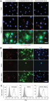
Figure 3. C5b-9 deposition on latently KSHV-infected endothelial cells is mediated by the alternative complement pathway.
(A) C3 protein was essential for C5b-9 deposition on TIME-KSHV cells. TIME or TIME-KSHV cells were incubated for 30 min with C3-depleted human serum or C3-depleted human serum reconstituted with purified C3 protein. Normal human serum was used as a positive control. Immunofluorescence staining was performed to detect C5b-9 deposition. The scale bar is 50 µm. (B–C) Activation of complement in TIME-KSHV cells was sensitive to EDTA but resistant to EGTA and MgCl2. TIME-KSHV cells were incubated with normal human serum with or without the presence of 10 mM EGTA and 20 mM MgCl2 or 20 mM EDTA. C5b-9 deposition was detected by immunofluorescence staining (B) and C3b was detected by flow cytometry analysis (C), respectively. The scale bar is 100 µm.
Figure 4
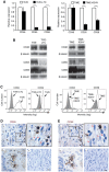
Figure 4. Complement regulatory proteins CD55 and CD59 but not CD46 are downregulated on latently KSHV-infected endothelial cells and on KS tumor cells.
(A) Relative expression levels of CD46, CD55 and CD59 mRNAs in uninfected and KSHV-infected endothelial cells. The level of mRNA expression was measured by RT-qPCR. GAPDH gene was used as a calibration control. The expression levels of uninfected cells were set as “1”. Results shown as means ± SD are representative from three independent experiments. * P<0.05, ** P<0.01 and *** P<0.001 by Student's t-test. (B) Total protein levels of CD46, CD55 and CD59 were examined by Western-blotting using antibodies to CD46, CD55 and CD59, respectively. (C) Surface protein levels of CD55 and CD59 on uninfected and KSHV-infected endothelial cells were analyzed by flow cytometry. (D–E) Representative illustration of detection of CD55 (D) and CD59 (E) in KS tumors. (a) Paraffin-embedded KS tumor sections with typical spindle cells were stained with antibodies to C55 and CD59. Representative areas are shown in the top right panel. Typical KS tumor spindle cells are indicated by red arrows while immune cells are indicated by black arrows. (b) Uninvolved adjacent tissues with endothelial cells and mononuclear cells were stained for CD55 and CD59 as positive controls. Representative mononuclear cells were indicated with black arrows. (c) Negative control staining with the isotype-matched control antibodies in the same KS tumors as (a). The scale bar is 10 µm.
Figure 5
Figure 5. Factor H is required for resistance to complement-mediated cytolysis of latently KSHV-infected endothelial cells.
(A–B) Most of latently KSHV-infected endothelial cells are resistant to complement-mediated cytolysis. TIME or TIME-KSHV cells were incubated with 10% normal human serum or heat-inactivated human serum for 1 h and 4 h, and the number of dead cells (A) and live cells (B) were determined. (C) Deposition of factor H but not factor I on TIME-KSHV cells following complement activation. TIME or TIME KSHV cells were incubated for 30 min with normal human serum, and stained for C5b-9 (green) and factor H (red in left panel) or factor I (red in right panel). Note the colocalization of factor H with C5b-9. The scale bar is 80 µm. (D) Factor H was required for resistance to complement-mediated cytolysis. TIME-KSHV cells were incubated for 1 h with heat-inactivated human serum, normal human serum, factor H-depleted human serum, factor H-depleted human serum reconstituted with purified factor H protein or factor I-depleted human serum, and the number of dead cells (left panel) and live cells (right panel) were determined. All the experiments were performed in 6 well plates with 6 repeats. Results are means ± SD from two to three independent experiments. * P<0.05 and ** P<0.01 by Student's t-test.
Figure 6
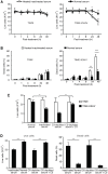
Figure 6. Complement activation promotes cell survival of latently KSHV-infected cells in growth factor-depleted medium.
(A–B) Complement activation promotes cell survival of latently KSHV-infected cells. Cells were cultured in endothelial cell medium depleted of growth factors and 10% of heat-inactivated or normal human serum, and live cells (A) and dead cells (B) were determined at the indicated times. (C) C3 was required for the enhanced cell survival of TIME-KSHV cells cultured in growth factor-depleted medium. TIME and TIME-KSHV cells were cultured for 48 h in 10% C3-depleted human serum or C3-depleted human serum reconstituted with purified C3 protein in endothelial cell medium without growth factors, and the numbers of live cells were determined. Cells cultured in heat-inactivated and normal human sera were used as controls. (D) Terminal complement complex was required for the enhanced survival of TIME-KSHV cells cultured in growth factor-depleted medium. TIME-KSHV cells were cultured for 48 h in 10% C6-depleted human serum or C6-depleted human serum reconstituted with purified C6 protein in endothelial cell medium depleted of growth factors, and the numbers of live cells (left panel) and dead cells (right panel) were determined. Cells cultured in heat-inactivated and normal human sera were used as controls. Results are means ± SD from three independent experiments with three repeats. * P<0.05, ** P<0.01 and *** P<0.001 by Student's t-test.
Figure 7
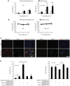
Figure 7. Activation of complement pathway induces STAT3 phosphorylation to enhance cell survival of latently KSHV-infected endothelial cells.
(A) The enhanced cell survival of KSHV-infected endothelial cells by complement was mediated by the STAT3 pathway. TIME-KSHV cells were cultured for 48 h in normal human serum in growth factor-depleted medium with and without JAK or STAT3 inhibitor, and the numbers of dead cells (left panel) and live cells (right panel) were determined. Cells cultured in heat-inactivated human serum were used as controls. Results are means ± SD from three independent experiments with three repeats. * P<0.05, ** P<0.01 and *** P<0.001 by Student's t-test. (B) Complement activation induced STAT3 tyrosine phosphorylation in TIME-KSHV cells. STAT3 tyrosine (Y705) and serine (S727) phosphorylation in TIME and TIME-KSHV cells switched from full endothelial cell medium with growth factors and 10% heat-inactivated human serum to endothelial cell medium depleted of growth factors with 10% heat-inactivated or normal human serum for the specified lengths of time. (C) Complement activation was required for the enhanced STAT3 tyrosine phosphorylation of TIME-KSHV cells. STAT3 tyrosine phosphorylation was examined in cells cultured for 24 h in 10% C3-depleted human serum or C3-depleted human serum reconstituted with purified C3 protein in endothelial cell medium depleted of growth factors. (D) Formation of C5b-9 complexes was required for the enhanced STAT3 tyrosine phosphorylation. STAT3 tyrosine phosphorylation was examined in cells cultured for 24 h in 10% C6-depleted human serum or C6-depleted human serum reconstituted with purified C6 protein in endothelial cell medium deprived of growth factors. (E) STAT3 tyrosine phosphorylation was unchanged without the continuous presence of normal human serum and growth factors. STAT3 tyrosine phosphorylation was examined in cells cultured in 10% heat-inactivated or normal human serum for 1 h, and then in endothelial cell medium without any serum and growth factors for additional 23 h. (F) JAK but not Src activation mediated STAT3 tyrosine phosphorylation. STAT3 tyrosine phosphorylations was examined in TIME-KSHV cells cultured in 10% normal human serum in endothelial cell medium depleted of growth factors for 24 h with or without the presence of STAT3, JAK or Src inhibitor.
Figure 8
Figure 8. Overexpression of CD55 and CD59 in latently KSHV-infected endothelial cells abolishes complement activation, induction of STAT3 phosphorylation, and enhancement of cell survival.
(A–B) Western-blot examination of CD55 (A) and CD59 (B) protein expression in TIME-KSHV cells with stable overexpression of CD55 and CD59. TIME cells were used as controls for the relative expression levels of CD55 and CD59. (C) Flow cytometry analysis of CD55 (left panel) and CD59 (right panel) cell surface expression on TIME-KSHV cells with stable overexpression of CD55 and CD59. TIME cells were used as controls for the relative expression levels of CD55 and CD59. (D) Quantification of C5b-9 deposition on TIME-KSHV cells with overexpression of CD55, CD59 or vector control. Box and whisker plots showing the mean (middle line) and 25–75th percentiles (lower and top box lines) of the average C5b-9 deposited areas on TIME cells (n = 10), TIME-KSHV cells (n = 10), and TIME-KSHV cells with overexpression of CD55 (n = 10), CD59 (n = 10) or vector control (n = 10) following exposure to 10% normal human serum for 1 h. Top and lower lines indicate the maximal and minimal values. Ten images from each cell type shown in Figure S14 were analyzed for C5b-9-positive areas using the ImageJ software. The average C5b-9 staining area per cell was calculated by dividing the total pixels of C5b-9 staining in a microscopic field by the cell number. (E) Flow cytometry analysis of C3b deposition on TIME-KSHV cells with overexpression of CD55 (left panel) or CD59 (right panel). TIME cells and TIME-KSHV cells transduced with the vector were used as controls. (F) Overexpression of CD55 or CD59 abolished complement activation of STAT3 tyrosine phosphorylation in TIME-KSHV cells. STAT3 tyrosine phosphorylation was examined in cells cultured in 10% normal human serum for 24 h. (G) Overexpression of CD55 or CD59 abolished the enhanced cell survival in TIME-KSHV cells cultured in medium depleted of growth factors. Cells were cultured for 48 h in heat-inactivated or normal human serum in endothelial cell medium depleted of growth factors in the presence of the indicated serum, and live cells were determined. Results are means ± SD from three independent experiments with three repeats. * P<0.05 and ** P<0.01 by Student's t-test.

Similar articles

Cited by

References

    1. Cesarman E, Chang Y, Moore PS, Said JW, Knowles DM (1995) Kaposi's sarcoma-associated herpesvirus-like DNA sequences in AIDS-related body-cavity-based lymphomas. New Engl J Med 332: 1186–1191. - PubMed
    1. Chang Y, Cesarman E, Pessin MS, Lee F, Culpepper J, et al. (1994) Identification of herpesvirus-like DNA sequences in AIDS-associated Kaposi's sarcoma. Science 266: 1865–1869. - PubMed
    1. Soulier J, Grollet L, Oksenhendler E, Cacoub P, Cazals-Hatem D, et al. (1995) Kaposi's sarcoma-associated herpesvirus-like DNA sequences in multicentric Castleman's disease. Blood 86: 1276–1280. - PubMed
    1. Cesarman E (2011) Gammaherpesvirus and lymphoproliferative disorders in immunocompromised patients. Cancer Lett 305: 163–174. - PMC - PubMed
    1. Ganem D (2010) KSHV and the pathogenesis of Kaposi's sarcoma: listening to human biology and medicine. J Clin Invest 120: 939–949. - PMC - PubMed

Publication types

MeSH terms