

doi: 10.1038/cr.2014.111.
Epub 2014 Aug 22.
Osteogenic fate of hypertrophic chondrocytes
Affiliations
- PMID: 25145361
- PMCID: PMC4185343
- DOI: 10.1038/cr.2014.111
Item in Clipboard
Osteogenic fate of hypertrophic chondrocytes
Cell Res.
2014 Oct.
No abstract available
Figures
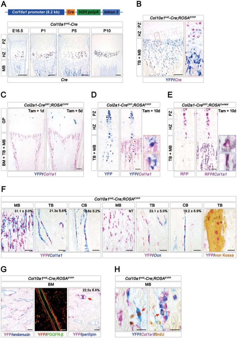
Multipotential fates of hypertrophic chondrocyte. (A) The transgenic vector contained an 8.2 kb promoter of mouse type X collagen (Col10a1), Cre cDNA, human growth hormone (hGH) polyadenylation signal and a 3.2 kb 2nd intron of Col10a1. Isotopic in situ hybridization assay was used to detect the transgenic Cre mRNA in tibia proximal growth plate from Col10a1int2-Cre transgenic mice at E16.5, P1, P5 and P10. (B) Double staining of YFP (blue) by immunohistochemistry (IHC) and Cre mRNA (fuchsia) by in situ hybridization in 10-day-old Col10a1int2-Cre;ROSAEYFP tibia. The right panel shows the high-magnification image of the area boxed in red on the left. (C) After 1 and 5 days following tamoxifen administration (50 mg/kg) at P5, Col2a1-CreERT;ROSAEYFP tibia was stained by YFP (blue) and Col1a1 (fuchsia) antibodies. The clonal columns of YFP+ cells were detectable in Col2a1-CreERT;ROSAEYFP chondrocyte zone. (D) After 10 days following tamoxifen administration at P5, Col2a1-CreERT;ROSAEYFP tibia was stained by YFP (blue) antibody, showing a column of YFP+ cells entering into the metaphysis. IHC double staining of YFP (blue) and type I collagen (Col1a1) (fuchsia) showed that some YFP+ cells underneath the growth plate were labeled with Col1a1 (red frames, high-magnification images of areas boxed on the left). (E) After 10 days following tamoxifen administration (50 mg/kg) at P5, Col2a1-CreERT;ROSAConfetti tibia was stained by RFP (fuchsia) antibody. Double staining of RFP (fuchsia) by IHC and Col1a1 (purple grey) mRNA by in situ hybridization showed that some RFP+ cells in metaphysis were Col1a1-positive (red frames, high-magnification images of areas boxed on the left). (F) Double staining of YFP (fuchsia) by IHC and the transcripts of Col1a1 or osteocalcin (Ocn) (blue) by in situ hybridization in metaphyseal, trabecular and cortical bones from 20-day-old Col10a1int2-Cre;ROSAEYFP tibia. The percentages of YFP+Col1a1+ and YFP+Ocn+osteogenic cells in total Col1a1+and Ocn+ cells from 3 images per mouse (n = 3) are shown in the top-right corner. Von Kossa staining showed that some YFP+cells (fuchsia) within the mineralized bone were osteocytes. (G) In 20-day-old Col10a1int2-Cre;ROSAEYFP bone marrow (BM), double immunostaining (left) of YFP (fuchsia) and endomucin (blue, marker for endothelial cells) as well as double immunofluorescence analysis (middle) using YFP (red) and PDGFR-β (green, marker for pericytes) antibodies indicated that some YFP+ cells were adjacent to the endothelial cells and vascular pericytes. Double immunostaining (right) of YFP (fuchsia) and perilipin (blue, marker for adipocytes) showed that some YFP+ cells were adipocytes (red arrow). (H) Triple staining of YFP (blue) and BrdU (brown) by IHC along with Col1a1 mRNA (fuchsia) by in situ hybridization in 20-day-old Col10a1int2-Cre;ROSAEYFP metaphyseal bones (MB). PZ, proliferation zone; HZ, hypertrophic zone; MB, metaphyseal bone; TB, trabecular bone; BM, bone marrow. Scale bars are 200 μm (A-C), 50 μm (D-F) and 25 μm (G, H).
Similar articles
-
A highly conserved enhancer in mammalian type X collagen genes drives high levels of tissue-specific expression in hypertrophic cartilage in vitro and in vivo.Matrix Biol. 2004 Aug;23(5):309-22. doi: 10.1016/j.matbio.2004.05.010. Matrix Biol. 2004. PMID: 15464363
-
BAC constructs in transgenic reporter mouse lines control efficient and specific LacZ expression in hypertrophic chondrocytes under the complete Col10a1 promoter.Histochem Cell Biol. 2007 Feb;127(2):183-94. doi: 10.1007/s00418-006-0236-8. Epub 2006 Oct 19. Histochem Cell Biol. 2007. PMID: 17051351 Free PMC article.
-
Tamoxifen-inducible gene deletion reveals a distinct cell type associated with trabecular bone, and direct regulation of PTHrP expression and chondrocyte morphology by Ihh in growth region cartilage.Dev Biol. 2007 Aug 1;308(1):93-105. doi: 10.1016/j.ydbio.2007.05.011. Epub 2007 May 18. Dev Biol. 2007. PMID: 17560974 Free PMC article.
-
Stimulation of type-X collagen gene transcription by retinoids occurs in part through the BMP signaling pathway.J Bone Joint Surg Am. 2003;85-A Suppl 3:29-33. doi: 10.2106/00004623-200300003-00006. J Bone Joint Surg Am. 2003. PMID: 12925606
-
Fate of growth plate hypertrophic chondrocytes: death or lineage extension?Dev Growth Differ. 2015 Feb;57(2):179-92. doi: 10.1111/dgd.12203. Epub 2015 Feb 24. Dev Growth Differ. 2015. PMID: 25714187 Review.
Cited by
-
Advances in Skeletal Dysplasia Genetics.Annu Rev Genomics Hum Genet. 2015;16:199-227. doi: 10.1146/annurev-genom-090314-045904. Epub 2015 Apr 22. Annu Rev Genomics Hum Genet. 2015. PMID: 25939055 Free PMC article. Review.
-
Dual pathways to endochondral osteoblasts: a novel chondrocyte-derived osteoprogenitor cell identified in hypertrophic cartilage.Biol Open. 2015 Apr 16;4(5):608-21. doi: 10.1242/bio.201411031. Biol Open. 2015. PMID: 25882555 Free PMC article.
-
Calcium-Sensing Receptors in Chondrocytes and Osteoblasts Are Required for Callus Maturation and Fracture Healing in Mice.J Bone Miner Res. 2020 Jan;35(1):143-154. doi: 10.1002/jbmr.3864. Epub 2019 Oct 18. J Bone Miner Res. 2020. PMID: 31498905 Free PMC article.
-
Fracture Healing in the Setting of Endocrine Diseases, Aging, and Cellular Senescence.Endocr Rev. 2022 Nov 25;43(6):984-1002. doi: 10.1210/endrev/bnac008. Endocr Rev. 2022. PMID: 35182420 Free PMC article. Review.
-
Wnt-associated adult stem cell marker Lgr6 is required for osteogenesis and fracture healing.Bone. 2023 Apr;169:116681. doi: 10.1016/j.bone.2023.116681. Epub 2023 Jan 25. Bone. 2023. PMID: 36708855 Free PMC article.
References
Publication types
MeSH terms
Substances
LinkOut - more resources
Full Text Sources
Other Literature Sources
