. 2014 Sep 2;111(35):12591-8.
doi: 10.1073/pnas.1413620111. Epub 2014 Aug 18.

Genetic and pharmacological reactivation of the mammalian inactive X chromosome

Affiliations

Genetic and pharmacological reactivation of the mammalian inactive X chromosome

Sanchita Bhatnagar et al. Proc Natl Acad Sci U S A. .

Abstract

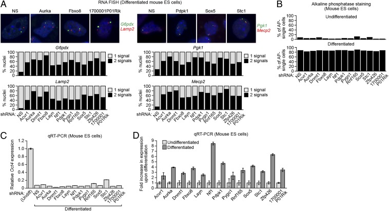
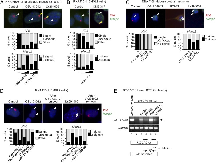
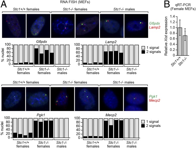
X-chromosome inactivation (XCI), the random transcriptional silencing of one X chromosome in somatic cells of female mammals, is a mechanism that ensures equal expression of X-linked genes in both sexes. XCI is initiated in cis by the noncoding Xist RNA, which coats the inactive X chromosome (Xi) from which it is produced. However, trans-acting factors that mediate XCI remain largely unknown. Here, we perform a large-scale RNA interference screen to identify trans-acting XCI factors (XCIFs) that comprise regulators of cell signaling and transcription, including the DNA methyltransferase, DNMT1. The expression pattern of the XCIFs explains the selective onset of XCI following differentiation. The XCIFs function, at least in part, by promoting expression and/or localization of Xist to the Xi. Surprisingly, we find that DNMT1, which is generally a transcriptional repressor, is an activator of Xist transcription. Small-molecule inhibitors of two of the XCIFs can reversibly reactivate the Xi, which has implications for treatment of Rett syndrome and other dominant X-linked diseases. A homozygous mouse knockout of one of the XCIFs, stanniocalcin 1 (STC1), has an expected XCI defect but surprisingly is phenotypically normal. Remarkably, X-linked genes are not overexpressed in female Stc1(-/-) mice, revealing the existence of a mechanism(s) that can compensate for a persistent XCI deficiency to regulate X-linked gene expression.

Keywords: MECP2; RNA FISH; RNA-seq.


Conflict of interest statement

The authors declare no conflict of interest.

Figures

Fig. 1.
Fig. 1.
Identification of factors required for mammalian XCI. (A) Schematic summary of the shRNA screen. The Xi is designated as such due to deletion of Xist on the Xa. (B) H4SV cells expressing an shRNA against 1 of the 13 candidates or, as a control, a nonsilencing (NS) shRNA were FACS sorted, and GFP-positive cells were isolated. For each KD cell line, the percentage of GFP-positive cells was expressed as the fold increase relative to that obtained with the NS shRNA, which was set to 1. (C) Two-color RNA FISH monitoring expression of G6pdx (red) and Lamp2 (green; Left) and Pgk1 (red) and Mecp2 (green; Right) in each of the 13 XCIF KD BMSL2 cell lines. DAPI staining is shown in blue. The experiment was performed at least twice, and representative images are shown (Upper) and the results quantified (Lower) from one experiment.
Fig. 2.
Fig. 2.
The XCIFs are required for initiation of XCI in mouse embryonic stem cells. (A) Two-color RNA FISH monitoring expression of G6pdx (green) and Lamp2 (red; Left) and Pgk1 (green) Mecp2 (red; Right) in the 13 XCIF KD ES cell lines following differentiation. DAPI staining is shown in blue. Representative images are shown (Upper), and the results quantified (Lower). (B) Percentage of alkaline phosphatase-negative single cells in the 13 XCIF KD ES cell lines before (Upper; undifferentiated) and after (Lower; differentiated) treatment with RA. (C) qRT-PCR analysis monitoring expression of Oct4 in the 13 XCIF KD ES cell lines following treatment with RA. As a control, expression of Oct4 in undifferentiated ES cells is shown and was set to 1. Error bars indicate SD. (D) qRT-PCR analysis of XCIFs in undifferentiated and differentiated mouse ES cells. Expression in differentiated ES cells was normalized to that observed in undifferentiated cells, which was set to 1. Error bars indicate SD.
Fig. 3.
Fig. 3.
XCIFs function by promoting Xist expression and/or localization, and DNMT1 is a transcriptional activator of Xist on the Xi. (A) qRT-PCR analysis monitoring Xist expression in the 13 XCIF KD ES cell lines following differentiation. Expression in differentiated ES cells was normalized to that obtained with the NS shRNA, which was set to 1. Error bars indicate SE. (B) RNA FISH monitoring localization of Xist in the 13 XCIF KD ES cell lines following differentiation. Cells were categorized as having either a typical Xist cloud or “other” pattern, which includes either the lack of a detectable Xist signal or presence of two small Xist signals, as in undifferentiated ES cells. (C) RNA FISH monitoring expression of Xist (Upper) and Mecp2 (Lower) in BMSL2 cells treated with an Xist locked nucleic acid antisense oligonucleotide (LNA ASO) or a control LNA ASO. (D) ChIP analysis monitoring binding of DNMT1 and POL2 to the Xist promoter and exon 2 in BMSL2 cells expressing a NS or Dnmt1 shRNA. Error bars indicate SD. (E) Nuclear run-on assay monitoring transcription of Xist, Hprt, and Tbp in BMSL2 cells expressing a NS or DNMT1 shRNA. (F) qRT-PCR analysis monitoring Xist levels in BMSL2 cells expressing a NS or Dnmt1 shRNA following treatment with actinomycin D. Actin mRNA was used as a normalization control. Error bars indicate SD. (G) qRT-PCR analysis monitoring Xist expression in MEFs isolated from female Dnmt1+/+ and Dnmt1−/− embryos. Four different litters were analyzed (n = 4 mice total per genotype), and the results were averaged. Expression was normalized to that observed in Dnmt1+/+ MEFs, which was set to 1. Error bars indicate SD. *P < 0.001 (Student t test). (H) qRT-PCR monitoring levels of Xist and Tsix in H4SV cells expressing a NS or DNMT1 shRNA. Expression was normalized to that obtained with the NS shRNA, which was set to 1. Error bars indicate SD. (I) qRT-PCR analysis monitoring Hprt and Xist expression in BMSL2 cells treated in the absence or presence of 5-azacytidine (5-AZA). Expression was normalized to that observed in the absence of 5-AZA, which was set to 1. Error bars indicate SD.
Fig. 4.
Fig. 4.
Reactivation of the Xi-linked Mecp2 gene by small-molecule XCIF inhibitors. (A and B) Two-color RNA FISH monitoring expression of Xist (red) and Mecp2 (green) in differentiated mouse ES cells treated with DMSO (control or –), OSU-03012, or LY294002 (A), and in BMSL2 cells treated with DMSO or GNE-317 (B). Representative images are shown (Upper) using the higher concentrations of the inhibitors, and the results quantified (Lower). The yellow arrowheads indicate colocalizing Xist and Mecp2 signals; the white arrowheads indicate Mecp2 signals not colocalizing with Xist. (C) Two-color RNA FISH monitoring Xist (red) and Mecp2 (green) expression in mouse cortical neurons treated with DMSO (control or –), OSU-03012, BX912, or LY294002. Representative images are shown (Upper), and the results quantified (Lower). The arrowheads indicate Mecp2 signals. (D) Two-color RNA FISH monitoring Xist (red) and Mecp2 (green) expression in BMSL2 cells treated with DMSO (control or –), LY294002, or OSU-03012, and at least 6 d following removal of the inhibitor. Representative images are shown (Upper), and the results quantified (Lower). The arrowheads indicate Mecp2 signals. (E) qRT-PCR monitoring Xi-linked wild-type MECP2 expression in human RTT fibroblasts treated with DMSO (–), 5-azacytidine (5-AZA), BX912, OSU-03012, or VX680. As a control, Xa-linked wild-type MECP2 expression was monitored in another clonal fibroblast cell line derived from the same RTT patient (lane 1). The arrowhead indicates the wild-type MECP2 qRT-PCR product. GAPDH was monitored as a loading control. (Lower) Schematic of the MECP2 wild-type (wt) and mutant (mut) alleles.
Fig. 5.
Fig. 5.
Defective XCI in female Stc1−/− MEFs. (A) Two-color RNA FISH monitoring expression of G6pdx (green) and Lamp2 (red; Upper) and Pgk1 (green) and Mecp2 (red; Lower) in female Stc1+/+ and Stc1−/− MEFs, and as a control male Stc1−/− MEFs. Representative images are shown (Upper), and the results quantified (Lower). (B) qRT-PCR analysis monitoring Xist expression in MEFs isolated from female Stc1+/+ and Stc1−/− embryos. Four different litters were analyzed (n = 4 mice total per genotype), and the results were averaged. Expression was normalized to that of the ribosomal gene RPL4, and Xist expression in Stc1+/+ MEFs was set to 1. Error bars indicate SD. *P < 0.001 (Student t test).
Fig. 6.
Fig. 6.
Defective XCI in female Stc1−/− mice is not accompanied by increased X-linked gene expression. (A) Schematic of the RNA-Seq analysis pipeline. (B) Distribution of log2-transformed ratio of X-linked gene expression in MEFs from female Stc1−/− (KO) and Stc1+/+ (WT) embryos (n = 3 per genotype). (C) Box plot of X-linked gene expression (log2-transformed FPKM) in MEFs from female Stc1−/− and Stc1+/+ embryos (n = 3 per genotype). The boxed areas span the first to the third quartile. The whiskers represent 15th and 85th percentiles. (D) qRT-PCR analysis monitoring expression of Mecp2 and Hprt in MEFs from two different litters of female Stc1−/− and Stc1+/+ embryos (n = 2 mice total per genotype). The results were normalized to those obtained in Stc1+/+ MEFs, which was set to 1. Error bars indicate SE. (E) Immunoblot showing MECP2 and STC1 levels in female Stc1+/+ and Stc1−/− MEFs (Left) or brain tissue female Stc1+/+ and Stc1−/− P1 mice (Right) (n = 3 per genotype). α-Tubulin (TUBA) was monitored as a loading control. (F) qRT-PCR analysis of Stc1, Xist, Mecp2, and Hprt expression in BMSL2 cells expressing a NS or STC1 shRNA. The results were normalized to those obtained with the NS shRNA, which was set to 1. Error bars indicate SE. (G) Immunoblot showing MECP2 and STC1 levels in BMSL2 cells expressing a NS or Stc1 shRNA.

Similar articles

Cited by

References

    1. Lyon MF. X-chromosome inactivation as a system of gene dosage compensation to regulate gene expression. Prog Nucleic Acid Res Mol Biol. 1989;36:119–130. - PubMed
    1. Brockdorff N. Chromosome silencing mechanisms in X-chromosome inactivation: Unknown unknowns. Development. 2011;138(23):5057–5065. - PubMed
    1. Plath K, Mlynarczyk-Evans S, Nusinow DA, Panning B. Xist RNA and the mechanism of X chromosome inactivation. Annu Rev Genet. 2002;36:233–278. - PubMed
    1. Leeb M, Steffen PA, Wutz A. X chromosome inactivation sparked by non-coding RNAs. RNA Biol. 2009;6(2):94–99. - PubMed
    1. Amir RE, et al. Rett syndrome is caused by mutations in X-linked MECP2, encoding methyl-CpG-binding protein 2. Nat Genet. 1999;23(2):185–188. - PubMed

Publication types

MeSH terms

Associated data