Hypertrophic chondrocytes can become osteoblasts and osteocytes in endochondral bone formation
- PMID: 25092332
- PMCID: PMC4143064
- DOI: 10.1073/pnas.1302703111
Hypertrophic chondrocytes can become osteoblasts and osteocytes in endochondral bone formation
Abstract
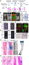
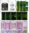
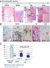
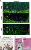
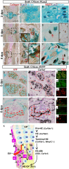
According to current dogma, chondrocytes and osteoblasts are considered independent lineages derived from a common osteochondroprogenitor. In endochondral bone formation, chondrocytes undergo a series of differentiation steps to form the growth plate, and it generally is accepted that death is the ultimate fate of terminally differentiated hypertrophic chondrocytes (HCs). Osteoblasts, accompanying vascular invasion, lay down endochondral bone to replace cartilage. However, whether an HC can become an osteoblast and contribute to the full osteogenic lineage has been the subject of a century-long debate. Here we use a cell-specific tamoxifen-inducible genetic recombination approach to track the fate of murine HCs and show that they can survive the cartilage-to-bone transition and become osteogenic cells in fetal and postnatal endochondral bones and persist into adulthood. This discovery of a chondrocyte-to-osteoblast lineage continuum revises concepts of the ontogeny of osteoblasts, with implications for the control of bone homeostasis and the interpretation of the underlying pathological bases of bone disorders.
Keywords: bone repair; chondrocyte lineage; osteoblast ontogeny.
Conflict of interest statement
The authors declare no conflict of interest.
Figures
Comment in
-
Developmental biology: Is there such a thing as a cartilage-specific knockout mouse?Nat Rev Rheumatol. 2014 Dec;10(12):702-4. doi: 10.1038/nrrheum.2014.168. Epub 2014 Sep 30. Nat Rev Rheumatol. 2014. PMID: 25266454 No abstract available.
Similar articles
-
Fate of growth plate hypertrophic chondrocytes: death or lineage extension?Dev Growth Differ. 2015 Feb;57(2):179-92. doi: 10.1111/dgd.12203. Epub 2015 Feb 24. Dev Growth Differ. 2015. PMID: 25714187 Review.
-
New morphological evidence of the 'fate' of growth plate hypertrophic chondrocytes in the general context of endochondral ossification.J Anat. 2020 Feb;236(2):305-316. doi: 10.1111/joa.13100. Epub 2019 Dec 9. J Anat. 2020. PMID: 31820452 Free PMC article.
-
Chondrocytes transdifferentiate into osteoblasts in endochondral bone during development, postnatal growth and fracture healing in mice.PLoS Genet. 2014 Dec 4;10(12):e1004820. doi: 10.1371/journal.pgen.1004820. eCollection 2014 Dec. PLoS Genet. 2014. PMID: 25474590 Free PMC article.
-
[Transdifferentiation of chondrocytes into osteogenic cells].Chir Narzadow Ruchu Ortop Pol. 2006;71(3):199-203. Chir Narzadow Ruchu Ortop Pol. 2006. PMID: 17131726 Review. Polish.
-
The extended chondrocyte lineage: implications for skeletal homeostasis and disorders.Curr Opin Cell Biol. 2019 Dec;61:132-140. doi: 10.1016/j.ceb.2019.07.011. Epub 2019 Sep 18. Curr Opin Cell Biol. 2019. PMID: 31541943 Review.
Cited by
-
Independent mesenchymal progenitor pools respectively produce and maintain osteogenic and chondrogenic cells in zebrafish.Dev Growth Differ. 2024 Feb;66(2):161-171. doi: 10.1111/dgd.12908. Epub 2024 Jan 9. Dev Growth Differ. 2024. PMID: 38193362 Free PMC article.
-
Advances in Skeletal Dysplasia Genetics.Annu Rev Genomics Hum Genet. 2015;16:199-227. doi: 10.1146/annurev-genom-090314-045904. Epub 2015 Apr 22. Annu Rev Genomics Hum Genet. 2015. PMID: 25939055 Free PMC article. Review.
-
Skeletal stem and progenitor cells in bone physiology, ageing and disease.Nat Rev Endocrinol. 2024 Oct 8. doi: 10.1038/s41574-024-01039-y. Online ahead of print. Nat Rev Endocrinol. 2024. PMID: 39379711 Review.
-
Sequential application of small molecule therapy enhances chondrogenesis and angiogenesis in murine segmental defect bone repair.J Orthop Res. 2023 Jul;41(7):1471-1481. doi: 10.1002/jor.25493. Epub 2022 Dec 23. J Orthop Res. 2023. PMID: 36448182 Free PMC article.
-
Osteoblast Differentiation at a Glance.Med Sci Monit Basic Res. 2016 Sep 26;22:95-106. doi: 10.12659/msmbr.901142. Med Sci Monit Basic Res. 2016. PMID: 27667570 Free PMC article.
References
-
- Karsenty G, Kronenberg HM, Settembre C. Genetic control of bone formation. Annu Rev Cell Dev Biol. 2009;25:629–648. - PubMed
-
- Day TF, Guo X, Garrett-Beal L, Yang Y. Wnt/beta-catenin signaling in mesenchymal progenitors controls osteoblast and chondrocyte differentiation during vertebrate skeletogenesis. Dev Cell. 2005;8(5):739–750. - PubMed
Publication types
MeSH terms
LinkOut - more resources
Full Text Sources
Other Literature Sources
Molecular Biology Databases

