

doi: 10.1128/JVI.01726-14.
Epub 2014 Jul 16.
Structural insights into the human metapneumovirus glycoprotein ectodomain
Affiliations
- PMID: 25031352
- PMCID: PMC4178817
- DOI: 10.1128/JVI.01726-14
Item in Clipboard
Structural insights into the human metapneumovirus glycoprotein ectodomain
J Virol.
2014 Oct.
Abstract
Human metapneumovirus is a major cause of respiratory tract infections worldwide. Previous reports have shown that the viral attachment glycoprotein (G) modulates innate and adaptive immune responses, leading to incomplete immunity and promoting reinfection. Using bioinformatics analyses, static light scattering, and small-angle X-ray scattering, we show that the extracellular region of G behaves as a heavily glycosylated, intrinsically disordered polymer. We discuss potential implications of these findings for the modulation of immune responses by G.
Copyright © 2014 Leyrat et al.
Figures
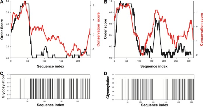
Computational analysis of HMPV (A and C) and HRSV (B and D) G sequence conservation, order/disorder propensity, and glycosylation sites. (A and B) The predicted disorder propensities and sequence conservation profiles are shown by black and red lines, respectively. Meta-disorder predictions were calculated following procedures described in reference . Sequence conservation was calculated using AL2CO (60) by applying a sliding average on a 20-residue window. (C and D) Location of predicted glycosylation sites along the amino acid sequence, shown as histogram bars, based on GlycoPred server output (61). HMPV sG (strain NL-1-00 [A1]) is predicted to contain 5 N-linked and 59 O-linked glycosylation sites, constituting an average of one site for every three residues.
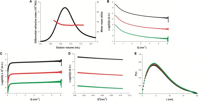
Biophysical characterization of HMPV sG. (A) Molecular mass determination of HMPV sG using SEC-MALLS-RI. The protein was purified by size exclusion chromatography on an S200 column equilibrated with 20 mM Tris (pH 7.5) and 150 mM NaCl prior to analysis. The black line shows the SEC elution profile as monitored by refractometry. The red line shows the molecular mass calculated from light scattering and refractometry data. (B, C, D, and E) Small-angle X-ray scattering (SAXS) experiments; (B) scattering curves of sG measured at concentrations of 4, 6, and 8 mg/ml are shown in green, red, and black, respectively; (C) Kratky plots showing linear behavior in the high Q range; (D) Guinier plots showing linear behavior in the low Q range; (E) distance distribution functions [P(r)] calculated using GNOM (62).
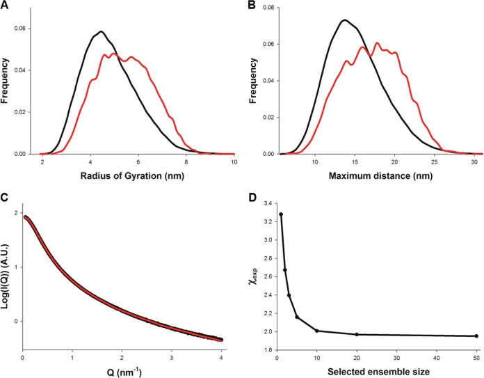
Analysis of sG flexibility using the ensemble optimization method (EOM). (A and B) Calculated radius of gyration (Rg) and maximal intramolecular distance (Dmax) distributions of the starting (black line) and optimized ensembles (red line). (C) Fitted SAXS profile of sG measured at 8 mg/ml. The experimental data are shown in black and the theoretical scattering from the optimized ensemble in red. (D) Variation of the goodness of fit (χexp) as a function of the optimized ensemble size.
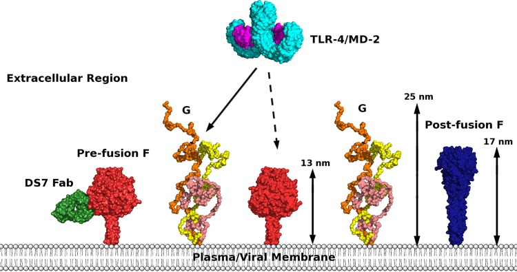
Comparison of the molecular dimensions of the extracellular regions of the HMPV F and G proteins, illustrating the G protein induced steric hindrance potentially hampering host factor-F protein interactions. The extracellular region of the Toll-like receptor 4–myeloid differentiation factor 2 (TLR-4/MD-2) complex (PDB identifier 3FXI) is represented as an example of such host factor. Homology models of the F protein trimers in the pre- and postfusion states form protrusions of 13 and 17 nm, respectively. Models were generated in HOMER (63) and are represented as red and blue surfaces. The homodimeric TLR-4 is shown in cyan and MD-2 in magenta, and three superimposed low-resolution models of sG are shown in orange, wheat color, and yellow. The anti-HMPV DS7 Fab bound to the prefusion model of F is shown in green based on PDB identifier 4DAG.
Similar articles
-
Structural dissection of human metapneumovirus phosphoprotein using small angle x-ray scattering.Sci Rep. 2017 Nov 1;7(1):14865. doi: 10.1038/s41598-017-14448-z. Sci Rep. 2017. PMID: 29093501 Free PMC article.
-
Human metapneumovirus glycoprotein G disrupts mitochondrial signaling in airway epithelial cells.PLoS One. 2013 Apr 23;8(4):e62568. doi: 10.1371/journal.pone.0062568. Print 2013. PLoS One. 2013. PMID: 23626834 Free PMC article.
-
Individual contributions of the human metapneumovirus F, G, and SH surface glycoproteins to the induction of neutralizing antibodies and protective immunity.Virology. 2006 Feb 20;345(2):492-501. doi: 10.1016/j.virol.2005.10.016. Epub 2005 Nov 21. Virology. 2006. PMID: 16300813
-
Human Metapneumovirus: Mechanisms and Molecular Targets Used by the Virus to Avoid the Immune System.Front Immunol. 2018 Oct 24;9:2466. doi: 10.3389/fimmu.2018.02466. eCollection 2018. Front Immunol. 2018. PMID: 30405642 Free PMC article. Review.
-
Modulation of Host Immunity by Human Respiratory Syncytial Virus Virulence Factors: A Synergic Inhibition of Both Innate and Adaptive Immunity.Front Cell Infect Microbiol. 2017 Aug 16;7:367. doi: 10.3389/fcimb.2017.00367. eCollection 2017. Front Cell Infect Microbiol. 2017. PMID: 28861397 Free PMC article. Review.
Cited by
-
Molecular characterization of human respiratory syncytial virus in Seoul, South Korea, during 10 consecutive years, 2010-2019.PLoS One. 2023 Apr 6;18(4):e0283873. doi: 10.1371/journal.pone.0283873. eCollection 2023. PLoS One. 2023. PMID: 37023101 Free PMC article.
-
Human metapneumovirus small hydrophobic (SH) protein downregulates type I IFN pathway signaling by affecting STAT1 expression and phosphorylation.Virology. 2016 Jul;494:248-56. doi: 10.1016/j.virol.2016.04.022. Epub 2016 Apr 27. Virology. 2016. PMID: 27131212 Free PMC article.
-
Structural basis for recognition of the central conserved region of RSV G by neutralizing human antibodies.PLoS Pathog. 2018 Mar 6;14(3):e1006935. doi: 10.1371/journal.ppat.1006935. eCollection 2018 Mar. PLoS Pathog. 2018. PMID: 29509814 Free PMC article.
-
Timing is everything: Fine-tuned molecular machines orchestrate paramyxovirus entry.Virology. 2015 May;479-480:518-31. doi: 10.1016/j.virol.2015.02.037. Epub 2015 Mar 12. Virology. 2015. PMID: 25771804 Free PMC article. Review.
-
Structure of the N-RNA/P interface indicates mode of L/P recruitment to the nucleocapsid of human metapneumovirus.Nat Commun. 2023 Nov 22;14(1):7627. doi: 10.1038/s41467-023-43434-5. Nat Commun. 2023. PMID: 37993464 Free PMC article.
References
-
- Boivin G, Abed Y, Pelletier G, Ruel L, Moisan D, Cote S, Peret TC, Erdman DD, Anderson LJ. 2002. Virological features and clinical manifestations associated with human metapneumovirus: a new paramyxovirus responsible for acute respiratory-tract infections in all age groups. J. Infect. Dis. 186:1330–1334. 10.1086/344319 - DOI - PubMed
Publication types
MeSH terms
Substances
Associated data
- Actions
- Actions
Grants and funding
LinkOut - more resources
Full Text Sources
Other Literature Sources
