. 2014 Jul;88(13):7367-78.
doi: 10.1128/JVI.00554-14. Epub 2014 Apr 16.

Kaposi's sarcoma-associated herpesvirus-encoded LANA can induce chromosomal instability through targeted degradation of the mitotic checkpoint kinase Bub1

Affiliations

Kaposi's sarcoma-associated herpesvirus-encoded LANA can induce chromosomal instability through targeted degradation of the mitotic checkpoint kinase Bub1

Zhiguo Sun et al. J Virol. 2014 Jul.

Abstract

Kaposi's sarcoma-associated herpesvirus (KSHV) has a significant contributory role in the development of three major human neoplastic or lymphoproliferative diseases: Kaposi's sarcoma (KS), primary effusion lymphoma (PEL), and multicentric Castleman's disease (MCD). These diseases are associated with chromosomal instability, a hallmark of human cancer. The latency-associated nuclear antigen (LANA) encoded by KSHV plays a key role in regulating a number of cellular pathways critical for oncogenesis. KSHV LANA alone can induce the development of B-cell hyperplasia and lymphoma in mice expressing LANA. LANA also induces chromosomal instability, thus promoting oncogenesis. However, the precise mechanism underlying LANA-mediated chromosomal instability remains uncharted. Here we report that LANA promoted the induction of chromosomal instability and the formation of micronuclei and multinucleation through its interaction with one of the critical spindle checkpoint proteins, Bub1, and the resulting degradation of Bub1. This interaction occurs through the Knl and kinase domains of Bub1, identified as important for stability and degradation. These results suggest that LANA can dysregulate Bub1 activity, which leads to aberrant chromosome replication and aneuploidy, thus contributing to KSHV-mediated oncogenesis.

Importance: This work represents the first set of results identifying a novel mechanism by which LANA, a latency-associated antigen encoded by KSHV, can induce the degradation of Bub1, a spindle checkpoint protein that is important for spindle checkpoint signaling and chromosome segregation. The downregulation of Bub1 mediated by LANA resulted in chromosomal instability, a hallmark of cancer. We further investigated the specific domains of Bub1 that are required for the interaction between LANA and Bub1. The results demonstrated that the Knl and kinase domains of Bub1 are required for the interaction between LANA and Bub1. In addition, we also investigated the mechanism by which LANA promoted Bub1 degradation. Our results showed that LANA interacted physically with the anaphase-promoting complex (APC/C), thus promoting the degradation of Bub1 in a ubiquitin-dependent process.


Figures

FIG 1
FIG 1
Bub1 levels are downregulated in cells latently infected with KSHV and expressing LANA. (A) Western blotting was used to detect the endogenous protein levels of Bub1 in KSHV-negative (BJAB and DG75) and KSHV-positive (BC-3, BCBL-1, JSC-1) cell lines. (B) Level of Bub1 protein in BJAB cells infected with KSHV. (C) LANA decreases the level of exogenous Bub1 protein in HEK-293 cells. HEK-293 cells were electroporated with increasing amounts of Flag-tagged LANA, Myc-tagged Bub1, and a GFP plasmid. Forty-eight hours later, the cells were collected for Western blot analysis, which was performed using the indicated antibodies. GFP served as a control for protein loading. (D) LANA knockdown increases Bub1 accumulation. Cell lysates from KSHV-positive B cells (BC-3 and JSC-1) in which LANA or a luciferase control had been stably knocked down (ShLANA or Shct, respectively) were subjected to Western blot analysis. (E) LANA decreases the level of endogenous Bub1 protein in HEK-293 cells and KSHV-negative B cells. HEK-293 and KSHV-negative B cells (BJAB, DG75) were electroporated with increasing amounts of Flag-tagged LANA. Forty-eight hours after electroporation, the cells were collected for Western blot analysis. (F) KSHV infection or LANA overexpression does not affect the mRNA level of Bub1. Total RNAs were isolated from KSHV-infected B cells (BJAB-KSHV, BJAB) and from control knockdown and LANA knockdown BC-3 cells (BC-3 Shct and BC-3 ShLANA, respectively), and the mRNA levels of Bub1 were analyzed by real-time PCR. The relative densities (RD) of Bub1 were quantified and plotted against the signal obtained from the control after normalization to glyceraldehyde-3-phosphate dehydrogenase (GAPDH) (for endogenous Bub1) or ectopically expressed GFP (for exogenous Bub1). Statistical significance was evaluated by using P values of <0.05 (*) and <0.01 (**).
FIG 2
FIG 2
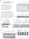
The Knl and kinase domains of Bub1 are necessary for its interaction with LANA. (A) (Left) Schematic diagram of Bub1 mutants. (Right) (Top) GST-tagged proteins purified from Escherichia coli. (Bottom) Eukaryotic expression of these deletion mutants (Myc tagged). ΔCSI and ΔCSII, Bub1 mutants with deletions of conserved domain I and conserved domain II, respectively. (B) Beads coated with GST or with fusion proteins consisting of GST and Bub1 domain deletion mutant proteins were incubated with lysates from HEK-293 cells electroporated with pA3F-LANA for GST pulldown assays. The pulldown of LANA was detected by Western blotting using antibody M2. (C) Bub1 interacts with LANA through its kinase and Knl binding domains. HEK-293 cells were electroporated with pA3F-LANA and pA3M-Bub1 or its related domain deletion mutants. Forty-eight hours after electroporation, the cell lysates were prepared for IP with antibody M2 and Western blotting with antibodies 9E10 and M2. (D) HEK-293 cells were electroporated with pA3F-LANA, pA3M-Bub1 or its related domain deletion mutants, and GFP. Forty-eight hours later, cells were collected for Western blot analysis. GFP served as a control for protein loading. The relative densities (RD) of Bub1 and its related deletion mutants were quantified and plotted against the signal obtained from the control after normalization to GFP. Statistical significance was evaluated by using P values of <0.05 (*) and <0.01 (**).
FIG 3
FIG 3
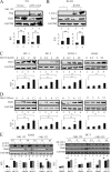
LANA-mediated Bub1 degradation is ubiquitin dependent. (A and B) LANA-transfected HEK-293 cells (A) and KSHV-infected BJAB cells (B) were either mock treated or treated with 20 μM MG132 for 12 h. The cells were then collected for Western blot analysis. (C) Cells were treated with MG132 for 12 h at 0, 0.2, 2, or 20 μM in a dose-dependent assay. The cells were then collected for Western blot analysis. (D) Cells were treated with MG132 at 20 μM for 0, 3, 6, or 12 h in a time course assay. The cells were then collected for Western blot analysis. (E) Cells were synchronized in the G1, S, or M phase as described in Materials and Methods. The cells were either mock treated or treated with MG132 (20 μM) for 12 h before they were harvested for Western blot analysis. The relative densities (RD) of Bub1 were quantified and plotted against the signal obtained from the control after normalization to GAPDH. Statistical significance was evaluated by using P values of <0.05 (*) and <0.01 (**).
FIG 4
FIG 4
LANA associates with the APC/C. (A) Bub1 does not interact with the EC5S complex. HEK-293 cells were electroporated with expression vectors as shown. Forty-eight hours postelectroporation, cells were harvested, and the cell lysates underwent IP with antibody M2 or an anti-GFP antibody. IP pellets and portions (10%) of lysates (input) were boiled, fractionated by SDS-PAGE, and subjected to Western blot analysis with the indicated antibodies. (B and C) LANA interacts with the APC/C. Cell lysates were used for IP with LANA- or CDC27-specific antibodies. IP pellets and portions (10%) of lysates were loaded onto SDS-PAGE gels, followed by Western blot analysis with the indicated antibodies. (D) LANA interacts with the activator protein of the APC/C. HEK-293 cells were electroporated with expression vectors as shown. Thirty-six hours postelectroporation, cells were treated with MG132 (20 μm) for 12 h before harvesting. The cell lysates were used for IP with an anti-Myc antibody. IP pellets and portions (10%) of lysates were boiled, fractionated by SDS-PAGE, and subjected to Western blot analysis with specific antibodies.
FIG 5
FIG 5
LANA promotes Bub1 ubiquitination. (A) HEK-293 cells were electroporated with expression vectors as shown. Forty-eight hours after electroporation, the cells were treated with MG132 for another 6 h. Cell lysates were then prepared for IP with 9E10, followed by Western blot analysis with the indicated antibodies. IB, immunoblot. (B) LANA ubiquitinates Bub1 in vitro. Bacterially expressed GST-Bub1, His-Ubca1 (E1), and His-Ubc5a (E2) were incubated with a purified LANA immune complex (E3) in kinase buffer for 1 h at 30°C. The sample was then resolved on an SDS-PAGE gel for Western blot analysis. (C) The Knl and kinase domains are required for LANA-mediated Bub1 ubiquitination in vitro. Bacterially expressed GST-Bub1 or its related deletion mutants, His-Ubca1 (E1), and His-Ubc5a (E2) were incubated with a purified LANA immune complex (E3) in kinase buffer for 1 h at 30°C. The samples were then resolved on an SDS-PAGE gel for Western blot analysis. (D) HEK-293 cells were electroporated with expression vectors as shown. Seventy-two hours after electroporation, the cells were treated with MG132 for another 12 h. Cell lysates were then prepared for IP with 9E10, followed by Western blot analysis with the indicated antibodies. (E) The endogenous Cdh1 of KSHV-positive cell lines (BC-3, BCBL-1, and JSC-1) was knocked down, and the protein levels of Bub1 were monitored. The relative densities (RD) of Bub1 and Cdh1 were quantified and plotted against the signals obtained from the control after normalization to GAPDH. Statistical significance was evaluated by using P values of <0.05 (*) and <0.01 (**).
FIG 6
FIG 6
Induction of micronuclei and multinucleation in various BC-3-derived cell clones. (A) (Left) BC-3-derived cell lines with stable expression of Bub1 or stable knockdown of Bub1 or LANA. The GFP signal appears in green in the bottom panels. (Right) (Top) Expression of LANA and Bub1 in the BC-3-derived cell lines. Lane numbers correspond to the numbering of the cell lines on the left. (Bottom) The relative densities (RD) of Bub1 were quantified and plotted against the signal obtained from the control after normalization to GAPDH. Statistical significance was evaluated by using P values of <0.05 (*) and <0.01 (**). (B) Examples of micronuclei and multinucleation induced in BC-3 cells in which either LANA or Bub1 was knocked down or in which Bub1 was stably expressed. Yellow arrows indicate micronuclei, and red arrows indicate multinucleation. (C) Quantitation of cells with multinucleation (left) or micronuclei (right) among BC-3-derived cell clones. All assays were carried out in triplicate.
FIG 7
FIG 7
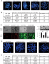
Metaphase chromosome spreads of BC-3- and BJAB-derived cell clones. (A) Representative metaphase spreads of the indicated BC-3-derived cell lines. (B) Distribution of chromosome numbers in the indicated BC-3-derived cell lines. Chromosomes were counted from metaphase spreads. (C) (Left) BJAB-derived cell lines with stable expression of LANA or stable knockdown of Bub1. (Right) (Top) Expression of LANA and Bub1 in the BJAB-derived cell lines. Lane numbers correspond to the numbering of the cell lines on the left. (Bottom) The relative densities (RD) of Bub1 were quantified and plotted against the signal obtained from the control after normalization to GAPDH. Statistical significance was evaluated by using P values of <0.05 (*) and <0.01 (**). (D) Representative metaphase spreads of the indicated BJAB-derived cell lines. (E) Distribution of chromosome numbers in the indicated BJAB-derived cell lines. Chromosomes were counted from metaphase spreads.

Similar articles

Cited by

References

    1. Chang Y, Cesarman E, Pessin MS, Lee F, Culpepper J, Knowles DM, Moore PS. 1994. Identification of herpesvirus-like DNA sequences in AIDS-associated Kaposi's sarcoma. Science 266:1865–1869. 10.1126/science.7997879 - DOI - PubMed
    1. Boshoff C, Schulz TF, Kennedy MM, Graham AK, Fisher C, Thomas A, McGee JO, Weiss RA, O'Leary JJ. 1995. Kaposi's sarcoma-associated herpesvirus infects endothelial and spindle cells. Nat. Med. 1:1274–1278. 10.1038/nm1295-1274 - DOI - PubMed
    1. Arvanitakis L, Mesri EA, Nador RG, Said JW, Asch AS, Knowles DM, Cesarman E. 1996. Establishment and characterization of a primary effusion (body cavity-based) lymphoma cell line (BC-3) harboring Kaposi's sarcoma-associated herpesvirus (KSHV/HHV-8) in the absence of Epstein-Barr virus. Blood 88:2648–2654 - PubMed
    1. Renne R, Zhong W, Herndier B, McGrath M, Abbey N, Kedes D, Ganem D. 1996. Lytic growth of Kaposi's sarcoma-associated herpesvirus (human herpesvirus 8) in culture. Nat. Med. 2:342–346. 10.1038/nm0396-342 - DOI - PubMed
    1. Sarid R, Flore O, Bohenzky RA, Chang Y, Moore PS. 1998. Transcription mapping of the Kaposi's sarcoma-associated herpesvirus (human herpesvirus 8) genome in a body cavity-based lymphoma cell line (BC-1). J. Virol. 72:1005–1012 - PMC - PubMed

Publication types

MeSH terms

LinkOut - more resources