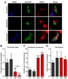
Evidence showing that tetraspanins inhibit HIV-1-induced cell-cell fusion at a post-hemifusion stage
- PMID: 24608085
- PMCID: PMC3970140
- DOI: 10.3390/v6031078
Evidence showing that tetraspanins inhibit HIV-1-induced cell-cell fusion at a post-hemifusion stage
Abstract
Human immunodeficiency virus type 1 (HIV-1) transmission takes place primarily through cell-cell contacts known as virological synapses. Formation of these transient adhesions between infected and uninfected cells can lead to transmission of viral particles followed by separation of the cells. Alternatively, the cells can fuse, thus forming a syncytium. Tetraspanins, small scaffolding proteins that are enriched in HIV-1 virions and actively recruited to viral assembly sites, have been found to negatively regulate HIV-1 Env-induced cell-cell fusion. How these transmembrane proteins inhibit membrane fusion, however, is currently not known. As a first step towards elucidating the mechanism of fusion repression by tetraspanins, e.g., CD9 and CD63, we sought to identify the stage of the fusion process during which they operate. Using a chemical epistasis approach, four fusion inhibitors were employed in tandem with CD9 overexpression. Cells overexpressing CD9 were found to be sensitized to inhibitors targeting the pre-hairpin and hemifusion intermediates, while they were desensitized to an inhibitor of the pore expansion stage. Together with the results of a microscopy-based dye transfer assay, which revealed CD9- and CD63-induced hemifusion arrest, our investigations strongly suggest that tetraspanins block HIV-1-induced cell-cell fusion at the transition from hemifusion to pore opening.
Figures


Similar articles
-
A role for tetraspanin proteins in regulating fusion induced by Burkholderia thailandensis.Med Microbiol Immunol. 2020 Aug;209(4):473-487. doi: 10.1007/s00430-020-00670-6. Epub 2020 Apr 6. Med Microbiol Immunol. 2020. PMID: 32253503 Free PMC article.
-
Formation of syncytia is repressed by tetraspanins in human immunodeficiency virus type 1-producing cells.J Virol. 2009 Aug;83(15):7467-74. doi: 10.1128/JVI.00163-09. Epub 2009 May 20. J Virol. 2009. PMID: 19458002 Free PMC article.
-
Human immunodeficiency virus type 1 assembly, budding, and cell-cell spread in T cells take place in tetraspanin-enriched plasma membrane domains.J Virol. 2007 Aug;81(15):7873-84. doi: 10.1128/JVI.01845-06. Epub 2007 May 23. J Virol. 2007. PMID: 17522207 Free PMC article.
-
Viruses and tetraspanins: lessons from single molecule approaches.Viruses. 2014 May 5;6(5):1992-2011. doi: 10.3390/v6051992. Viruses. 2014. PMID: 24800676 Free PMC article. Review.
-
Tetraspanins, Another Piece in the HIV-1 Replication Puzzle.Front Immunol. 2018 Aug 3;9:1811. doi: 10.3389/fimmu.2018.01811. eCollection 2018. Front Immunol. 2018. PMID: 30127789 Free PMC article. Review.
Cited by
-
Distinct regions of the large extracellular domain of tetraspanin CD9 are involved in the control of human multinucleated giant cell formation.PLoS One. 2014 Dec 31;9(12):e116289. doi: 10.1371/journal.pone.0116289. eCollection 2014. PLoS One. 2014. PMID: 25551757 Free PMC article.
-
CD9 Tetraspanin: A New Pathway for the Regulation of Inflammation?Front Immunol. 2018 Oct 9;9:2316. doi: 10.3389/fimmu.2018.02316. eCollection 2018. Front Immunol. 2018. PMID: 30356731 Free PMC article. Review.
-
Rab3a, a small GTP-binding protein, is required for the stabilization of the murine leukaemia virus Gag protein.Small GTPases. 2022 Jan;13(1):162-182. doi: 10.1080/21541248.2021.1939631. Epub 2021 Jun 27. Small GTPases. 2022. PMID: 34180342 Free PMC article.
-
HIV-1 adaptation studies reveal a novel Env-mediated homeostasis mechanism for evading lethal hypermutation by APOBEC3G.PLoS Pathog. 2018 Apr 20;14(4):e1007010. doi: 10.1371/journal.ppat.1007010. eCollection 2018 Apr. PLoS Pathog. 2018. PMID: 29677220 Free PMC article.
-
Meeting Review: 2018 International Workshop on Structure and Function of the Lentiviral gp41 Cytoplasmic Tail.Viruses. 2018 Nov 7;10(11):613. doi: 10.3390/v10110613. Viruses. 2018. PMID: 30405009 Free PMC article.
References
-
- Koenig S., Gendelman H.E., Orenstein J.M., dal Canto M.C., Pezeshkpour G.H., Yungbluth M., Janotta F., Aksamit A., Martin M.A., Fauci A.S. Detection of AIDS virus in macrophages in brain tissue from AIDS patients with encephalopathy. Science. 1986;233:1089–1093. - PubMed
-
- Frankel S.S., Wenig B.M., Burke A.P., Mannan P., Thompson L.D., Abbondanzo S.L., Nelson A.M., Pope M., Steinman R.M. Replication of HIV-1 in dendritic cell-derived syncytia at the mucosal surface of the adenoid. Science. 1996;272:115–117. - PubMed
Publication types
MeSH terms
Substances
Grants and funding
LinkOut - more resources
Full Text Sources
Other Literature Sources
Research Materials
Miscellaneous

