

MiR-26a promotes ovarian cancer proliferation and tumorigenesis
- PMID: 24466274
- PMCID: PMC3899311
- DOI: 10.1371/journal.pone.0086871
MiR-26a promotes ovarian cancer proliferation and tumorigenesis
Abstract
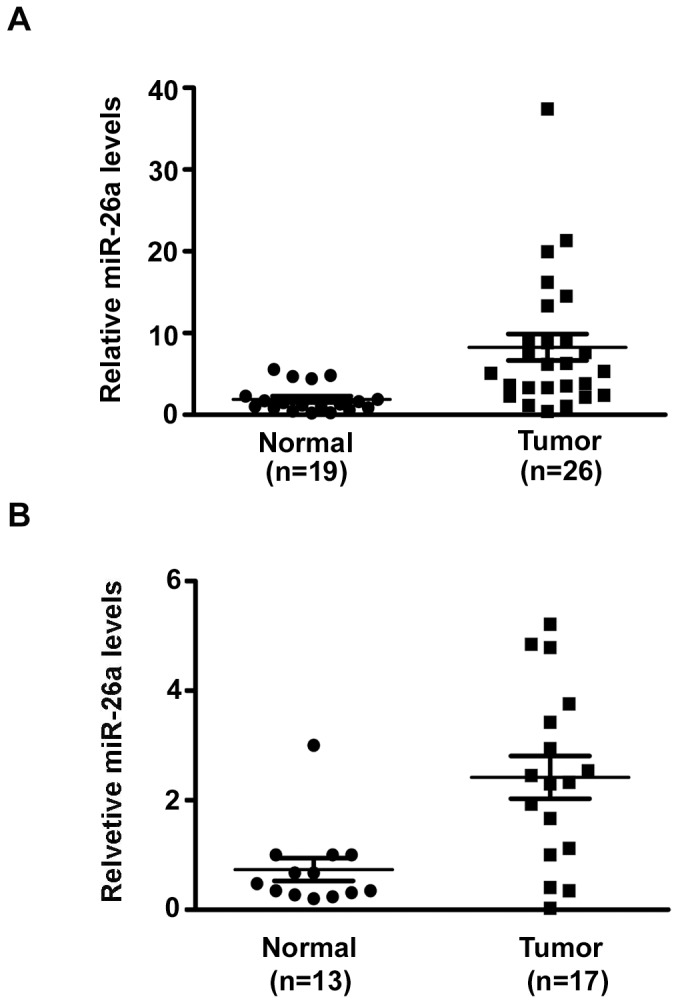
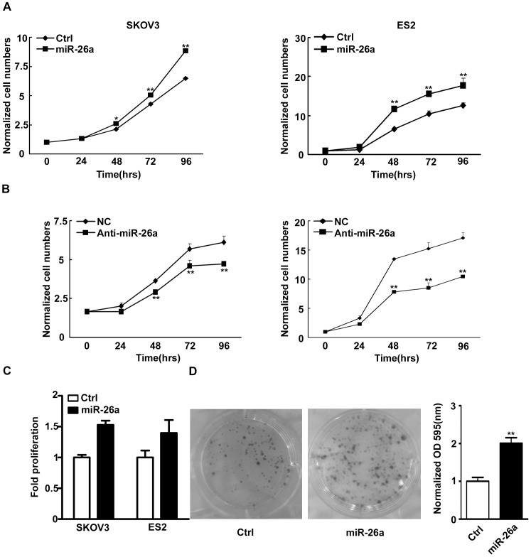
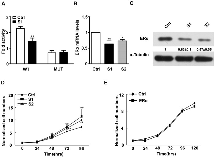
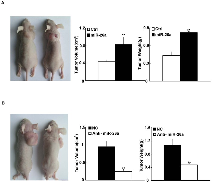
MicroRNAs (miRNAs) important for posttranscriptional gene expression are involved in the initiation and progression of human cancer. In this study, we reported that miR-26a was over-expressed in human EOC specimens and the expression level of extracellular miR-26a in plasma can distinguish patients from healthy controls in EOC. Ectopic expression of miR-26a in ovarian cancer (OC) cells increased cell proliferation and clonal formation. This growth promoting effect of OC cell growth was mediated by miR-26a inhibition of the posttranscription of ER-α. Furthermore, inhibition of miR-26a suppressed the tumor formation generated by injecting OC cells in nude mice. Our results suggest that aberrantly expressed miR-26a may contribute to OC development.
Conflict of interest statement
Figures
Similar articles
-
MiR-125b targets BCL3 and suppresses ovarian cancer proliferation.Int J Cancer. 2011 May 15;128(10):2274-83. doi: 10.1002/ijc.25575. Int J Cancer. 2011. PMID: 20658525
-
Inhibition of Ovarian Epithelial Carcinoma Tumorigenesis and Progression by microRNA 106b Mediated through the RhoC Pathway.PLoS One. 2015 May 1;10(5):e0125714. doi: 10.1371/journal.pone.0125714. eCollection 2015. PLoS One. 2015. PMID: 25933027 Free PMC article.
-
MicroRNA-26a inhibits angiogenesis by down-regulating VEGFA through the PIK3C2α/Akt/HIF-1α pathway in hepatocellular carcinoma.PLoS One. 2013 Oct 23;8(10):e77957. doi: 10.1371/journal.pone.0077957. eCollection 2013. PLoS One. 2013. PMID: 24194905 Free PMC article.
-
MiR-26a inhibits cell growth and tumorigenesis of nasopharyngeal carcinoma through repression of EZH2.Cancer Res. 2011 Jan 1;71(1):225-33. doi: 10.1158/0008-5472.CAN-10-1850. Cancer Res. 2011. PMID: 21199804
-
The loss of miR-26a-mediated post-transcriptional regulation of cyclin E2 in pancreatic cancer cell proliferation and decreased patient survival.PLoS One. 2013 Oct 8;8(10):e76450. doi: 10.1371/journal.pone.0076450. eCollection 2013. PLoS One. 2013. PMID: 24116110 Free PMC article.
Cited by
-
Circulating miR-20a and miR-26a as Biomarkers in Prostate Cancer.Asian Pac J Cancer Prev. 2019 May 25;20(5):1453-1456. doi: 10.31557/APJCP.2019.20.5.1453. Asian Pac J Cancer Prev. 2019. PMID: 31127907 Free PMC article.
-
Cell migration and proliferation are regulated by miR-26a in colorectal cancer via the PTEN-AKT axis.Cancer Cell Int. 2019 Apr 2;19:80. doi: 10.1186/s12935-019-0802-5. eCollection 2019. Cancer Cell Int. 2019. PMID: 30983885 Free PMC article.
-
A combination of circulating miRNAs for the early detection of ovarian cancer.Oncotarget. 2017 Sep 6;8(52):89811-89823. doi: 10.18632/oncotarget.20688. eCollection 2017 Oct 27. Oncotarget. 2017. PMID: 29163790 Free PMC article.
-
An overview of long non-coding RNAs in ovarian cancers.Oncotarget. 2016 Jul 12;7(28):44719-44734. doi: 10.18632/oncotarget.8089. Oncotarget. 2016. PMID: 26992233 Free PMC article. Review.
-
[MicroRNA-26a and Tumor].Zhongguo Fei Ai Za Zhi. 2017 Nov 20;20(11):769-774. doi: 10.3779/j.issn.1009-3419.2017.11.08. Zhongguo Fei Ai Za Zhi. 2017. PMID: 29167007 Free PMC article. Review. Chinese.
References
-
- Hoskins WJ (1995) Prospective on ovarian cancer: why prevent? J Cell Biochem Suppl 23: 189–199. - PubMed
-
- Filipowicz W, Bhattacharyya SN, Sonenberg N (2008) Mechanisms of post-transcriptional regulation by microRNAs: are the answers in sight? Nat Rev Genet 9: 102–114. - PubMed
-
- Calin GA, Croce CM (2006) MicroRNA signatures in human cancers. Nat Rev Cancer 6: 857–866. - PubMed
-
- Takamizawa J, Konishi H, Yanagisawa K, Tomida S, Osada H, et al. (2004) Reduced expression of the let-7 microRNAs in human lung cancers in association with shortened postoperative survival. Cancer Res 64: 3753–3756. - PubMed
Publication types
MeSH terms
Substances
Grants and funding
LinkOut - more resources
Full Text Sources
Other Literature Sources
Medical
