. 2014 Jan 16;156(1-2):317-331.
doi: 10.1016/j.cell.2013.12.010.

Regulation of ferroptotic cancer cell death by GPX4

Affiliations

Regulation of ferroptotic cancer cell death by GPX4

Wan Seok Yang et al. Cell. .

Abstract

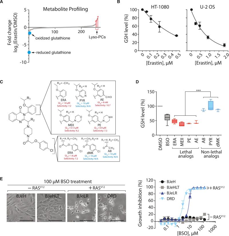
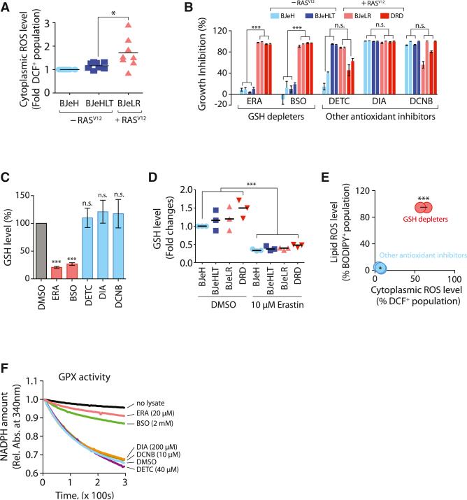
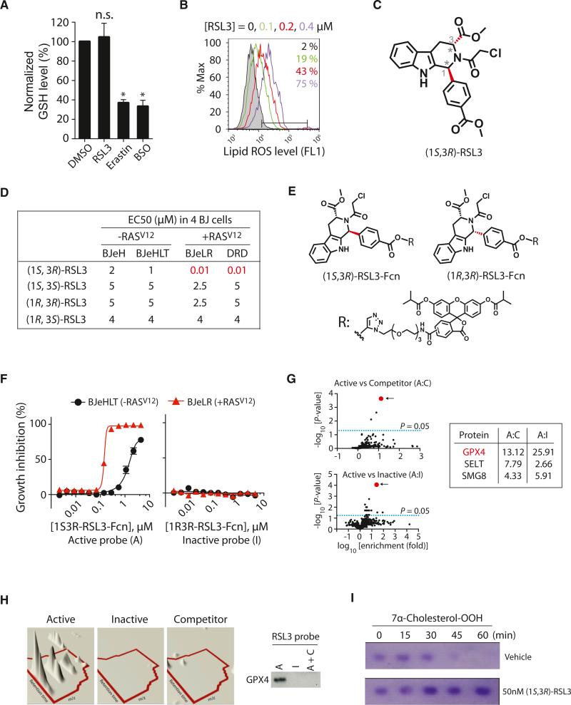
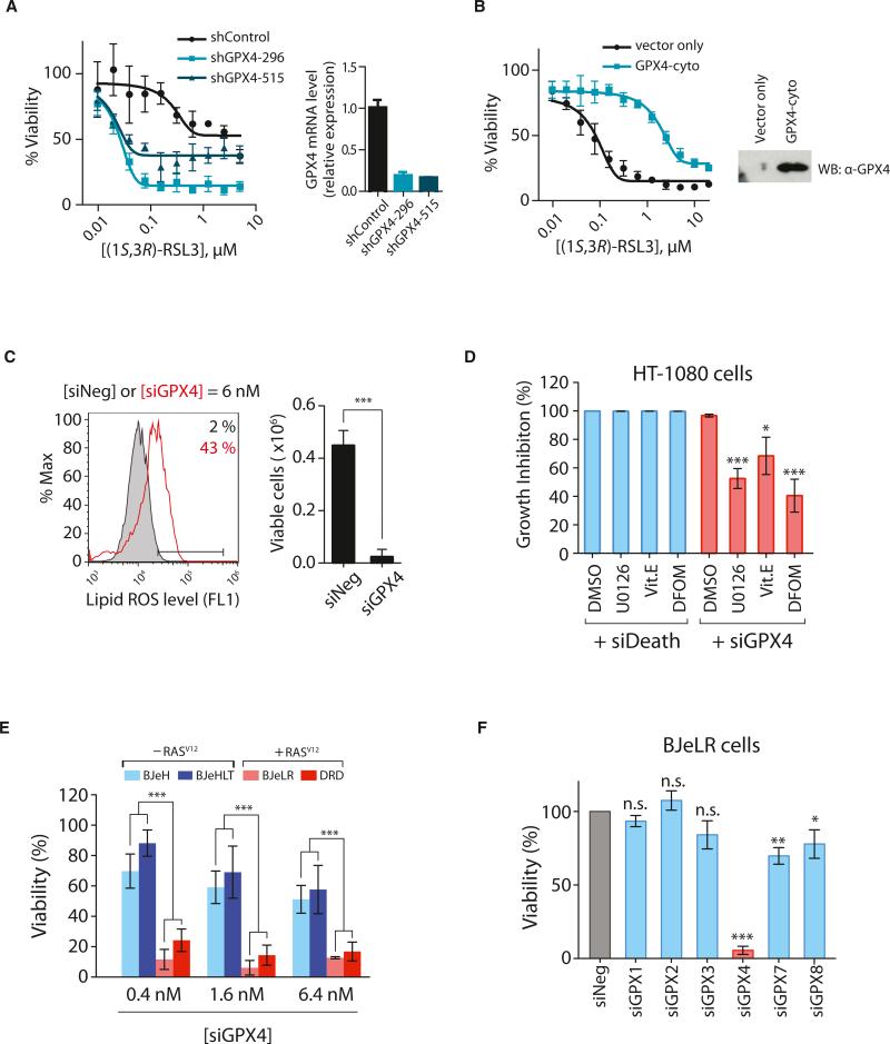
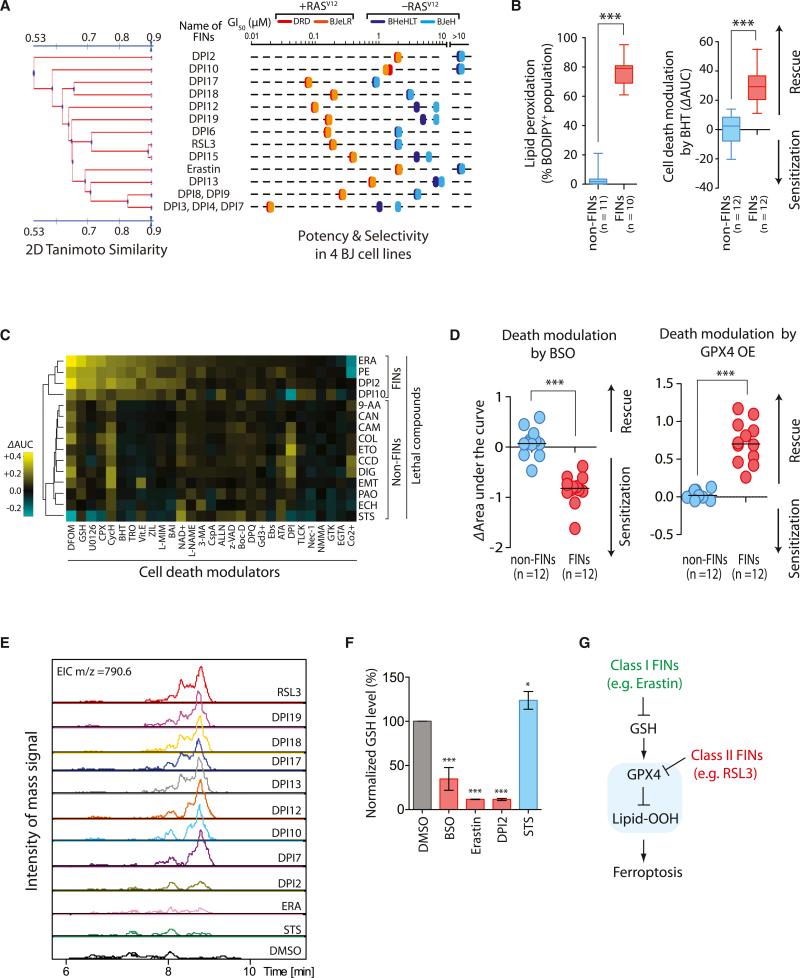
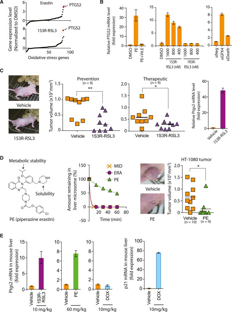
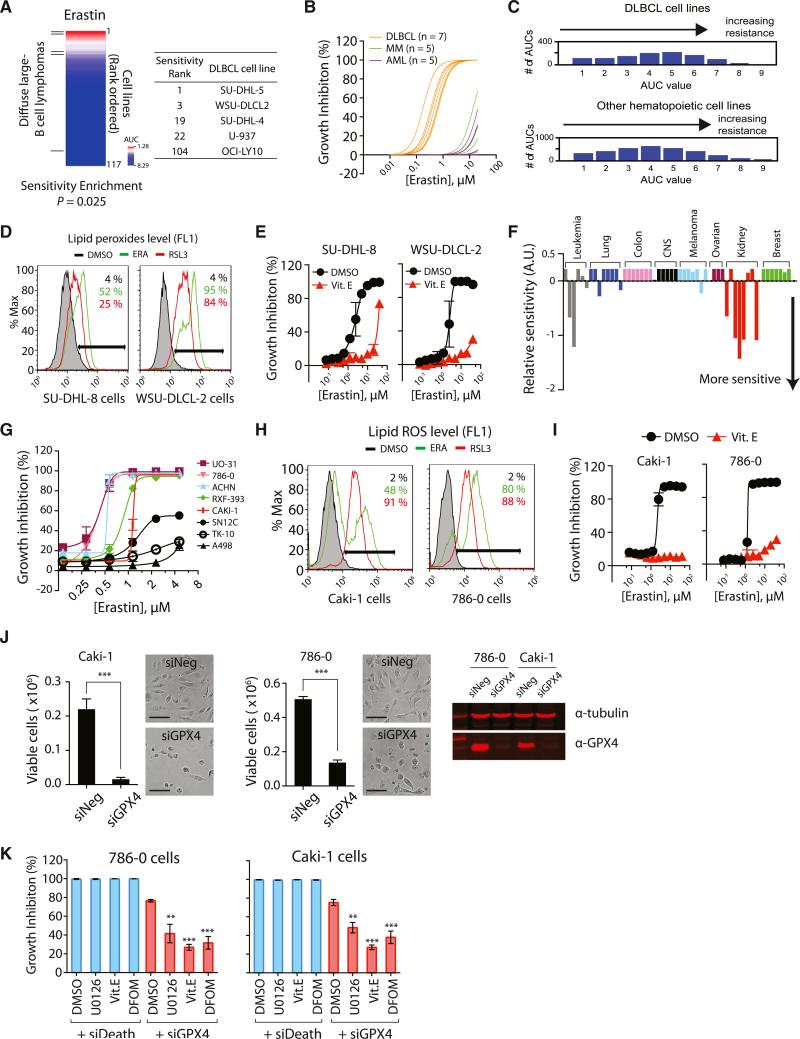
Ferroptosis is a form of nonapoptotic cell death for which key regulators remain unknown. We sought a common mediator for the lethality of 12 ferroptosis-inducing small molecules. We used targeted metabolomic profiling to discover that depletion of glutathione causes inactivation of glutathione peroxidases (GPXs) in response to one class of compounds and a chemoproteomics strategy to discover that GPX4 is directly inhibited by a second class of compounds. GPX4 overexpression and knockdown modulated the lethality of 12 ferroptosis inducers, but not of 11 compounds with other lethal mechanisms. In addition, two representative ferroptosis inducers prevented tumor growth in xenograft mouse tumor models. Sensitivity profiling in 177 cancer cell lines revealed that diffuse large B cell lymphomas and renal cell carcinomas are particularly susceptible to GPX4-regulated ferroptosis. Thus, GPX4 is an essential regulator of ferroptotic cancer cell death.


Figures

Figure 1
Figure 1. Ferroptosis Involves Generation of Lyso-PC and Depletion of Glutathione
(A) Changes in metabolites upon erastin treatment. (B) Dose-dependent depletion of GSH by erastin in HT-1080 cells and U-2 OS cells. (C) Structure and activity of erastin (ERA) analogs. Potency (GI50; concentration required for 50% growth inhibition) and selectivity (ratio of GI50 in HRAS wild-type cells divided by GI50 in HRAS mutant cells) of each analog are shown. PYR, pyridine erastin; AE, aldehyde erastin; MEII, morpholine erastin II; PE, piperazine erastin. (D) GSH depletion by erastin analogs. HT-1080 cells were incubated with 10 μM erastin analogs for 5 hr or 100 μM BSO for 12 hr. BSO was used as a positive control for GSH depletion. Data were normalized to the DMSO sample. Box-and-whisker plots (n = 3–8) are as follows: midline represents median, box is the 25th– 75th percentiles, and whiskers are minimum and maximum. ***p < 0.001. (E) BSO induces selective lethality in BJ-derived tumorigenic cells expressing oncogenic HRAS. Scale bars, 60 μm. In (B) and (E), data are presented as mean ± SD (n = 3). See also Figure S1 and Table S1.
Figure 2
Figure 2. GSH Depletion Inactivates GPX Enzymes to Induce Ferroptosis
(A) Basal ROS levels among BJ-derived cell lines were compared (n = 8). (B) The growth inhibition effect of antioxidant-targeting compounds was determined in the four BJ-derived cells (n = 3). The bar graph indicates growth inhibition at two different concentrations (2× GI50 and 4× GI50 for each compound in BJeLR cells). (C) Other antioxidant inhibitors do not deplete GSH during cell death (n = 3). (D) Erastin depletes cellular GSH equally in the four BJ-derived cell lines. (E) GSH-depleting reagents elevated both cytosolic and lipid ROS level, whereas other antioxidant inhibitors did not (n = 3). (F) GSH-depleting reagents (ERA and BSO) inhibited GPX activity. Rel. Abs., relative absorbance. n.s., not significant; *p < 0.05; ***p < 0.001. Error bars in (B), (C), and (E) represent mean ± SD. See also Figure S2.
Figure 3
Figure 3. RSL3 Binds to and Inhibits GPX4
(A) RSL3 does not deplete GSH. The level of GSH was determined after treating with 2 μM RSL3, 10 μM erastin, or 1 mM BSO (n = 3; *p < 0.05). Error bars indicate ± SD. (B) RSL3 treatment increased lipid ROS level, as erastin did. (C) The structure of (1S, 3R)-RSL3 is shown. (D) Only the (1S, 3R) diastereomer displayed selective lethality in HRASV12-expressing cells in the four BJ-derived cell lines. (E) Structure of RSL3 affinity probes used in the chemoproteomics experiments is shown. Fcn, fluorescein affinity tag. (F) The active affinity probe with the (1S, 3R) stereochemistry exhibited selective lethality against cells with HRASV12, whereas an affinity probe with the (1R, 3R) stereochemistry was not lethal. (G) Affinity-based chemoproteomics identified GPX4 (red dot) as the most likely binding protein for (1S, 3R)-RSL3. Fold enrichment values of peptides at the indicated condition and their FDR-adjusted p values were represented as volcano plots. The top three candidates are shown. (H) Confirmation of GPX4 binding to active (1S, 3R)-RSL3 affinity probe. Left panel is a 3D visualization of isotopic clusters of peptide ILAFPCNQFGK from GPX4 as rendered by TransOmics software. Right panel: cell lysates prepared from BJeLR cells treated with active probe (A), inactive probe (I), or active probe in the presence of competitor (A+C) that were affinity purified by α-fluorescein antibodies. Then, the purified protein samples were probed for GPX4 by western blot using GPX4-specific antibody. (I) (1S, 3R)-RSL3 inhibits enzyme activity of GPX4. See also Figure S3 and Table S2.
Figure 4
Figure 4. RSL3 Targets GPX4 to Induce Ferroptosis
(A) Knockdown of GPX4 using shRNAs rendered HT-1080 cells hypersensitive to (1S, 3R)-RSL3 lethality. (B) Overexpression of GPX4 rendered HT-1080 cells resistant to (1S, 3R)-RSL3 lethality. (C) HT-1080 cells transfected with a pool of siRNAs targeting GPX4 showed increased lipid ROS level as assessed by BODIPY-C11 staining. siNeg has no homology to any known mammalian genes and was used as a negative control. (D) Known inhibitors of ferroptosis, 10 μM U0126, 100 μM Vit. E, or 100 μM DFOM, were able to suppress siGPX4-induced cell death, whereas they could not suppress cell death induced by siDeath. (E) Knockdown of GPX4 displayed selective lethality in the four BJ-derived isogenic cell lines. (F) Other GPX isoforms are not relevant to ferroptotic cell death. The values in (D)–(F) were normalized to control samples transfected with siNeg. Bar graphs in (C)–(F) are mean ± SD (n = 3). *p < 0.05; **p < 0.01; ***p < 0.001. Data in (A) and (B) are presented as mean ± SD (n = 3). See also Figure S4.
Figure 5
Figure 5. Ferroptosis Occurs through a GPX4-Regulated Pathway
(A) Discovery of additional FINs based on selective lethality in the four BJ cell lines. (B) FINs (red) are distinct from non-FINs (blue) in accompanying lipid ROS generation during the cell death process and in death suppression by an antioxidant, BHT. (C) Modulatory profiling (Wolpaw et al., 2011) with erastin, PE, DPI2, DPI10, and other lethal molecules confirmed that PE, DPI2, and DPI10 induced a similar form of cell death as erastin in HT-1080 cells. ΔAUC with a positive sign indicates suppression of cell death, whereas a negative sign indicates sensitization by cell death modulators upon lethal compound treatment. (D) Inhibition of GPX4 by BSO sensitized cells to death induced by 12 FIN compounds, whereas activation of GPX4 by cDNA overexpression rescued cells from the lethality of FIN compounds. (E) Eight structurally diverse FIN compounds inhibited GPX4, whereas two FIN compounds, DPI2 and erastin, and the negative control staurosporine (STS), a non-FIN compound, did not show direct GPX4 inhibition in this LC-MS-based assay. (F) The two FIN compounds, DPI2 and erastin, depleted cellular GSH, which inhibits GPX4 indirectly, whereas staurosporine did not deplete GSH. Bar graph indicates mean ± SD (n = 3). (G) Model of GPX4-regulated ferroptosis pathway. Ferroptosis inducers can be categorized into two classes based on the mode of GPX4 inhibition. *p < 0.05; ***p < 0.001. See also Figure S5 and Table S3.
Figure 6
Figure 6. Ferroptosis Suppresses Tumor Growth in a Xenograft Mouse Model
(A) Upregulation of PTGS2 expression upon erastin and (1S, 3R)-RSL3 treatments. (B) PTGS2 expression was induced by PE, (1S, 3R)-RSL3, and siGPX4, but not by PE with Vit. E, (1R, 3R)-RSL3, and siDeath. (C) (1S, 3R)-RSL3 inhibited tumor formation and tumor progression through induction of ferroptosis as demonstrated by upregulation of PTGS2 in the tumors. (1S, 3R)-RSL3 was administered s.c. twice a week for 2 weeks. (D) PE showed efficacy in preventing HT-1080 tumor formation in a mouse xenograft model. The left view shows the structure of PE. The middle view is a mouse liver microsome assay demonstrating improved metabolic stability of PE over erastin. Midazolam was used as a positive control for metabolic degradation. The right view shows images representative of tumors in live mice from each treatment group. PE was delivered s.c. twice a week for 1 week and then delivered through tail vein injection once every other day for 6 days. (E) Pharmacodynamics of PE and (1S, 3R)-RSL3 in the mouse liver tissue. Bar graphs in (B), (C), and (E) represent mean ± SD (n = 3). In (C) and (D), the lines in the tumor volume plots indicate mean of nine data points. *p < 0.05; **p < 0.01. See also Figure S6 and Table S4.
Figure 7
Figure 7. DLBCLs and RCCs Are Sensitive to GPX4-Regulated Ferroptosis
(A) Testing erastin in 117 cancer cell lines revealed DLBCLs as a cancer subtype susceptible to ferroptosis. DLBCL cell lines are marked with lines on the left. The table shows the name of DLBCL cell lines along with the sensitivity rank. (B) DLBCLs were more sensitive to erastin than AML and MM cells. (C) DLBCL cell lines are no more sensitive to lethal compounds than other hematopoietic cell lines. The total number of AUCs in the analysis was 3,883 (972 for DLBCL and 2,911 for other hematopoietic cell lines). (D and E) DLBCL cells died through a mechanism characteristic of ferroptosis, as determined by lipid peroxide generation and death rescue by Vit. E. (F) Sensitivity profile of 53 cancer cell lines in the “NCI60” cell panel against erastin. The cell lines were grouped based on their tissue origins. (G) The eight RCC cell lines were retested with erastin to confirm their sensitivity against erastin. (H) Erastin and RSL3 generated lipid ROS in the two RCC cell lines. (I) Cell death was rescued by a lipophilic antioxidant, Vit. E. (J) GPX4 depletion by siGPX4 induced cell death in RCC cell lines. The western blot (right) confirmed expression of GPX4 protein in these RCC cell lines and knockdown of GPX4 by siRNAs. Scale bars, 30 μm. (K) Ferroptosis inhibitors suppressed cell death induced by GPX4 knockdown but could not suppress cell death induced by the control siRNAs (siDeath) that kill cells via a nonferroptotic pathway. Data points in (E), (G), and (I)–(K) represent mean ± SD (n = 3). **p < 0.01; ***p < 0.001. See also Figure S7 and Table S5.

Similar articles

Cited by

References

    1. Basu A, Bodycombe NE, Cheah JH, Price EV, Liu K, Schaefer GI, Ebright RY, Stewart ML, Ito D, Wang S, et al. An interactive resource to identify cancer genetic and lineage dependencies targeted by small molecules. Cell. 2013;154:1151–1161. - PMC - PubMed
    1. Brigelius-Flohé R, Maiorino M. Glutathione peroxidases. Biochim. Biophys. Acta. 2013;1830:3289–3303. - PubMed
    1. Cotter TG. Apoptosis and cancer: the genesis of a research field. Nat. Rev. Cancer. 2009;9:501–507. - PubMed
    1. Degterev A, Huang Z, Boyce M, Li Y, Jagtap P, Mizushima N, Cuny GD, Mitchison TJ, Moskowitz MA, Yuan J. Chemical inhibitor of nonapoptotic cell death with therapeutic potential for ischemic brain injury. Nat. Chem. Biol. 2005;1:112–119. - PubMed
    1. Dixon SJ, Lemberg KM, Lamprecht MR, Skouta R, Zaitsev EM, Gleason CE, Patel DN, Bauer AJ, Cantley AM, Yang WS, et al. Ferroptosis: an iron-dependent form of nonapoptotic cell death. Cell. 2012;149:1060–1072. - PMC - PubMed

Publication types