. 2014 Mar 15;127(Pt 6):1263-78.
doi: 10.1242/jcs.140087. Epub 2014 Jan 14.

Differential roles of the ubiquitin proteasome system and autophagy in the clearance of soluble and aggregated TDP-43 species

Affiliations

Differential roles of the ubiquitin proteasome system and autophagy in the clearance of soluble and aggregated TDP-43 species

Emma L Scotter et al. J Cell Sci. .

Abstract

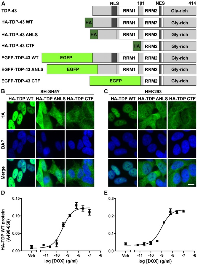
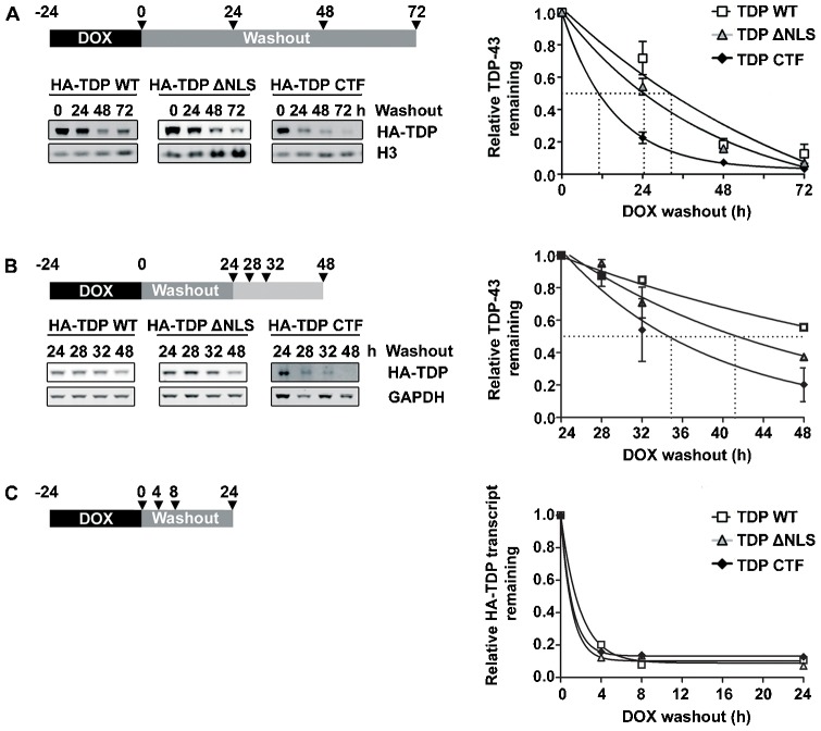
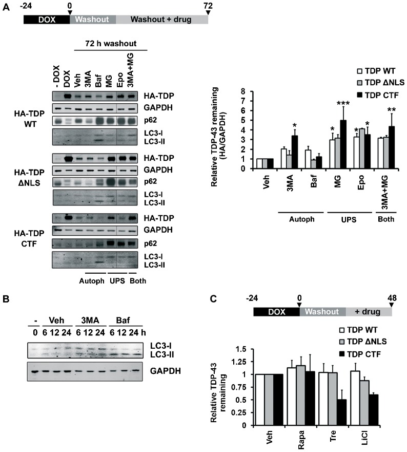
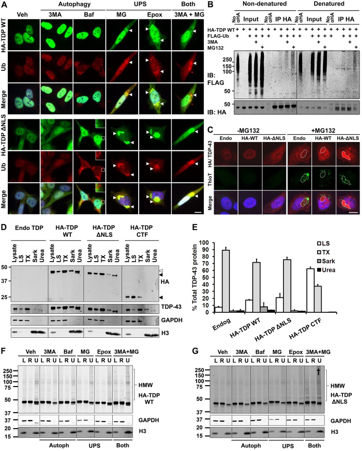
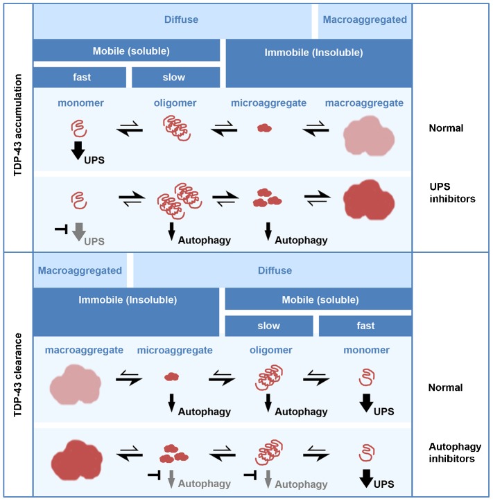
TAR DNA-binding protein (TDP-43, also known as TARDBP) is the major pathological protein in amyotrophic lateral sclerosis (ALS) and frontotemporal dementia (FTD). Large TDP-43 aggregates that are decorated with degradation adaptor proteins are seen in the cytoplasm of remaining neurons in ALS and FTD patients post mortem. TDP-43 accumulation and ALS-linked mutations within degradation pathways implicate failed TDP-43 clearance as a primary disease mechanism. Here, we report the differing roles of the ubiquitin proteasome system (UPS) and autophagy in the clearance of TDP-43. We have investigated the effects of inhibitors of the UPS and autophagy on the degradation, localisation and mobility of soluble and insoluble TDP-43. We find that soluble TDP-43 is degraded primarily by the UPS, whereas the clearance of aggregated TDP-43 requires autophagy. Cellular macroaggregates, which recapitulate many of the pathological features of the aggregates in patients, are reversible when both the UPS and autophagy are functional. Their clearance involves the autophagic removal of oligomeric TDP-43. We speculate that, in addition to an age-related decline in pathway activity, a second hit in either the UPS or the autophagy pathway drives the accumulation of TDP-43 in ALS and FTD. Therapies for clearing excess TDP-43 should therefore target a combination of these pathways.

Keywords: ALS; Aggrephagy; Autophagy; Proteasome; TDP-43; UPS.


Figures

Fig. 1.
Fig. 1.
Establishing cellular models of TDP-43 proteinopathy. (A) A schematic of TDP-43 constructs used in this study. NES, nuclear export signal; RRM, RNA recognition motif. (B,C) Immunocytochemical analysis of TDP-43 expression and localisation in stable SH-SY5Y (B) and HEK293 (C) cell lines induced to express HA–TDP-43 constructs by a 48-h induction with DOX, showing nuclear localisation (WT), cytoplasmic localisation (CTF) or both (ΔNLS). Scale bar: 10 µm. (D,E) Cell-based ELISA validation of DOX-dose-dependent expression of HA–TDP WT in stable SH-SY5Y (D) and HEK293 (E) lines after a 48-h induction with DOX (EC50; HEK293, 0.97 ng/ml; SH-SY5Y, 0.35 ng/ml).
Fig. 2.
Fig. 2.
Degradation of TDP-43. (A) Degradation of HA–TDP-43 proteins in stable HEK293 cells as assessed by DOX pulse–chase experiments. TDP-43 CTF was degraded far more rapidly than TDP-43 ΔNLS or TDP-43 WT (t1/2; HA–TDP-43 WT, 32.5 h; HA–TDP-43 ΔNLS, 24.4 h; HA–TDP-43 CTF, 11 h). (B) Degradation of HA–TDP-43 proteins in stable SH-SY5Y cells over a shorter timecourse, and allowing a delay after washout for the clearance of mRNA transcripts. HA–TDP-43 protein-degradation rates closely reflected those seen in HEK293 cells (t1/2; HA–TDP-43 WT, 29.2 h; HA–TDP-43 ΔNLS, 16.6 h; HA–TDP-43 CTF, 10.2 h). (C) The degradation of HA–TDP-43 mRNA transcripts in stable SH-SY5Y cells as assessed by quantitative reverse transcriptase PCR. Transcripts for all three constructs decayed rapidly after DOX washout and had reached minima by 8 h (a representative experiment is shown, n = 2). (A–C) Arrowheads on schematics show the timepoints at which samples were taken.
Fig. 3.
Fig. 3.
Involvement of the UPS and autophagy in the degradation of TDP-43. (A) The relative HA–TDP-43 remaining in stable HEK293 cells at 72 h post-DOX washout, with inhibitors of autophagy or the UPS included during the last 48 h. 3MA, 3-methyladenine; Baf, bafilomycin; Epo, epoxomicin; MG, MG132; Veh, vehicle. The −DOX control lane represents the amount of HA–TDP-43 present at the 72-h washout point without expression having been induced (‘leakage’). All lanes for each construct are from the same blot, spliced as shown. (B) Western blot of LC3 levels after short treatments with the autophagy inhibitors 3MA and bafilomycin showing LC3-I and II accumulation with 3MA and LC3-II accumulation with bafilomycin. (C) The relative HA–TDP-43 remaining in stable HEK293 cells subjected to DOX pulse–chase with activators of autophagy included during the last 24 h of the 48-h chase period. LiCl, lithium chloride; Rapa, rapamycin; Tre, trehalose; Veh, vehicle. The bars represent means±s.e.m. *P≤0.05, **P≤0.01, ***P≤0.001 between vehicle and inhibitor-treated cells (two-way ANOVA, Bonferroni post-test).
Fig. 4.
Fig. 4.
Inhibiton of the UPS, but not autophagy, induces the formation of detergent-resistant cytoplasmic aggregates of TDP-43. (A) Immunocytochemical analysis of stable SH-SY5Y lines induced to express HA–TDP-43 constructs by a 48-h induction with DOX in the presence of various inhibitors of the UPS or autophagy. When used alone, UPS inhibitors, but not autophagy inhibitors, induced the formation of ubiquitin (Ub)-positive TDP-43 macroaggregates (arrowheads). Scale bar: 10 µm. (B) Immunoprecipitation of HA–TDP-43 WT from HEK293 stable lines transfected with FLAG–Ub then induced with 1 µg/ml DOX alone or together with 3MA or MG132 for 24 h. Under non-denaturing conditions that preserve complexes (left) HA–TDP-43 WT co-immunoprecipitated with an increased amount of FLAG–Ub (smear >100 kDa) under treatment with 3MA and a greater amount again with MG132. Immunoprecipitation of denatured lysate (right) showed that HA–TDP-43 that was directly and covalently linked to FLAG–Ub only accumulated following MG132 treatment. IB, immunoblot. (C) TDP-43 aggregates are non-amyloid. Stable SH-SY5Y lines were induced to express HA–TDP-43 constructs by a 48-h induction with DOX in the absence or presence of MG132. In MG132-treated cells, thioflavin T staining detected amyloid aggregates that were independent of macroaggregates of HA–TDP-43 (dashed lines). MG132 treatment of parent SH-SY5Y cells induced macroaggregates (dashed line) with low levels of endogenous (Endo) TDP-43, likely owing to autoregulation of TDP-43 protein levels. Scale bar: 10 µm. (D,E) Four-step fractionation of stable SH-SY5Y lines induced to express HA–TDP-43 constructs by a 48-h induction with DOX. WT, upper white arrowhead; ΔNLS, middle black arrowhead; CTF, lower black arrowhead. The CTF was largely soluble in the low-salt (LS) fraction (TX, Triton X-100; Sark, sarkosyl). Note that the pellets from each step were resuspended in different volumes to ensure the detection of all fractions. Relative concentrations of the fractions are: Lys, 1; LS, 1; TX, 0.5; Sark, 3.33; urea 2.5. The bars represent means±s.e.m. (F,G) Solubility fractionation of the cells treated as in A, showing the lysate (L), RIPA-soluble (R) and urea-soluble (U) fractions of HA–TDP-43 WT (F) and ΔNLS (G). Urea fractions are concentrated 10-fold. Combined treatment with UPS and autophagy inhibitors (†) increased the amount of insoluble high-molecular-weight (HMW) TDP-43, particularly for TDP-43 ΔNLS (G). 3MA, 3-methyladenine; Baf, bafilomycin; Epox, epoxomicin; MG, MG132; Veh, vehicle.
Fig. 5.
Fig. 5.
TDP-43 aggregates induced by UPS inhibition or dual inhibition of the UPS and autophagy resemble those seen in human disease. (A) Immunocytochemical analysis of untreated SH-SY5Y cells showing the diffuse localisation of the degradation pathway proteins ubiquilin-1 or -2 (UBQLN) and p62, and K48- and K63-linked forms of ubiquitin (Ub). Scale bar: 25 µm. (B–E) Immunocytochemical analysis of stable SH-SY5Y lines induced to express HA–TDP-43 by a 48-h induction with DOX in the presence of UPS inhibition or combined inhibition of the UPS and autophagy. (B) HA–TDP-43 WT formed cytoplasmic macroaggregates (arrowheads) following treatment with DOX plus the proteasome inhibitor MG132 in stable SH-SY5Y cells. These aggregates were positive for post-translational modifications (PTMs) including K48- and K63-linked ubiquitin, ubiquilin-1 and -2 and p62. (C) Combined treatment with DOX, MG132 and the autophagy inhibitor 3MA enhanced the recruitment of HA–TDP-43 WT to macroaggregates, which were positive for the same markers as with MG132 treatment alone. (D) HA–TDP-43 ΔNLS also formed cytoplasmic aggregates following a 48-h treatment with DOX plus MG132 in stable SH-SY5Y cells. As was seen for HA–TDP-43 WT, these aggregates were positive for K48- and K63-linked ubiquitin, ubiquilin-1 and -2 and p62. (E) Combined treatment with DOX plus MG132 and 3MA did not markedly alter the recruitment of HA–TDP-43 ΔNLS to macroaggregates. Arrows on the bottom row of images in B–E represent the section taken for the intensity profiles shown below the images. Scale bars: 10 µm.
Fig. 6.
Fig. 6.
TDP-43 macroaggregates are cleared upon the restoration of UPS function. Immunocytochemical analysis of stable SH-SY5Y lines induced to form HA–TDP-43 WT (A) or HA–TDP-43 ΔNLS (B) aggregates (arrowheads) by a 48 h induction with DOX plus MG132 then fixed or subjected to MG132 washout (wo) after which the cells were left for a further 48 h. Scale bar: 10 µm. (C,D) Solubility fractionation of stable HA–TDP-43 WT (C) or HA–TDP-43 ΔNLS (D) SH-SY5Y lines either induced with DOX, induced to form aggregates by 48-h treatment with DOX plus MG132 or induced to form aggregates then subjected to MG132 washout after which the cells were left for a further 48 h. Lysate (L), RIPA-soluble (R) and urea-soluble (U) fractions are shown. Urea fractions are concentrated 10-fold. MG132 (*) increased the proportion of high-molecular-weight insoluble TDP-43 (HMW) and this was reversible upon MG132 washout (48 h). (E) Validation that EGFP-tagged TDP-43 WT is degraded over the same timecourse as HA–TDP-43 WT in stable SH-SY5Y cells (t1/2; HA–TDP-43 WT, 29.2 h; EGFP–TDP-43 WT, 29.3 h). (F) Validation that both HA- and EGFP-tagged TDP-43 WT (grey arrowheads) are functional and can regulate the level of endogenous TDP-43 (black arrowhead) in stable SH-SY5Y cells. Black bars represent endogenous TDP-43 and grey bars represent exogenous TDP-43. UT, untransfected. (G) Live-cell imaging of stable SH-SY5Y lines induced to form EGFP–TDP-43 ΔNLS aggregates by 48 h induction with DOX plus MG132 then subjected to washout and followed for a further 15 h. In cells that cleared aggregates, the aggregates fragmented before clearance. Scale bar: 10 µm.
Fig. 7.
Fig. 7.
TDP-43 aggregate clearance is due to enhanced mobility of lower-order aggregated species, rather than mobility of macroaggregated TDP-43. (A) Confocal images acquired during FRAP. EGFP–TDP-43-ΔNLS showed rapid fluorescence recovery for diffuse cytosolic forms (Cyt TDP) but no recovery for aggregated forms (Agg TDP), indicating that macroaggregated TDP-43 is immobile and unlikely to readily exchange with the diffuse pool. The white dashed line encloses the bleached area. Scale bar: 10 µm. (B) FRAP recovery curves, with bleaching at t = 0 and 100% representing the pre-bleach fluorescence intensity. The fraction of cytosolic TDP-43 that was mobile was slightly decreased in cells that were treated with MG132 (MG) but were devoid of aggregates (Cyt TDP −Agg) and was greatly decreased in cells containing an aggregate (Cyt TDP +Agg). (C) The fraction of TDP-43 that was mobile under various conditions. The decrease in the mobile fraction in cells treated with MG132 and containing an aggregate (Cyt TDP +Agg) was rescued 8–10 h after washout (wash). There was no mobilisation of macroaggregated TDP-43 upon washout. (D) The rate of FRAP recovery [given as K = ln(2)/t1/2] for mobile species under various conditions. The FRAP rate was significantly decreased in cells that were treated with MG132 and contained an aggregate (Cyt TDP +Agg), which was reversed after washout. The bars represent means±s.e.m.; n.s., not significant. **P≤0.01, ***P≤0.001 between control and treated cells or as shown (one-way ANOVA, Bonferroni post-test).
Fig. 8.
Fig. 8.
TDP-43 aggregate clearance requires autophagy. (A) Autophagosomes in cells containing TDP-43 aggregates. Stable SH-SY5Y lines were transfected with mCherry–GFP–LC3 and then induced to form HA–TDP-43 WT or ΔNLS aggregates (arrowheads) by a 48-h induction with DOX plus MG132. (B) Quantification of the number of GFP–LC3-positive autophagosomes per cell in cells treated as in A. The bars represent means±s.e.m. *P≤0.05, **P≤0.01 between control and treated cells (unpaired Student’s t-tests with Welch's correction). (C) Live-cell imaging of stable SH-SY5Y lines that were induced to form EGFP–TDP-43 ΔNLS aggregates (arrowheads) by a 48-h induction with DOX plus MG132, then subjected to washout and followed for a further 15 h in the presence of bafilomycin (Baf). Aggregates fragmented but were not cleared. (D) Regression analysis of the relationship between the aggregate load (integrated intensity of aggregate) and clearance time in the presence or absence of autophagy inhibitors during washout (wo) [3MA, 3-methyladenine; Baf, bafilomycin; Veh, vehicle; Pearson's r = 0.72 (Veh wo); 0.70 (3MA wo); 0.23 (Baf wo)]. Small aggregates were cleared rapidly in the absence, but not presence, of autophagy inhibitors. In Baf wo cells a distinct population of aggregates that were not cleared by the end of imaging (scored as 15 h) can be seen (dashed line). (E) Relative rates of clearance of EGFP–TDP-43 ΔNLS aggregates according to the slopes of the lines shown in D. Note that the slope for Baf wo includes the ‘non-clearing’ population. The results are not significant by one-way ANOVA. Scale bars: 10 µm.
Fig. 9.
Fig. 9.
A working model for the differential degradation of TDP-43 species. Upper panel; under normal conditions, TDP-43 exists as several different species with variable appearance (diffuse or macroaggregated), solubility and mobility (fast or slow). When the UPS is functional, the removal of soluble monomer drives the equilibrium in favour of small soluble species and precludes the formation of macroaggregates. By contrast, UPS blockade drives the accumulation of oligomers and insoluble species, where insoluble species include ‘microaggregated’ and macroaggregated TDP-43; microaggregates appear visually diffuse but are immobile in FRAP experiments and macroaggregates are visible by immunofluorescence and immobile. Lower panel; a working model for TDP-43 clearance after macroaggregate formation. Under normal conditions macroaggregates are cleared by the induction of autophagy, which removes oligomeric and microaggregated species. Monomer is cleared by the UPS. Autophagy blockade prevents the removal of oligomer and microaggregates, although the fragmentation of macroaggregates still occurs.

Similar articles

Cited by

References

    1. Arai T., Nonaka T., Hasegawa M., Akiyama H., Yoshida M., Hashizume Y., Tsuchiya K., Oda T., Ikeda K. (2003). Neuronal and glial inclusions in frontotemporal dementia with or without motor neuron disease are immunopositive for p62. Neurosci. Lett. 342, 41–44 10.1016/S0304-3940(03)00216-7 - DOI - PubMed
    1. Ayala Y. M., De Conti L., Avendaño-Vázquez S. E., Dhir A., Romano M., D’Ambrogio A., Tollervey J., Ule J., Baralle M., Buratti E. et al. (2011). TDP-43 regulates its mRNA levels through a negative feedback loop. EMBO J. 30, 277–288 10.1038/emboj.2010.310 - DOI - PMC - PubMed
    1. Bar J., Cohen-Noyman E., Geiger B., Oren M. (2004). Attenuation of the p53 response to DNA damage by high cell density. Oncogene 23, 2128–2137 10.1038/sj.onc.1207325 - DOI - PubMed
    1. Ben-Gedalya T., Lyakhovetsky R., Yedidia Y., Bejerano-Sagie M., Kogan N. M., Karpuj M. V., Kaganovich D., Cohen E. (2011). Cyclosporin-A-induced prion protein aggresomes are dynamic quality-control cellular compartments. J. Cell Sci. 124, 1891–1902 10.1242/jcs.077693 - DOI - PubMed
    1. Bigio E. H., Wu J. Y., Deng H. X., Bit-Ivan E. N., Mao Q., Ganti R., Peterson M., Siddique N., Geula C., Siddique T. et al. (2013). Inclusions in frontotemporal lobar degeneration with TDP-43 proteinopathy (FTLD-TDP) and amyotrophic lateral sclerosis (ALS), but not FTLD with FUS proteinopathy (FTLD-FUS), have properties of amyloid. Acta Neuropathol. 125, 463–465 10.1007/s00401-013-1089-6 - DOI - PMC - PubMed

Publication types

Substances