Neonatal Fc receptor promotes immune complex-mediated glomerular disease
- PMID: 24357670
- PMCID: PMC4005300
- DOI: 10.1681/ASN.2013050498
Neonatal Fc receptor promotes immune complex-mediated glomerular disease
Abstract
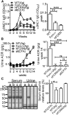
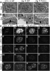
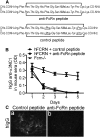
The neonatal Fc receptor (FcRn) is a major regulator of IgG and albumin homeostasis systemically and in the kidneys. We investigated the role of FcRn in the development of immune complex-mediated glomerular disease in mice. C57Bl/6 mice immunized with the noncollagenous domain of the α3 chain of type IV collagen (α3NC1) developed albuminuria associated with granular capillary loop deposition of exogenous antigen, mouse IgG, C3 and C5b-9, and podocyte injury. High-resolution imaging showed abundant IgG deposition in the expanded glomerular basement membrane, especially in regions corresponding to subepithelial electron dense deposits. FcRn-null and -humanized mice immunized with α3NC1 developed no albuminuria and had lower levels of serum IgG anti-α3NC1 antibodies and reduced glomerular deposition of IgG, antigen, and complement. Our results show that FcRn promotes the formation of subepithelial immune complexes and subsequent glomerular pathology leading to proteinuria, potentially by maintaining higher serum levels of pathogenic IgG antibodies. Therefore, reducing pathogenic IgG levels by pharmacologic inhibition of FcRn may provide a novel approach for the treatment of immune complex-mediated glomerular diseases. As proof of concept, we showed that a peptide inhibiting the interaction between human FcRn and human IgG accelerated the degradation of human IgG anti-α3NC1 autoantibodies injected into FCRN-humanized mice as effectively as genetic ablation of FcRn, thus preventing the glomerular deposition of immune complexes containing human IgG.
Copyright © 2014 by the American Society of Nephrology.
Figures


Similar articles
-
Aliskiren Regulates Neonatal Fc Receptor and IgG Metabolism with Attenuation of Anti-GBM Glomerulonephritis in Mice.Nephron. 2016;134(4):272-282. doi: 10.1159/000448789. Epub 2016 Aug 25. Nephron. 2016. PMID: 27560625
-
Proteolysis breaks tolerance toward intact α345(IV) collagen, eliciting novel anti-glomerular basement membrane autoantibodies specific for α345NC1 hexamers.J Immunol. 2013 Feb 15;190(4):1424-32. doi: 10.4049/jimmunol.1202204. Epub 2013 Jan 9. J Immunol. 2013. PMID: 23303673 Free PMC article.
-
Murine membranous nephropathy: immunization with α3(IV) collagen fragment induces subepithelial immune complexes and FcγR-independent nephrotic syndrome.J Immunol. 2012 Apr 1;188(7):3268-77. doi: 10.4049/jimmunol.1103368. Epub 2012 Feb 27. J Immunol. 2012. PMID: 22371398 Free PMC article.
-
FcRn: from molecular interactions to regulation of IgG pharmacokinetics and functions.Curr Top Microbiol Immunol. 2014;382:249-72. doi: 10.1007/978-3-319-07911-0_12. Curr Top Microbiol Immunol. 2014. PMID: 25116104 Review.
-
The Neonatal Fc Receptor (FcRn): A Misnomer?Front Immunol. 2019 Jul 10;10:1540. doi: 10.3389/fimmu.2019.01540. eCollection 2019. Front Immunol. 2019. PMID: 31354709 Free PMC article. Review.
Cited by
-
The fate of immune complexes in membranous nephropathy.Front Immunol. 2024 Aug 8;15:1441017. doi: 10.3389/fimmu.2024.1441017. eCollection 2024. Front Immunol. 2024. PMID: 39185424 Free PMC article. Review.
-
Exploiting the neonatal crystallizable fragment receptor to treat kidney disease.Kidney Int. 2024 Jan;105(1):54-64. doi: 10.1016/j.kint.2023.09.024. Epub 2023 Oct 29. Kidney Int. 2024. PMID: 38707675 Review.
-
The therapeutic age of the neonatal Fc receptor.Nat Rev Immunol. 2023 Jul;23(7):415-432. doi: 10.1038/s41577-022-00821-1. Epub 2023 Feb 1. Nat Rev Immunol. 2023. PMID: 36726033 Free PMC article. Review.
-
Anti-phospholipase A2 receptor antibodies directly induced podocyte damage in vitro.Ren Fail. 2022 Dec;44(1):304-313. doi: 10.1080/0886022X.2022.2039705. Ren Fail. 2022. PMID: 35333675 Free PMC article.
-
The Collagen Receptor Discoidin Domain Receptor 1b Enhances Integrin β1-Mediated Cell Migration by Interacting With Talin and Promoting Rac1 Activation.Front Cell Dev Biol. 2022 Mar 3;10:836797. doi: 10.3389/fcell.2022.836797. eCollection 2022. Front Cell Dev Biol. 2022. PMID: 35309920 Free PMC article.
References
-
- Roopenian DC, Akilesh S: FcRn: The neonatal Fc receptor comes of age. Nat Rev Immunol 7: 715–725, 2007 - PubMed
-
- Sesarman A, Sitaru AG, Olaru F, Zillikens D, Sitaru C: Neonatal Fc receptor deficiency protects from tissue injury in experimental epidermolysis bullosa acquisita. J Mol Med (Berl) 86: 951–959, 2008 - PubMed
-
- Haymann JP, Levraud JP, Bouet S, Kappes V, Hagège J, Nguyen G, Xu Y, Rondeau E, Sraer JD: Characterization and localization of the neonatal Fc receptor in adult human kidney. J Am Soc Nephrol 11: 632–639, 2000 - PubMed
Publication types
MeSH terms
Substances
Grants and funding
LinkOut - more resources
Full Text Sources
Other Literature Sources
Molecular Biology Databases
Research Materials
Miscellaneous

