. 2014 Jan 15;74(2):460-70.
doi: 10.1158/0008-5472.CAN-13-1713. Epub 2013 Nov 26.

MUC1 in macrophage: contributions to cigarette smoke-induced lung cancer

Affiliations

MUC1 in macrophage: contributions to cigarette smoke-induced lung cancer

Xiuling Xu et al. Cancer Res. .

Abstract

Expression of the pro-oncogenic mucin MUC1 is elevated by inflammation in airway epithelial cells, but the contributions of MUC1 to the development of lung cancer are uncertain. In this study, we developed our finding that cigarette smoke increases Muc1 expression in mouse lung macrophages, where we hypothesized MUC1 may contribute to cigarette smoke-induced transformation of bronchial epithelial cells. In human macrophages, cigarette smoke extract (CSE) strongly induced MUC1 expression through a mechanism involving the nuclear receptor PPAR-γ. CSE-induced extracellular signal-regulated kinase (ERK) activation was also required for MUC1 expression, but it had little effect on MUC1 transcription. RNA interference-mediated attenuation of MUC1 suppressed CSE-induced secretion of TNF-α from macrophages, by suppressing the activity of the TNF-α-converting enzyme (TACE), arguing that MUC1 is required for CSE-induced and TACE-mediated TNF-α secretion. Similarly, MUC1 blockade after CSE induction through suppression of PPAR-γ or ERK inhibited TACE activity and TNF-α secretion. Conditioned media from CSE-treated macrophages induced MUC1 expression and potentiated CSE-induced transformation of human bronchial epithelial cells in a TNF-α-dependent manner. Together, our results identify a signaling pathway involving PPAR-γ, ERK, and MUC1 for TNF-α secretion induced by CSE from macrophages. Furthermore, our results show how MUC1 contributes to smoking-induced lung cancers that are driven by inflammatory signals from macrophages.


Conflict of interest statement

Note: The authors have no conflict of interest to declare.

Figures

Figure 1
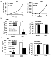
Figure 1. CS increases Muc1 expression in lung macrophages in vivo and human macrophage cell lines
A), Quantitative representation of the macrophages expressing Muc1 after CS or FA treatment. Bars show the averages of macrophage numbers of 10 randomly selected fields. Data shown are mean ± S.D; * p<0.05. B), THP-1 cells were differentiated for 24 h in the presence of PMA (50 ng/ml) before the cells were treated with CSE (20 μg/ml TPM) at different time points. MUC1 expression was detected by Western blot with antibody Muc1 GP1.4 against the extracellular domain (MUC1-N) and antibody MUC1Ab-5 recognizing the C-terminal domain (MUC1-CT). GAPDH was detected as an input control. C), U937 cells were differentiated for 72 h in the presence of PMA (50ng/ml) before the cells were treated with CSE for 24h. MUC1 expression was detected by Western blot. β-Actin was detected as an input control. D), Differentiated THP-1 and U937 cells were transfected with MUC1 siRNA or negative control siRNA for 48 h. MUC1 expression was detected by Western blot. β-Actin was detected as an input control.
Figure 2
Figure 2. MUC1 is required for CSE-induced TNF-α secretion from human macrophages
A), THP-1 and U937 cells were differentiated for 24 h and 72 h, respectively, in the presence of PMA (50 ng/ml). THP-1 and U937 cells were treated with CSE (20 μg/ml TPM) at the indicated time points. Conditioned media were collected for detection of TNF-α secretion by ELISA assay. B) and C), Inserts are the confirmations of MUC1 knockdown by Western blot. THP-1 cells B) and U937 cells C) were transfected with MUC1 siRNA or negative control siRNA for 24 h. The cells were then treated with CSE (20 μg/ml TPM) for 1h. At 24h post-treatment, conditioned media were collected for detection of TNF-α by ELISA assay. Meanwhile, cell viability was detected by MTT assay. Data shown are mean ± S.D; ** p< 0.01, * p<0.05.
Figure 3
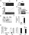
Figure 3. CSE induces MUC1 expression in macrophages depending on PPAR-γ
A), THP-1 cells were differentiated for 24 h in the presence of PMA (50 ng/ml). THP-1 cells were treated with CSE (20 μg/ml TPM) at the indicated time points. MUC1 mRNA level was detected by RT-PCR. β-Actin was detected as an input control. B), U937 cells were differentiated for 72 h in the presence of PMA (50 ng/ml). U937 cells were treated with CSE (20 μg/ml TPM) for 1h. MUC1 mRNA level was detected by RT-PCR. β-Actin was detected as an input control. C), THP-1 cells were treated with CSE (20 μg/ml TPM) with or without different inhibitors (U0126, SP, SB, SC-514, and BHA) for 30 min. MUC1 mRNA level was detected by RT-PCR. β-Actin was detected as an input control. D), THP-1 cells were pre-treated with BADGE (10 μM) for 30 min, followed by CSE treatment (20 μg/ml TPM) for 24h for Western blot or 30 min for RT-PCR. E), U937 cells were transfected with PPARγ siRNA or negative control siRNA for 24 h. After transfection, cells were exposed to CSE (40 μg/ml TPM) for 24h. MUC1 and PPARγ expression were detected by Western blot. β-Actin was detected as an input control. F), ChIP assay shows the enrichment of PPARγ at the hMUC1 promoter. U937 cells were exposure to CSE (40 μg/ml TPM) for 1h. Data shown are mean ± S.D. G), THP-1 cells were pre-treated with BADGE (10 μM) for 30 min before the cells were treated with CSE (20 μg/ml TPM) for 1h. At 24h post-treatment, conditioned media were collected for detection of TNF-α by ELISA assay. BHA and R031 served as negative controls. Cell viability was detected by MTT assay. Data shown are mean ± S.D; ** p< 0.01.
Figure 4
Figure 4. ERK pathway is involved in CSE-induced MUC1 expression and TNF-α secretion in macrophages
A), THP-1 cells were pre-treated with U0126 (5 μM) for 30 min, followed by CSE treatment (20 μg/ml TPM) for 24 h. U0124 served as the negative control. MUC1expression was detected by Western blot. β-Actin was detected as an input control. B), THP-1 cells were treated with CSE (20 μg/ml TPM) for the indicated time points. ERK activation was detected by Western blot. β-Tubulin was detected as an input control. C), U937 cells were treated with cycloheximide (CHX, 10 μM) and CSE (40 μg/ml TPM) with or without U0126 (10 μM) for the indicated time periods. MUC1 expression was detected by Western blot. β-Actin was detected as an input control. The intensity of the individual bands was quantified by densitometry (Image J) and normalized to the corresponding input control bands. MUC1 expression changes were calculated with the control taken as 100%. D), THP-1 cells were pre-treated with U0126 (5 μM) for 30 min before the cells were treated with CSE (20 μg/ml TPM) for 1h. At 24h post-treatment, conditioned media were collected for detection of TNF-α by ELISA assay. SB and SC-514 served as negative controls. Cell viability was detected by MTT assay. Data shown are mean ± S.D, ** p< 0.01.
Figure 5
Figure 5. MUC1 mediates CSE-induced TACE activation
A), THP-1 were treated with CSE (20 μg/ml TPM) for the indicated time points. TACE activity was detected using SensoLyte® 520 TACE (α-Secretase) Activity Assay Kit. B), THP-1 cells were transfected with MUC1 siRNA or negative control siRNA for 24 h. After transfection, the cells were treated with CSE (20 μg/ml TPM) for 8 h for TACE activity assay. C), Upper, TACE expression was detected by Western blot. GAPDH was detected as a loading control. Lower, THP-1 cells were exposed to CSE (20 μg/ml TPM) for 8 h with or without U0126 (10 μM). TACE expression was detected by Western blot. β-Actin was detected as a loading control. D), THP-1 cells were pre-treated with BADGE (10 μM) or U0126 (5 μM) for 30 min before CSE treatment for 8 h. Cells were lysed for TACE activity assay. Data shown are mean ± S.D; ** p< 0.01, * P<0.05.
Figure 6
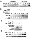
Figure 6. TNF-α secreted from macrophages induces MUC1 expression in human bronchial epithelial cells
A), HBEC-13 cells were treated with conditioned medium from CSE or DMSO-treated macrophages for the indicated time points. B), HBEC-13 cells were treated as described in the figure legend. MUC1 expression was detected by Western blot. The intensity of the individual bands was quantified by densitometry (Image J) and normalized to the corresponding input control bands. C), HBEC-13 cells were treated with conditioned medium from CSE-treated macrophages with or without a TNF-α neutralizing antibody (50 ng/ml) for 24 h. D), HBEC-13 cells were exposed to recombinant TNF-α (1 ng/ml) for the indicated time points. The expression of MUC1 was detected by Western blot. β-Tubulin and GAPDH were detected as loading controls.
Figure 7
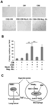
Figure 7. TNF-α in conditioned medium from CSE-treated macrophages facilitates CSE-induced HBEC-13 cell transformation
A) Representative images of and B) quantitative representation of colony formation of HBEC-13 cells in soft agar. HBEC-13 cells were pre-treated with the conditioned medium from CSE or DMSO-treated macrophages with or without a TNF-α neutralizing antibody (50 ng/ml) for 24 h and followed by CSE treatment (10 μg/ml TPM) for 1h. The cells were treated identically twice a week for 2 continuous weeks before they were seeded in soft agar. Colonies were allowed to develop for 3 weeks before they were photographed and counted. Bars show the average colony numbers of 6 randomly selected fields. Data shown are mean ± S.D; ** p< 0.01. C), A model of MUC1 in CS-induced and inflammation-associated lung cancer development. Cigarette smoke (CS) triggers MUC1 expression in macrophages, which facilitates TNF-α secretion from macrophages. TNF-α secreted from macrophages enhances MUC1 expression in bronchial epithelial cells, potentiating transformation of HBECs.

Similar articles

Cited by

References

    1. Lin WW, Karin M. A cytokine-mediated link between innate immunity, inflammation, and cancer. The Journal of clinical investigation. 2007;117:1175–83. - PMC - PubMed
    1. Cho WC, Kwan CK, Yau S, So PP, Poon PC, Au JS. The role of inflammation in the pathogenesis of lung cancer. Expert opinion on therapeutic targets. 2011;15:1127–37. - PubMed
    1. Milara J, Cortijo J. Tobacco, inflammation, and respiratory tract cancer. Current pharmaceutical design. 2012;18:3901–38. - PubMed
    1. Hecht SS. Tobacco smoke carcinogens and lung cancer. Journal of the National Cancer Institute. 1999;91:1194–210. - PubMed
    1. Belinsky SA. Gene-promoter hypermethylation as a biomarker in lung cancer. Nat Rev Cancer. 2004;4:707–17. - PubMed

Publication types