. 2014 Jan 7;110(1):83-93.
doi: 10.1038/bjc.2013.695. Epub 2013 Nov 26.

Gemcitabine enhances the efficacy of reovirus-based oncotherapy through anti-tumour immunological mechanisms

Affiliations

Gemcitabine enhances the efficacy of reovirus-based oncotherapy through anti-tumour immunological mechanisms

S A Gujar et al. Br J Cancer. .

Abstract

Background: Reovirus preferentially infects and kills cancer cells and is currently undergoing clinical trials internationally. While oncolysis is the primary mode of tumour elimination, increasing evidence illustrates that reovirus additionally stimulates anti-tumour immunity with a capacity to target existing and possibly relapsing cancer cells. These virus-induced anti-tumour immune activities largely determine the efficacy of oncotherapy. On the other hand, anti-viral immune responses can negatively affect oncotherapy. Hence, the strategic management of anti-tumour and anti-viral immune responses through complementary therapeutics is crucial to achieve the maximum anti-cancer benefits of oncotherapy.

Methods: Intra-peritoneal injection of mouse ovarian surface epithelial cells (ID8 cells) into wild-type C57BL/6 mice was treated with a therapeutic regimen of reovirus and/or gemcitabine and then analysed for prolonged survival, disease pathology, and various immunological parameters. Furthermore, in vitro analyses were conducted to assess apoptosis, viral spread, and viral production during reovirus and/or gemcitabine treatment.

Results: We demonstrate that reovirus and gemcitabine combination treatment postpones peritoneal carcinomatosis development and prolongs the survival of cancer-bearing hosts. Importantly, these anti-cancer benefits are generated through various immunological mechanisms, including: (1) inhibition of myeloid-derived suppressor cells recruitment to the tumour microenvironment, (2) downmodulation of pro-MDSC factors, and (3) accelerated development of anti-tumour T-cell responses.

Conclusion: The complementation of reovirus with gemcitabine further potentiates virus-initiated anti-cancer immunity and enhances the efficacy of oncotherapy. In the context of ongoing clinical trials, our findings represent clinically relevant information capable of enhancing cancer outcomes.


Figures

Figure 1
Figure 1
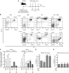
Reovirus and gemcitabine combination treatment significantly increases the survival of OC-bearing mice. (A) Female C57BL/6 mice were implanted i.p. with 3 × 10⁶ MOSE ID8 cells, injected with a regimen of PBS/GEM/Reo alone or in combination, and then monitored for the development of ascites (B) and survival (C). Survival in respective experimental groups was calculated with the Kaplan–Meier survival method. Results were analysed with two-tailed, Student's t-tests with 95% confidence interval (CI); ns=P>0.05; *P⩽0.05; ***P⩽0.001. Asterisks shown immediately on top of the bars represent the P-values obtained by comparing the respective data against PBS control, whereas asterisks shown above the horizontal lines display the P-values obtained through comparison between Reo-treated group. Error bars are defined as mean+s.d. and data are representative of three independent experiments.
Figure 2
Figure 2
Increased gemcitabine-induced cell death negatively affects the spread and propagation of reovirus in vitro. MOSE ID8 cells were infected in vitro with 0.1 or 1 MOI of Reo in the presence or absence of 1 μM of gemcitabine and then harvested at 24, 48, and 72 h, stained with annexin-V/7-AAD (detection of apoptotic cells) (A). (B) ID8 cells were infected with Reovirus (0.1 MOI) and treated with various GEM concentrations (0.01, 0.1, 1, 10, or 100 μM). Next, the intracellular and extracellular fractions were collected after 24 h and assessed by standard plaque assay to quantify viral titers (PFU ml−1). (C) Cumulative data on intracellular staining of MOSE ID8 cells with anti-reovirus antibodies to visualise reovirus-infected cells are illustrated. The cumulative data for all conditions tested as noted. The asterisks shown above the horizontal lines display the P-values obtained through comparison between Reo and Reo+GEM groups at the respective time points. Asterisks shown immediately on top of the bars represent the P-values obtained by comparing the respective GEM-treated group against PBS control. Statistical analysis was performed with two-tailed, Student's t-test with 95% CI; ns=P>0.05; *P⩽0.05; **P⩽0.01; ***P⩽0.001. Error bars are defined as mean+s.d. (D) Abundance of reovirus protein in the ID8 cells collected after 24, 48, and 72 h were analysed by western blot. Data are representative of three independent experiments.
Figure 3
Figure 3
Gemcitabine blocks reovirus-induced early recruitment of cancer-associated MDSCs in cancer environment through means other than direct oncolysis. (A) Female C57BL/6 mice were implanted i.p. with ID8 cells and then monitored for the development of PC. Then, these mice were injected with regimen of PBS/GEM/Reo alone or in combination as shown in the schematic. (B) The ascites were harvested and stained to detect the percentage of MDSCs (Gr-1+/CD11b+ cells) by flow cytometry at the respective time points and as shown in a representative example. The events were collected with same settings of the acquisition mode in FACSCalibur, demonstrating the gradient of CD11b expression between various samples; however, the statistical data include all relevant events in the analysis. The cumulative data from both ascites and spleen shown in (C) represent the fold increase/decrease in the percentages of MDSCs after normalising against PBS alone-treated control at the respective day post first injection (d.p.f.i.) or day post last injection (d.p.l.i.). The data are representative of n=3 except for Reo+GEM at 3 d.p.l.i., which is n=1. MDSCs were isolated from the ascites of OC-bearing mice and cultured in 50% cell-free ascites fluid and complete RPMI-1640 (D), while those isolated from the bone marrow of naive mice were cultured in complete RPMI-1640 (E). Isolated MDSCs were then treated with Reo (1 MOI) and GEM (1 μM) alone or in combination as indicated. Cells were harvested at 24 h and stained with GR-1+/CD11b+ antibodies along with apoptosis marker (Annexin-V) followed by flow cytometry. Data are representative of three independent experiments. Asterisks shown immediately on top of the bars represent the P-values obtained by comparing the respective Reo+PBS, GEM+PBS, and Reo+GEM-treated groups against PBS control. Statistical analysis was performed with two-tailed, Student's t-test with 95% CI; ns=P>0.05; *P⩽0.05; **P⩽0.01; ***P⩽0.001. Error bars are defined as mean+s.d.
Figure 4
Figure 4
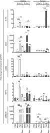
Gemcitabine impairs the expression of reovirus-induced pro-MDSC factors in ovarian tumours in vivo. Female C57Bl/6 mice were injected as per protocol shown in Figure 3A, and then killed at the indicated time points to obtain respective tissues. The samples for ‘ascites' were collected from the cellular contents of ascitic fluid, whereas the samples for ‘tumour' were collected from the solid tumour masses attached to the vasculature in the peritoneum. These samples were processed, RNA was extracted, purified, and reverse transcribed using random primers. Quantitative real-time PCR was conducted with the gene-specific primers for IL-1β, IDO1, COX2, TGF-β, and GM-CSF followed by analysis using the Livak and Schmittgen's 2-ΔΔCT method (Livak and Schmittgen, 2001). Bar graphs illustrate the linear fold change of the indicated mRNA replicates normalised to GAPDH and compared against PBS control. Gene expression data are representative of three independent experiments. Statistical analysis was performed with two-tailed, Student's t-test as described; ns=P>0.05; *P⩽0.05; **P⩽0.01; ***P⩽0.001. Asterisks shown immediately on top of the bars represent the P-values obtained by comparing the respective data against PBS control, whereas asterisks shown above the horizontal lines display the P-values obtained through comparison between Reo alone-treated animals and GEM alone or Reo+ GEM-treated animals at respective time points. Error bars are defined as mean+s.d. and data are representative of 3–5 mice in each group and the qRT-PCR was completed in duplicates.
Figure 5
Figure 5
Effect of Reo+GEM combination on T-cell functionality and survival, and tumour antigen presentation capacity. Splenocytes isolated from naive C57BL/6 mice were stimulated with Con A (2.5 μg ml−1) and treated with Reo (1 MOI) and GEM (1 μM) alone or in combination for 24 h. (A) Splenocytes were harvested and stained with anti-CD3, anti-CD4, and anti-CD8 antibodies along with annexin-V and analysed by flow cytometry. (BD) Alternatively, cells were stained for anti-CD4 (B) and anti-CD8 (C) antibodies along with anti-CD69 and anti-CD71 antibodies and analysed by flow cytometry. Bar graph represents a representative data from one of four independent experiments (D). Asterisks shown immediately on top of the bars signify the P-values obtained by comparing the Con A-stimulated groups against their respective Con A-stimulated counterparts, whereas asterisks shown above the horizontal lines display the P-values obtained through comparison between Con A-stimulated PBS control and other Con A-stimulated experimental groups as indicated. Asterisks represent the P-values obtained by comparing the PBS control groups at indicated time points with that of various experimental conditions at respective time points. Statistical analysis was performed with two-tailed, Student's t-test; ns=P>0.05; *P⩽0.05; **P⩽0.01; ***P⩽0.001. Error bars are defined as mean+s.d. and data are representative of four independent experiments.
Figure 6
Figure 6
Gemcitabine potentiates reovirus-induced anti-tumour immune responses. C57BL/6 mice were implanted with MOSE ID8-OVA cells and then treated with a therapeutic regimen of Reo and GEM alone or in combination as per protocol shown in Figure 3A. (A) Splenocytes were obtained on 3 d.p.f.i and 3 d.p.l.i., stained with CFSE and then stimulated with SIINFEKL peptide. After 72 h, cells were stained with anti-CD3 antibodies and then analysed by flow cytometry for the CFSE fluorescence halving in CD3+ cells to define cell division index. (B) Alternatively, splenocytes were stimulated with SIINFEKL for 18 h, stained with anti-CD3, anti-CD8, and anti-INF-γ antibodies and then analysed by flow cytometry. Statistical analysis was performed with two-tailed, Student's t-test; ns=P>0.05; *P⩽0.05; **P⩽0.01; ***P⩽0.001. Asterisks shown immediately on top of the bars signify the P-values obtained by comparing the respective data against PBS-treated control group, whereas asterisks shown above the horizontal lines display the P-values obtained through comparison between Reo alone-treated animals and GEM alone or Reo+GEM-treated animals at respective time points.
Figure 7
Figure 7
An illustration of various mechanisms through which gemcitabine enhances the efficacy of reovirus-based oncotherapy. Reovirus oncotherapy is known to target cancer through two distinct prongs: direct oncolysis (prong I) and virus-induced anti-tumour immune response (prong II). Gemcitabine also directly kills cancer cells, but also enhances the efficacy of reovirus-based therapy through immunological mechanisms, including: (1) inhibition of reovirus-induced MDSCs recruitment to the tumour microenvironment, (2) downmodulation of pro-MDSC factors, and (3) accelerated development of anti-tumour immunity.

Similar articles

Cited by

References

    1. Antonarakis ES, Drake CG. Current status of immunological therapies for prostate cancer. Curr Opin Urol. 2010;20 (3:241–246. - PMC - PubMed
    1. Bellmunt J, von der MH, Mead GM, Skoneczna I, De SM, Daugaard G, Boehle A, Chevreau C, Paz-Ares L, Laufman LR, Winquist E, Raghavan D, Marreaud S, Collette S, Sylvester R, de Wit R. Randomized phase III study comparing paclitaxel/cisplatin/gemcitabine and gemcitabine/cisplatin in patients with locally advanced or metastatic urothelial cancer without prior systemic therapy: EORTC Intergroup Study 30987. J Clin Oncol. 2012;30 (10:1107–1113. - PMC - PubMed
    1. Bunt SK, Sinha P, Clements VK, Leips J, Ostrand-Rosenberg S. Inflammation induces myeloid-derived suppressor cells that facilitate tumor progression. J Immunol. 2006;176 (1:284–290. - PubMed
    1. Coffey MC, Strong JE, Forsyth PA, Lee PW. Reovirus therapy of tumors with activated Ras pathway. Science. 1998;282 (5392:1332–1334. - PubMed
    1. Drake CG, Antonarakis ES. Update: immunological strategies for prostate cancer. Curr Urol Rep. 2010;11 (3:202–207. - PMC - PubMed

Publication types

MeSH terms