. 2014 Feb;34(3):452-63.
doi: 10.1128/MCB.00279-13. Epub 2013 Nov 25.

Psy2 targets the PP4 family phosphatase Pph3 to dephosphorylate Mth1 and repress glucose transporter gene expression

Affiliations

Psy2 targets the PP4 family phosphatase Pph3 to dephosphorylate Mth1 and repress glucose transporter gene expression

Hui Ma et al. Mol Cell Biol. 2014 Feb.

Abstract

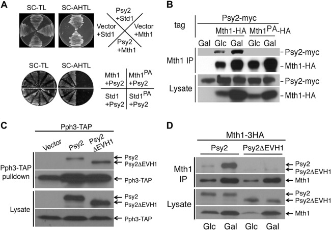
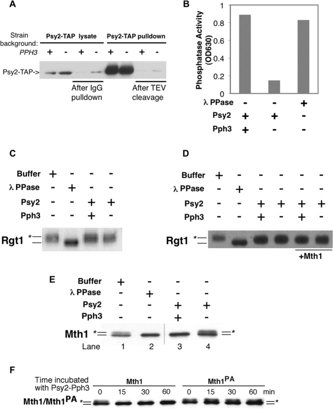
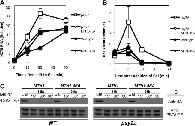
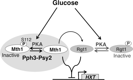
The reversible nature of protein phosphorylation dictates that any protein kinase activity must be counteracted by protein phosphatase activity. How phosphatases target specific phosphoprotein substrates and reverse the action of kinases, however, is poorly understood in a biological context. We address this question by elucidating a novel function of the conserved PP4 family phosphatase Pph3-Psy2, the yeast counterpart of the mammalian PP4c-R3 complex, in the glucose-signaling pathway. Our studies show that Pph3-Psy2 specifically targets the glucose signal transducer protein Mth1 via direct binding of the EVH1 domain of the Psy2 regulatory subunit to the polyproline motif of Mth1. This activity is required for the timely dephosphorylation of the downstream transcriptional repressor Rgt1 upon glucose withdrawal, a critical event in the repression of HXT genes, which encode glucose transporters. Pph3-Psy2 dephosphorylates Mth1, an Rgt1 associated corepressor, but does not dephosphorylate Rgt1 at sites associated with inactivation, in vitro. We show that Pph3-Psy2 phosphatase antagonizes Mth1 phosphorylation by protein kinase A (PKA), the major protein kinase activated in response to glucose, in vitro and regulates Mth1 function via putative PKA phosphorylation sites in vivo. We conclude that the Pph3-Psy2 phosphatase modulates Mth1 activity to facilitate precise regulation of HXT gene expression by glucose.


Figures

FIG 1
FIG 1
Psy2 interacts with Mth1 through a polyproline motif. (A) In the upper panel, the initial results from yeast two-hybrid screening using the EVH1 domain of Psy2 were confirmed with full-length Psy2 and Mth1/Std1 constructs. Yeast strains expressing Mth1/Std1 fused to the Gal4 DNA-binding domain (DBD) and Psy2 fused to Gal4 transcriptional activation domain or empty vector were plated on synthetic medium lacking either (i) Trp and Leu for growth or (ii) Ade, His, Trp, and Leu for selecting interactions. The lower panel shows that proline-to-alanine mutations of the MPPP motif of Mth1 (Mth1PA) and Std1 (Std1PA) abolished the interaction with Psy2. (B) Yeast cells expressing endogenous Myc-tagged Psy2 and HA-tagged wild-type Mth1 (WT) or Mth1PA were grown in glucose (Glc)- or galactose (Gal)-containing medium till log phase before being processed for coimmunoprecipitation analysis. (C) Psy2ΔEVH1 maintained the interaction with Pph3. psy2Δ strains harboring a genomic PPH3-TAP allele (MAY205) and expressing Psy2-6Myc or Psy2 ΔEVH1-6Myc from the endogenous promoter were subjected to Pph3-TAP pulldown, followed by immunoblot analysis for both Myc-tagged Psy2 variants and Pph3-TAP. (D) Psy2ΔEVH1 abolished interaction with Mth1. psy2Δ strains were transformed with the WT Psy2 and Psy2 ΔEVH1 constructs as described in panel C, together with pMT52. Cell extracts were made from cells grown in Glc-containing medium and 1 h after a shift to Gal medium. Immunoprecipitation was performed with anti-HA antibody coupled to protein A-Sepharose, followed by immunoblot analysis for both Myc-tagged Psy2 variants and Mth1-3HA.
FIG 2
FIG 2
Psy2 promotes HXT gene repression in the absence of glucose. (A) The phosphorylation state of Rgt1 was analyzed by immunoblotting using wild-type (WT) and psy2Δ mutant cells grown in Gal-containing medium and shifted to Glc-containing medium. The position of unphosphorylated Rgt1 is indicated by a line, whereas the lines with asterisks denote the most highly phosphorylated species of Rgt1. The membrane was stained for protein prior to development for the loading control. (B) Similar analysis was performed with WT and psy2Δ mutant cells shifted from Glc- to Gal-containing medium for the time points indicated. An immunoblot with anti-PSTAIRE antibody was used for the loading control. (C) Quantitative RT-PCR analysis of HXT3 RNA with samples collected as described in panel B at the indicated time points. (D) Chromatin immunoprecipitation assay was performed using wild-type (WT) and psy2Δ mutant cells treated as in panel B, after cross-linking and immunoprecipitation of the Rgt1-HA-DNA complex using anti-HA monoclonal antibodies. The qPCR data represent the average of HXT3 RNA samples analyzed in triplicate and normalized to ACT1 RNA. HXT3 RNA is plotted relative to the wild-type time zero sample.
FIG 3
FIG 3
The integrity of the Pph3-Psy2-Mth1 complex is critical for maintaining timely repression of HXT3 in response to glucose depletion. (A) Yeast cells expressing TAP-tagged Pph3 and HA-tagged Mth1/Mth1PA were subjected to coimmunoprecipitation analysis with anti-HA antibodies coupled to protein A-beads under the indicated conditions. The samples were then analyzed by immunoblotting with anti-HA to detect Mth1/Mth1PA-3HA and rabbit anti-mouse antibodies conjugated to horseradish peroxidase to detect Pph3-TAP, respectively. (B) Wild-type (WT) and psy2Δ mutant strains expressing Myc-tagged Pph3 and HA-tagged Mth1 from the endogenous loci were analyzed by coimmunoprecipitation. Pph3 expressed in the WT control is tagged with 13×Myc tag, whereas the other strains express Pph3 with a 6×Myc tag, which has more rapid gel mobility. (C) The repression of HXT3 gene upon glucose depletion in pph3Δ, psy2Δ, and pph3Δ psy2Δ cells was analyzed by quantitative RT-PCR using samples collected as described in Fig. 2B. (D and E) Yeast cells expressing the mth1PA mutant from the endogenous locus were analyzed by quantitative RT-PCR for HXT3 expression (D) and immunoblotting for Rgt1-HA (E) in comparison to WT cells at the times indicated.
FIG 4
FIG 4
Mth1, but not Rgt1, is targeted by the Pph3-Psy2 phosphatase in vitro. (A) Psy2-TAP complex was purified from wild-type or pph3Δ strains by incubating the lysate with IgG-Sepharose, followed by TEV protease cleavage. The level of Psy2-TAP was detected by using rabbit anti-mouse antibody conjugated to horseradish peroxidase. The presence of Psy2-TAP before/after IgG pulldown and before and after TEV cleavage are shown sequentially from left to right. (B) Purified Psy2-Pph3 complex exhibited robust phosphatase activity. The results from an in vitro phosphate release assay using malachite green and a phosphopeptide substrate are shown. (C) FLAG-tagged Rgt1 purified from yeast was incubated with Psy2-TAP complex purified from wild-type (WT) or pph3Δ cells. Lambda phosphatase was used as a positive control. (D) The same experiment presented in panel C was repeated with the addition of the Mth1 protein. (E) Mth1 purified from a grr1-AAA yeast strain, which sequesters phosphorylated form of Mth1, was incubated with Psy2-TAP complex purified from WT or pph3Δ mutant cells as described in panel A. The position of the unphosphorylated Mth1 protein is indicated by a line, and the line marked with the asterisk denotes phosphorylated Mth1. The hairline marks the position of a splice between lanes from same gel. (F) Wild-type Mth1 and Mth1PA purified from grr1-AAA mutant cells were incubated with Pph3-Psy2 complex and samples were collected at the indicated time points. The positions of Mth1 and Mth1PA are labeled as in panel E.
FIG 5
FIG 5
Mth1 is a bona fide substrate for PKA. (A) Pph3-Psy2 deficiency does not change the stability of Mth1. Yeast extracts from WT, psy2Δ, and pph3Δ strains expressing Mth1-HA from the endogenous promoter were collected under the conditions indicated and analyzed by immunoblotting using anti-HA antibody. The anti-PSTAIRE antibody immunoblots were used as loading controls. (B) The top panel is an autoradiograph showing that Mth1-4SA, with mutation of four serines (Ser 111, 112, 161, and 358) to alanines abolishes the GST-Mth1 phosphorylation by PKA in vitro. The bottom panel depicts Coomassie staining showing the equal amount of GST-Mth1 and Ser-to-Ala mutant substrates. (C) At the top is an autoradiograph showing that the mutation of four serines also abolishes the phosphorylation of immunoprecipitated Mth1 by PKA. At the bottom is an immunoblot using anti-HA antibody showing the protein level of WT Mth1 and Mth1-4SA. (D) AT the top left, an autoradiograph shows that PKA-phosphorylated Mth1 is dephosphorylated by Pph3-Psy2 complex in vitro. At the bottom left, an immunoblot shows the Mth1 protein levels used in the assay. Quantification of the phosphorylation levels of Mth1 (shown at the left) is depicted on the right side of the panel. (E) MS analysis identified phosphopeptides containing a single phosphoserine at either S111 or S112 in both immunoprecipitated Mth1 and in vitro PKA-phosphorylated Mth1. (F) Immunoblot with anti-HA antibody showing the comparison of the phosphorylation levels of Mth1, Mth1PA, and Mth1-4SA mutant proteins expressed from pMT52 in a grr1-AAA strain after shifting cells from galactose (Gal)- to glucose (Glc)-containing medium. Immunoblotting with anti-PSTAIRE antibody was used as a loading control.
FIG 6
FIG 6
Mth1-4SA mutant counteracts the effect of psy2 deletion on HXT3 expression. (A) Mth1-4SA mutant restores the induction of HXT3 gene in psy2Δ cells to wild-type level, as determined by quantitative RT-PCR. (B) Mth1-4SA mutant rescues the delayed repression of HXT3 gene in psy2Δ cells in the absence of glucose, as determined by quantitative RT-PCR. (C) Mth1-4SA mutation does not lead to significant changes in the Mth1 degradation pattern. An anti-PSTAIRE antibody immunoblot was used as a loading control.
FIG 7
FIG 7
Model showing how the coordinated action of Pph3-Psy2 and PKA determines the precise expression of HXT genes in response to glucose availability. PKA phosphorylation of Mth1 regulates the repressive activity of Mth1 without affecting its abundance, which is instead a consequence of its glucose-induced phosphorylation by Yck1/2 and resulting ubiquitylation. PKA phosphorylation of Mth1 is counteracted by the Pph3-Psy2 phosphatase. The dynamic interplay between Pph3-Psy2 and PKA determines the functional state of Mth1 (possibly through spatiotemporal regulation), leading to the precise regulation of HXT gene expression in response to changes in glucose availability.

Similar articles

Cited by

References

    1. Johnston M, Kim JH. 2005. Glucose as a hormone: receptor-mediated glucose sensing in the yeast Saccharomyces cerevisiae. Biochem. Soc. Trans. 33:247–252. 10.1042/BST0330247 - DOI - PubMed
    1. Flick KM, Spielewoy N, Kalashnikova TI, Guaderrama M, Zhu Q, Chang HC, Wittenberg C. 2003. Grr1-dependent inactivation of Mth1 mediates glucose-induced dissociation of Rgt1 from HXT gene promoters. Mol. Biol. Cell 14:3230–3241. 10.1091/mbc.E03-03-0135 - DOI - PMC - PubMed
    1. Schmidt MC, McCartney RR, Zhang X, Tillman TS, Solimeo H, Wolfl S, Almonte C, Watkins SC. 1999. Std1 and Mth1 proteins interact with the glucose sensors to control glucose-regulated gene expression in Saccharomyces cerevisiae. Mol. Cell. Biol. 19:4561–4571 - PMC - PubMed
    1. Schulte F, Wieczorke R, Hollenberg CP, Boles E. 2000. The HTR1 gene is a dominant-negative mutant allele of MTH1 and blocks Snf3- and Rgt2-dependent glucose signaling in yeast. J. Bacteriol. 182:540–542. 10.1128/JB.182.2.540-542.2000 - DOI - PMC - PubMed
    1. Lakshmanan J, Mosley AL, Ozcan S. 2003. Repression of transcription by Rgt1 in the absence of glucose requires Std1 and Mth1. Curr. Genet. 44:19–25. 10.1007/s00294-003-0423-2 - DOI - PubMed

Publication types

MeSH terms

Substances

LinkOut - more resources