. 2014 Jan 31;289(5):2497-514.
doi: 10.1074/jbc.M113.514018. Epub 2013 Nov 21.

Genetic signatures of HIV-1 envelope-mediated bystander apoptosis

Affiliations

Genetic signatures of HIV-1 envelope-mediated bystander apoptosis

Anjali Joshi et al. J Biol Chem. .

Abstract

The envelope (Env) glycoprotein of HIV is an important determinant of viral pathogenesis. Several lines of evidence support the role of HIV-1 Env in inducing bystander apoptosis that may be a contributing factor in CD4(+) T cell loss. However, most of the studies testing this phenomenon have been conducted with laboratory-adapted HIV-1 isolates. This raises the question of whether primary Envs derived from HIV-infected patients are capable of inducing bystander apoptosis and whether specific Env signatures are associated with this phenomenon. We developed a high throughput assay to determine the bystander apoptosis inducing activity of a panel of primary Envs. We tested 38 different Envs for bystander apoptosis, virion infectivity, neutralizing antibody sensitivity, and putative N-linked glycosylation sites along with a comprehensive sequence analysis to determine if specific sequence signatures within the viral Env are associated with bystander apoptosis. Our studies show that primary Envs vary considerably in their bystander apoptosis-inducing potential, a phenomenon that correlates inversely with putative N-linked glycosylation sites and positively with virion infectivity. By use of a novel phylogenetic analysis that avoids subtype bias coupled with structural considerations, we found specific residues like Arg-476 and Asn-425 that were associated with differences in bystander apoptosis induction. A specific role of these residues was also confirmed experimentally. These data demonstrate for the first time the potential of primary R5 Envs to mediate bystander apoptosis in CD4(+) T cells. Furthermore, we identify specific genetic signatures within the Env that may be associated with the bystander apoptosis-inducing phenotype.

Keywords: AIDS; Apoptosis; Genetics; HIV; Infectious Diseases; Retrovirus; Virology.


Figures

FIGURE 1.
FIGURE 1.
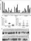
Development and validation of high throughput assay for HIV-1 Env-mediated bystander apoptosis. A, flow chart depicting the procedure for detection of apoptosis mediated by different HIV Envs via the caspase-3-based apoptosis detection assay. B, optimization of the high throughput assay for bystander apoptosis. Different numbers of HeLa cells were transfected with the YU2-Env clone expressing the Env and Rev proteins in a 96-well plate. The next day, the indicated numbers of SupT-R5-H6 were added to the transfected wells. After an overnight incubation, the cells were analyzed for caspase-3 activity using a luminescence substrate-based assay (Caspase-Glo 3/7 assay, Promega). Bars represent means of duplicate wells. One representative of three independent experiments is shown. C, comparison of the high throughput caspase-3 based assay for bystander apoptosis detection with the classical method of apoptosis detection via annexin V staining or by 7-aminoactinomycin D staining. 7500 HeLa cells were transfected with HIV-1 Envs from different laboratory-adapted strains of HIV-1 like YU-2, AD-8, JRCSF, Bal, and 89.6. The next day, 40,000 SupT-R5-H6 cells were added to each well. After an overnight incubation, the cells were analyzed for caspase-3 activity using the Caspase-Glo 3/7 assay. In parallel, HeLa cells were transfected with the above Envs in a 6-well plate. After an overnight incubation, the target cells were harvested and stained for surface phosphatidylserine expression using the annexin V staining kit (BD Biosciences) or by use of cell viability stain 7-aminoactinomycin D (7AAD) (BD Biosciences) followed by flow cytometry. Data are mean ± S.D. (error bars) from triplicate observations.
FIGURE 2.
FIGURE 2.
Suitability of H6 cells for the high throughput assay. A, PBMCs were obtained from Stem Cell Technologies, and CD4+ T cells were isolated using the Human CD4+ T Cell Enrichment Kit (Stem Cell Technologies). HeLa cells were transfected with the indicated Env clones or pcDNA3.1 control vector. The next day, phytohemagglutinin-activated purified CD4+ PBMCs were added to the transfected wells, incubated overnight, and analyzed for caspase-3 activity using the Caspase-Glo 3/7 assay as described in the legend to Fig. 1. B, in parallel, the assay described in A was also conducted with the H6 cell line. C, bright field analysis of syncytia formation in cultures in A and its inhibition via use of AMD-3100 or maraviroc. D, purified CD4+ T cells isolated from PBMCs were stained using anti-CXCR4 and anti-CCR5 antibodies followed by flow cytometry. RLU, relative light units. Error bars, S.D.
FIGURE 3.
FIGURE 3.
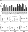
Analysis of bystander apoptosis-inducing activity of primary Envs derived from HIV-infected patients. A high throughput assay was used to determine bystander apoptosis induction of a panel of HIV-1 Envs as described in the legend to Fig. 1. The YU-2 Env (100%) was used as a normalizing control, whereas cells transfected with empty pcDNA3.1 vector were used as background, which was subtracted from all wells for data analysis. A, the graph represents pooled data from three independent experiments with each well assayed in triplicate in each experiment. B, classification of the apoptotic potential of each primary Env into high, medium, or low apoptosis inducers based on the caspase assay data from A. C, classification of the apoptotic potential of each primary Env based on the virus subtype. D, detection of Env expression via HIV Ig-mediated immunoprecipitation. Error bars, S.D.
FIGURE 4.
FIGURE 4.
Single cycle infectivity of primary Envs derived from HIV-infected patients. Different Envs were used to generate pseudotyped virus particles that were used to determine relative infectivity in the TZM-bl cell line. A, bars represent pooled data (mean ± S.E. (error bars)) of duplicate or triplicate observations from three independent experiments. B, classification of infectivity of each primary Env based on high, medium, or low apoptosis inducers (*, p < 0.05). C, classification of infectivity of each primary Env based on the virus subtype. D, graph depicting variation in apoptosis and infectivity of selected Envs.
FIGURE 5.
FIGURE 5.
Bystander apoptosis-inducing activity of primary Envs correlates negatively with PNGS. PNGs were estimated using the sequence information for each Env and the N-Glycosite software available at the LANL database. The variable loop regions of gp120 are V1(6615–6692), V2(6693–6812), V3(7110–7217), V4(7377–7478), and V5(7596–7637), numbered according to the HxB2 nucleotide sequence. V1V2 length is the combined peptide length of the V1 and V2 loop regions. A site with the sequence motif NX(S/T)X, where X represents a non-proline amino acid, is used to predict PNGS. A, representation of PNGS of all of the Envs used in the study. Distribution of PNGS in gp160 (B and E), gp120 (C and F), or gp41 (D and G) is based on low, medium, or high apoptosis-inducing groups (B–D) or virus subtype (E–G) (*, p < 0.05).
FIGURE 6.
FIGURE 6.
Determination of neutralizing antibody sensitivity of primary Envs. Virus stocks pseudotyped with various Envs were used to infect TZM-bl cells in the presence of different concentrations of the TriMab antibody mix as described under “Experimental Procedures.” Infection was determined 48 h postinfection by measuring luciferase activity. A, classification of IC50 concentrations of the TriMab antibody for different Envs based on high, medium, or low apoptosis inducers. B, classification of IC50 concentrations of the TriMab antibody for different Envs based on virus subtype. Due to availability of the TriMab antibody mix in limited amounts, the experiment was conducted once. For analysis based on apoptosis groups (A) and virus subtypes (B), Envs with an IC50 of >25 were excluded.
FIGURE 7.
FIGURE 7.
Sequence analysis of primary Envs. Sequences for the HIV Env proteins used in the study were downloaded from NCBI and aligned with MAFFT, and the respective motif regions were visualized in Jalview using ClustalX-like coloring based on physicochemical properties and conservation. Virus names are shown on the left with the NCBI accession numbers. This analysis revealed a number of amino acid differences between the high and low apoptosis inducers, which led to apoptosis-determining sites like those at positions 425, 462, and 476.
FIGURE 8.
FIGURE 8.
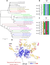
Phylogenetic analysis of primary Envs to identify sequences that share genetic signatures but vary in apoptosis induction. A, phylogeny of primary Envs by a maximum likelihood method based on the HKY model conducted in MEGA5 (51). γ-Distribution (five categories) with shape parameter (0.4327) was used to model evolutionary rate differences among the sites. Pairs of high-low bystander apoptosis are colored with the same color. Shown are sequence signatures of high and low apoptosis inducers at amino acid positions 425 (B) and 476 (C). The high apoptosis-inducing Envs predominantly bear an Asn at position 425 and an Arg at position 476, whereas low apoptosis-inducing Envs bear an Arg at position 425 and a Lys at position 476. D, sequence harmony score coloring of GP120 in complex with CD4. The red colored residue is sequence harmony's best estimate of an important residue distinguishing high/low apoptosis, but based on its structural position away from CD4, such an effect is not intuitively clear. The further ranking can be seen by color transition from red to white to blue.
FIGURE 9.
FIGURE 9.
Structure (Protein Data Bank entry 2B4C) of GP120 (gray ribbon) binding to CD4 (yellow ribbon) rendered using Yasara (55). Arg-476 (red residue) from gp120 is in close proximity with Gln-25 and Ser-23 (pink residues) from CD4. Asn-425 (blue residue) from GP120 is also within 5 Å of Phe-43 and Arg-59 (light blue residues) from CD4.
FIGURE 10.
FIGURE 10.
Mutation of residues Arg-476 and Asn-425 to R476K and N425R, respectively, leads to a reduction in Env-mediated bystander apoptosis-inducing activity. HeLa cells transfected with WT Env clones or their corresponding N425R (A) or R476K (B) counterparts were assayed for bystander apoptosis induction as described in Fig. 1. C, gain of function analysis for the 11034 and 11035 Env pairs. HeLa cells transfected with the indicated WT DNAs or their gain of function counterpart mutants were analyzed for bystander apoptosis induction. Data represent mean ± S.D. (error bars) from triplicate observations. Shown is pairwise analysis of the nearest neighbors 11034 and 11035 for various Env characteristics like apoptosis (D), virus infectivity (E), antibody neutralization (F), and PNGs (G). This pair represents the perfect case scenario for all of the parameters studied. H, detection of Env expression for various WT and mutant Env pairs used in the study by HIV Ig-mediated immunoprecipitation.
FIGURE 11.
FIGURE 11.
Bystander apoptosis induction by primary Envs requires CCR5 binding as well as gp41 function. HeLa cells were transfected with the indicated Envs. The next day, SupT-R5-H6 cells were added to each well in the presence of medium alone, enfuvirtide (2 μm), or maraviroc (1 μm). After an overnight incubation, the cells were analyzed for caspase-3 activity using the Caspase-Glo 3/7 assay as described in the legend to Fig. 1. Data represent mean ± S.D. (error bars) of triplicate observations from three independent experiments.

Similar articles

Cited by

References

    1. Gandhi R. T., Chen B. K., Straus S. E., Dale J. K., Lenardo M. J., Baltimore D. (1998) HIV-1 directly kills CD4+ T cells by a Fas-independent mechanism. J. Exp. Med. 187, 1113–1122 - PMC - PubMed
    1. Garg H., Mohl J., Joshi A. (2012) HIV-1 induced bystander apoptosis. Viruses 4, 3020–3043 - PMC - PubMed
    1. Douek D. (2007) HIV disease progression. Immune activation, microbes, and a leaky gut. Top. HIV Med. 15, 114–117 - PubMed
    1. Meythaler M., Martinot A., Wang Z., Pryputniewicz S., Kasheta M., Ling B., Marx P. A., O'Neil S., Kaur A. (2009) Differential CD4+ T-lymphocyte apoptosis and bystander T-cell activation in rhesus macaques and sooty mangabeys during acute simian immunodeficiency virus infection. J. Virol. 83, 572–583 - PMC - PubMed
    1. van Gils M. J., Edo-Matas D., Bowles E. J., Burger J. A., Stewart-Jones G. B., Schuitemaker H. (2011) Evolution of human immunodeficiency virus type 1 in a patient with cross-reactive neutralizing activity in serum. J. Virol. 85, 8443–8448 - PMC - PubMed

MeSH terms

Substances