Molecular basis for oligomeric-DNA binding and episome maintenance by KSHV LANA
- PMID: 24146617
- PMCID: PMC3798644
- DOI: 10.1371/journal.ppat.1003672
Molecular basis for oligomeric-DNA binding and episome maintenance by KSHV LANA
Abstract
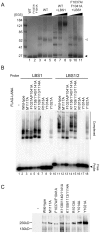
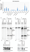
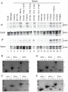
LANA is the KSHV-encoded terminal repeat binding protein essential for viral replication and episome maintenance during latency. We have determined the X-ray crystal structure of LANA C-terminal DNA binding domain (LANADBD) to reveal its capacity to form a decameric ring with an exterior DNA binding surface. The dimeric core is structurally similar to EBV EBNA1 with an N-terminal arm that regulates DNA binding and is required for replication function. The oligomeric interface between LANA dimers is dispensable for single site DNA binding, but is required for cooperative DNA binding, replication function, and episome maintenance. We also identify a basic patch opposite of the DNA binding surface that is responsible for the interaction with BRD proteins and contributes to episome maintenance function. The structural features of LANADBD suggest a novel mechanism of episome maintenance through DNA-binding induced oligomeric assembly.
Conflict of interest statement
The authors have declared that no competing interests exist.
Figures


Similar articles
-
The Kaposi Sarcoma Herpesvirus Latency-associated Nuclear Antigen DNA Binding Domain Dorsal Positive Electrostatic Patch Facilitates DNA Replication and Episome Persistence.J Biol Chem. 2015 Nov 20;290(47):28084-28096. doi: 10.1074/jbc.M115.674622. Epub 2015 Sep 29. J Biol Chem. 2015. PMID: 26420481 Free PMC article.
-
The 3D structure of Kaposi sarcoma herpesvirus LANA C-terminal domain bound to DNA.Proc Natl Acad Sci U S A. 2015 May 26;112(21):6694-9. doi: 10.1073/pnas.1421804112. Epub 2015 May 6. Proc Natl Acad Sci U S A. 2015. PMID: 25947153 Free PMC article.
-
Kaposi's Sarcoma-Associated Herpesvirus LANA-Adjacent Regions with Distinct Functions in Episome Segregation or Maintenance.J Virol. 2019 Mar 5;93(6):e02158-18. doi: 10.1128/JVI.02158-18. Print 2019 Mar 15. J Virol. 2019. PMID: 30626680 Free PMC article.
-
The latency-associated nuclear antigen, a multifunctional protein central to Kaposi's sarcoma-associated herpesvirus latency.Future Microbiol. 2011 Dec;6(12):1399-413. doi: 10.2217/fmb.11.137. Future Microbiol. 2011. PMID: 22122438 Free PMC article. Review.
-
Kaposi's Sarcoma-Associated Herpesvirus Latency-Associated Nuclear Antigen: Replicating and Shielding Viral DNA during Viral Persistence.J Virol. 2017 Jun 26;91(14):e01083-16. doi: 10.1128/JVI.01083-16. Print 2017 Jul 15. J Virol. 2017. PMID: 28446671 Free PMC article. Review.
Cited by
-
Kaposi's sarcoma-associated herpesvirus terminal repeat regulates inducible lytic gene promoters.J Virol. 2024 Feb 20;98(2):e0138623. doi: 10.1128/jvi.01386-23. Epub 2024 Jan 19. J Virol. 2024. PMID: 38240593 Free PMC article.
-
LANA-Mediated Recruitment of Host Polycomb Repressive Complexes onto the KSHV Genome during De Novo Infection.PLoS Pathog. 2016 Sep 8;12(9):e1005878. doi: 10.1371/journal.ppat.1005878. eCollection 2016 Sep. PLoS Pathog. 2016. PMID: 27606464 Free PMC article.
-
Latently KSHV-Infected Cells Promote Further Establishment of Latency upon Superinfection with KSHV.Int J Mol Sci. 2021 Nov 5;22(21):11994. doi: 10.3390/ijms222111994. Int J Mol Sci. 2021. PMID: 34769420 Free PMC article.
-
The Kaposi Sarcoma Herpesvirus Latency-associated Nuclear Antigen DNA Binding Domain Dorsal Positive Electrostatic Patch Facilitates DNA Replication and Episome Persistence.J Biol Chem. 2015 Nov 20;290(47):28084-28096. doi: 10.1074/jbc.M115.674622. Epub 2015 Sep 29. J Biol Chem. 2015. PMID: 26420481 Free PMC article.
-
Kaposi sarcoma herpesvirus pathogenesis.Philos Trans R Soc Lond B Biol Sci. 2017 Oct 19;372(1732):20160275. doi: 10.1098/rstb.2016.0275. Philos Trans R Soc Lond B Biol Sci. 2017. PMID: 28893942 Free PMC article. Review.
References
-
- Cesarman E, Chang Y, Moore PS, Said JW, Knowles DM (1995) Kaposi's sarcoma-associated herpesvirus-like DNA sequences in AIDS-related body-cavity-based lymphomas. The New England Journal of Medicine 332: 1186–1191. - PubMed
-
- Chang Y, Cesarman E, Pessin MS, Lee F, Culpepper J, et al. (1994) Identification of herpesvirus-like DNA sequences in AIDS-associated Kaposi's sarcoma. Science (New York, NY) 266: 1865–1869. - PubMed
-
- Soulier J, Grollet L, Oksenhendler E, Cacoub P, Cazals-Hatem D, et al. (1995) Kaposi's sarcoma-associated herpesvirus-like DNA sequences in multicentric Castleman's disease. Blood 86: 1276–1280. - PubMed
-
- Ballestas ME, Chatis PA, Kaye KM (1999) Efficient persistence of extrachromosomal KSHV DNA mediated by latency-associated nuclear antigen. Science 284: 641–644. - PubMed
-
- Cotter MA 2nd, Robertson ES (1999) The latency-associated nuclear antigen tethers the Kaposi's sarcoma- associated herpesvirus genome to host chromosomes in body cavity-based lymphoma cells [In Process Citation]. Virology 264: 254–264. - PubMed
Publication types
MeSH terms
Substances
Grants and funding
LinkOut - more resources
Full Text Sources
Other Literature Sources

