. 2013;9(10):e1003640.
doi: 10.1371/journal.ppat.1003640. Epub 2013 Oct 17.

A structural basis for BRD2/4-mediated host chromatin interaction and oligomer assembly of Kaposi sarcoma-associated herpesvirus and murine gammaherpesvirus LANA proteins

Affiliations

A structural basis for BRD2/4-mediated host chromatin interaction and oligomer assembly of Kaposi sarcoma-associated herpesvirus and murine gammaherpesvirus LANA proteins

Jan Hellert et al. PLoS Pathog. 2013.

Abstract

Kaposi sarcoma-associated herpesvirus (KSHV) establishes a lifelong latent infection and causes several malignancies in humans. Murine herpesvirus 68 (MHV-68) is a related γ2-herpesvirus frequently used as a model to study the biology of γ-herpesviruses in vivo. The KSHV latency-associated nuclear antigen (kLANA) and the MHV68 mLANA (orf73) protein are required for latent viral replication and persistence. Latent episomal KSHV genomes and kLANA form nuclear microdomains, termed 'LANA speckles', which also contain cellular chromatin proteins, including BRD2 and BRD4, members of the BRD/BET family of chromatin modulators. We solved the X-ray crystal structure of the C-terminal DNA binding domains (CTD) of kLANA and MHV-68 mLANA. While these structures share the overall fold with the EBNA1 protein of Epstein-Barr virus, they differ substantially in their surface characteristics. Opposite to the DNA binding site, both kLANA and mLANA CTD contain a characteristic lysine-rich positively charged surface patch, which appears to be a unique feature of γ2-herpesviral LANA proteins. Importantly, kLANA and mLANA CTD dimers undergo higher order oligomerization. Using NMR spectroscopy we identified a specific binding site for the ET domains of BRD2/4 on kLANA. Functional studies employing multiple kLANA mutants indicate that the oligomerization of native kLANA CTD dimers, the characteristic basic patch and the ET binding site on the kLANA surface are required for the formation of kLANA 'nuclear speckles' and latent replication. Similarly, the basic patch on mLANA contributes to the establishment of MHV-68 latency in spleen cells in vivo. In summary, our data provide a structural basis for the formation of higher order LANA oligomers, which is required for nuclear speckle formation, latent replication and viral persistence.


Conflict of interest statement

The authors have declared that no competing interests exist.

Figures

Figure 1
Figure 1. Crystal Structure of the KSHV LANA CTD with Its Orthologs.
A: Crystal structure of the dimeric kLANA CTD, front view. B: Surface electrostatic potential of kLANA, mLANA, and the EBV EBNA-1 CTD in front view (bottom) and top view (above). Red represents negative charge and blue represents positive charge. C: Residues at the sequence specific DNA binding site of kLANA, bottom view. Residues mutated in this study are bold faced. D: 3D superpositions of the kLANA CTD (white) with mLANA (blue), EBV EBNA-1 (red), and HPV-16 E2 (green) CTD monomers. E: Structure-based sequence alignment of kLANA, mLANA, EBV EBNA-1, and HPV-16 E2 CTDs. Acidic residues are red and basic residues are blue. See also Figure S1.
Figure 2
Figure 2. Oligomerization of the KSHV and MHV-68 LANA CTDs.
A: Oligomeric assemblies of kLANA (top) and mLANA (below) CTD dimers as found in the respective crystals. Inter-chain contact areas within and between dimers are indicated. B: Details on the oligomerization sites of kLANA (left) and mLANA CTDs (right), viewed from the center of the ring (kLANA, monoclinic crystal) and the top of the linear chain (mLANA). Color scheme corresponds to Figure 1A. C: Flow profiles (black graph) and molecular weight (red graph) of kLANA(1013-1149) (top) and mLANA(124-260) (bottom) in asymmetric field flow fractionation. D: Oligomerization assay with kLANA mutants. Top left: Western blot detecting FL kLANA wt or mutants bound to GST-fused kLANA wt or mutant CTDs. Bottom left: Ponceau S –stained WB membrane showing GST-LANA(934–1162) used in this assay. (e.v.) empty vector, (-) GST only, (MUT) mutant GST-LANA CTD always corresponding to the FL LANA mutant indicated above. Right: Expression of FL LANA proteins in eukaryotic cells. The aberrant running behavior of some mutants might be due to different posttranslational modification. E: EMSA with LBS1+2 oligonucleotide and GST-LANA(934-1162) oligomerization deficient mutants. (wt+comp.) control with 10× excess of unlabeled probe. Right: Expression of GST-LANA CTD proteins; Coomassie stained SDS PAGE gel. F: Transient replication assay with oligomerization mutants and pGTR4 vector in HeLa cells. Panel I: Southern blot of replicated DNA, remaining after digest with MfeI and DpnI. Panel II: Southern blot of input DNA linearized with MfeI; pEGFP does not replicate and serves as an internal control. Assay was performed in duplicates. Panel III: LANA expression. Panel IV: Actin loading control. (-) empty vector control.
Figure 3
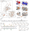
Figure 3. Interaction of the KSHV LANA CTD with ET Domains in Solution.
A: [1H,15N]-TROSY spectra of 0.48 mM [2H,13C,15N]-kLANA(1013–1149) in 30 mM NaCl in the absence (black) and presence (orange) of 0.48 mM unlabeled BRD4(600–680). Prominent chemical shift perturbations are encircled in red. Bottom left: Chemical shift perturbation of H1126 upon titration of BRD4 ET. Bottom right: Backbone amide assignment of the central spectral region. B: Magnitude of chemical shift perturbations from (A) over the sequence of kLANA(1013–1149). Unassigned residues are indicated (*). Acidic residues are red and basic residues are blue, prolines are in bold face. C: Top: Surface electrostatic potential of BRD2 ET (left) and the kLANA CTD in bottom view (right). Below: Chemical shift perturbations mapped on the structures of BRD2 ET (200 mM NaCl) and the kLANA CTD (30 mM NaCl). Prolines and other unassigned residues are grey. D: Details of [1H,15N]-TROSY spectra of 0.48 mM 15N-BRD4(600–680) (top) and 15N-BRD2(632–713) (bottom) at different NaCl concentrations in the absence (black) and presence (orange) of 0.96 mM unlabeled kLANA(996–1153). Perturbation of S619 (BRD4) and S651 (BRD2) is indicated by an arrow. E: Magnitude of chemical shift perturbations from (D) over an alignment of BRD4 (top) and BRD2 (bottom) ET domains at different NaCl concentrations. Histogram bars are given for 200 mM NaCl. A region of strong chemical shift perturbation only at 50 mM NaCl is indicated (*); compare Figure 4D.
Figure 4
Figure 4. Interaction of the KSHV LANA CTD with ET Domains in vivo.
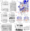
A: kLANA wt was co-immunoprecipitated with GFP-tagged full-length BRD2 wt or mutants. Left: Panel I: Immunoblot of co-IP samples detecting LANA. Panel II: Blot of the same samples detecting GFP-BET proteins. Panel III: Expression of LANA in all of the samples. Panel IV: Actin loading control. Right: Immunoblot of co-IP samples with αLANA antibody (panel I), blot of the expression of the GFP-BRD2 proteins (panel II) and of LANA (panel IV) in all of the samples. Panels III and V: Actin loading control for GFP-BRD2 and LANA expression, respectively. ‘82A/83A/85A’: Triple mutant E682A/E683A/E685A, ‘87A/89A’: Double mutant D687A/E689A. B: kLANA ‘ET binding site’ mutants were co-immunoprecipitated with GFP-tagged BRD4 (HUNK; left) and BRD2 (right). Panels as described for (A) left side. (*) nonspecific bands appearing with some αGFP antibody lots. C: Detail of the complex ‘model II’ of kLANA (surface electrostatic potential representation) and BRD2 ET (cartoon representation). D: DNA-bound EBNA-1 CTD (left) and ‘model II’ of the kLANACTD:ET complex (right) in comparison. The acidic DNA backbone and the acidic loop region of the ET domains are shown in red. The region of strong chemical shift perturbation only at 50 mM NaCl is indicated (*). E: Transient replication assay with ‘ET binding site’ mutants and pGTR4 vector in HeLa cells. Panels I–IV as described in Figure 2F. F: EMSA with LBS1+2 oligonucleotide and GST-LANA(934–1162) wt or ‘ET binding site’ mutants. (wt+comp.) control with wt LANA and 10× excess of unlabeled probe. Right: Expression of GST-LANA CTD proteins; Coomassie stained SDS PAGE gel. See also Figure S2.
Figure 5
Figure 5. Role of the ‘Basic top’ of LANA in BET Protein Interaction and Oligomerization.
A: Co-IP of kLANA ‘basic top’ mutants with GFP-BRD4 (left) and GFP-BRD2 (right). Panels I–IV as in Figure 4B. ‘09A/38A’: K1109A/K1138A mutant. B: Oligomerization assay with kLANA ‘basic top’ mutants. Upper top: WB detecting FL kLANA wt or mutants bound to GST-kLANA wt or mutant CTDs. Lower top: same WB membrane as above detecting GST-LANA CTD proteins. (e.v.) empty vector, (-) GST alone, (MUT) mutant GST-LANA CTD corresponding to the FL LANA mutant indicated above. Bottom: Expression of FL LANA proteins and the actin control. C: EMSA with LBS1+2 oligonucleotide and GST-LANA(934-1162) ‘basic top’ mutants. (wt+comp.) control with 10× excess of unlabeled probe. Below: Expression of the GST-LANA CTD proteins (Coomassie stain). D: Transient replication assay with kLANA ‘basic top’ mutants and pTR1 vector in HeLa cells (performed in duplicates). Panel I: Southern blot of replicated DNA, remaining after digest with KpnI and DpnI. Panel II: Southern blot of input DNA linearized with KpnI; pBluescript (pBS) does not replicate (internal control). (-) empty vector control. Panel III: LANA expression. Panel IV: Actin control. E: Top view of kLANA and mLANA CTDs. Mutated residues are labeled. F: Co-IP of GFP-BRD4 (left) and GFP-BRD2 (right) with HA-mLANA wt or 4A mutant. Panels I: Immunoblot of co-IP samples detecting GFP-BET proteins. Panels II: Blot of the same samples detecting HA-mLANA. Panels III: Expression of BET proteins. Panels IV: Actin loading control. G: C57BL/6 mice were infected i.n. with the MHV68 wt, the mLANA: 4A (‘4A’) and STOP (‘73-STOP’) mutant viruses and respective revertants (‘4A rev’), (‘73-STOP rev’). DNA isolated from splenocytes was used for qPCR analysis. Marks represent individual mice and the bars the means. Data include two independent experiments. (***) P<0.001. H: Ex vivo reactivation assay with the splenocyte samples from (G). Dashed line: the point of 63.2% reactivation (MOI = 1).
Figure 6
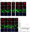
Figure 6. Contribution of Different Sites in KSHV LANA CTD to the Formation of LANA Nuclear ‘Speckles’.
A: Speckle formation assay with kLANA mutants of different sites. HeLa cells were transfected with vector containing 4xTR and kLANA wt or mutant vectors. kLANA was stained with a mouse αLANA antibody and DNA was stained with DAPI. Expression of GFP confirms the presence of the TR containing vector in the cells. Close up of an exemplary single cell from each of the images is shown in the top row. Images taken at 63× magnification, a scale bar of 20 µm is shown in the DAPI image of the first sample (wt+GFP). B: Quantification of the number of LANA speckles per nucleus performed with Cell Profiler software. The mean LANA speckle number per nucleus is plotted on the graph for each of the LANA proteins. A total of 80–110 cells were analyzed per sample in two independent experiments. The error bars indicate SEM. Speckle numbers in the samples marked in red are significantly different from those obtained with wt LANA.
Figure 7
Figure 7. Hypothetical model of KSHV LANA oligomers.
Dark blue: kLANA CTD dimers. Cyan: kLANA acidic internal repeat regions and N-terminal domains. Grey: DNA. Orange: BET proteins.

Similar articles

Cited by

References

    1. Schulz TF (2006) The pleiotropic effects of Kaposi's sarcoma herpesvirus. J Pathol 208: 187–198. - PubMed
    1. Dupin N, Fisher C, Kellam P, Ariad S, Tulliez M, et al. (1999) Distribution of human herpesvirus-8 latently infected cells in Kaposi's sarcoma, multicentric Castleman's disease, and primary effusion lymphoma. Proc Natl Acad Sci U S A 96: 4546–4551. - PMC - PubMed
    1. Rainbow L, Platt GM, Simpson GR, Sarid R, Gao SJ, et al. (1997) The 222- to 234-kilodalton latent nuclear protein (LNA) of Kaposi's sarcoma-associated herpesvirus (human herpesvirus 8) is encoded by orf73 and is a component of the latency-associated nuclear antigen. J Virol 71: 5915–5921. - PMC - PubMed
    1. Garber AC, Hu J, Renne R (2002) Latency-associated nuclear antigen (LANA) cooperatively binds to two sites within the terminal repeat, and both sites contribute to the ability of LANA to suppress transcription and to facilitate DNA replication. JBiolChem 277: 27401–27411. - PubMed
    1. Hu J, Garber AC, Renne R (2002) The latency-associated nuclear antigen of Kaposi's sarcoma-associated herpesvirus supports latent DNA replication in dividing cells. JVirol 76: 11677–11687. - PMC - PubMed

Publication types

MeSH terms

Grants and funding

This work was supported by the DFG (http://www.dfg.de/) Collaborative Research Centre SFB900 ‘Chronic Infections: Microbial Persistence and its Control’ and the European Union Integrated project INCA (http://cordis.europa.eu/search/index.cfm?fuseaction=proj.document&PJ_RCN=8323620) - LSHC-CT-2005-018704 - to TFS, the Emmy Noether Young Investigator Grant LU1471/3-1 (http://www.dfg.de/foerderung/programme/einzelfoerderung/emmy_noether/) to TL, a Helmholtz (HGF) ‘Impuls und Vernetzungsfonds’ (http://www.helmholtz.de/en/about_us/initiating_and_networking/) grant to CR, by HGF grant VH-GS-202 to the HZI Grad School, by BMBF (http://www.bmbf.de/en/index.php) grants (NGFNplus, FKZ PIM-01GS0802-3) and Wilhelm Sander-Stiftung (http://www.wilhelm-sander-stiftung.de/cms/front_content.php) grant (2009.046.2) to HA. The funders had no role in study design, data collection and analysis, decision to publish, or preparation of the manuscript.