. 2013 Nov;123(11):4821-35.
doi: 10.1172/JCI68523.

NF-κB-mediated Pax7 dysregulation in the muscle microenvironment promotes cancer cachexia

NF-κB-mediated Pax7 dysregulation in the muscle microenvironment promotes cancer cachexia

Wei A He et al. J Clin Invest. 2013 Nov.

Abstract

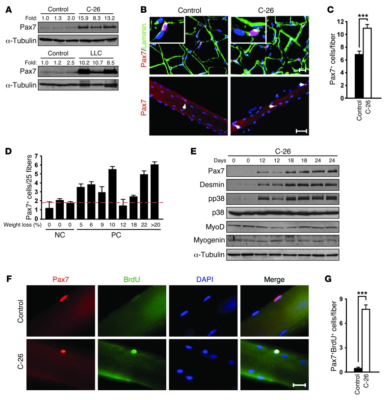
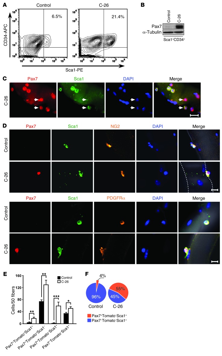
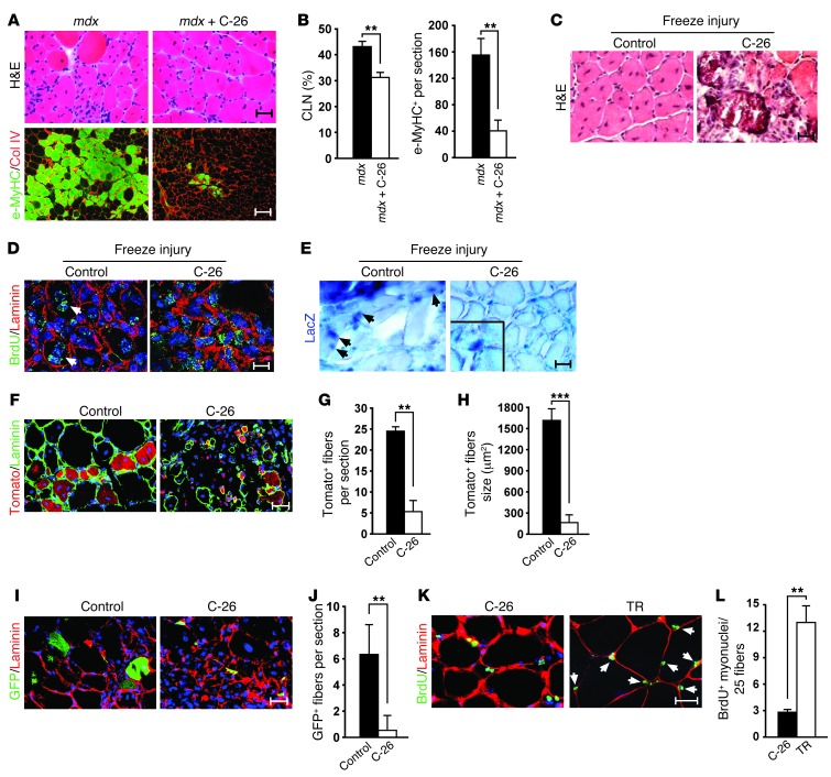
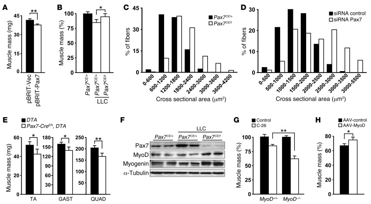
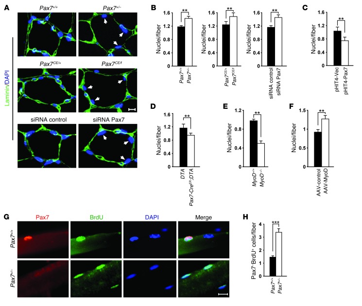
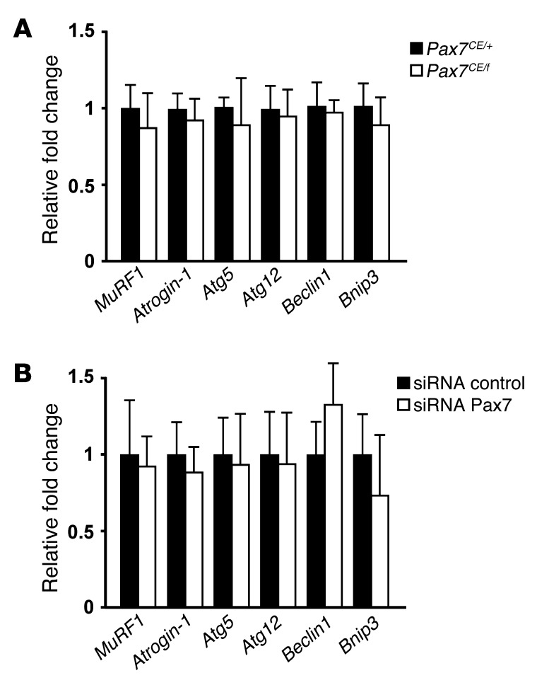
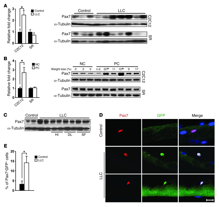
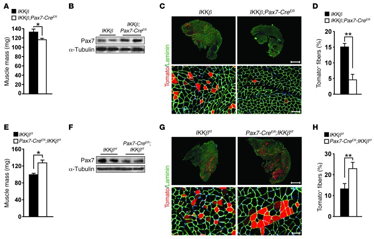
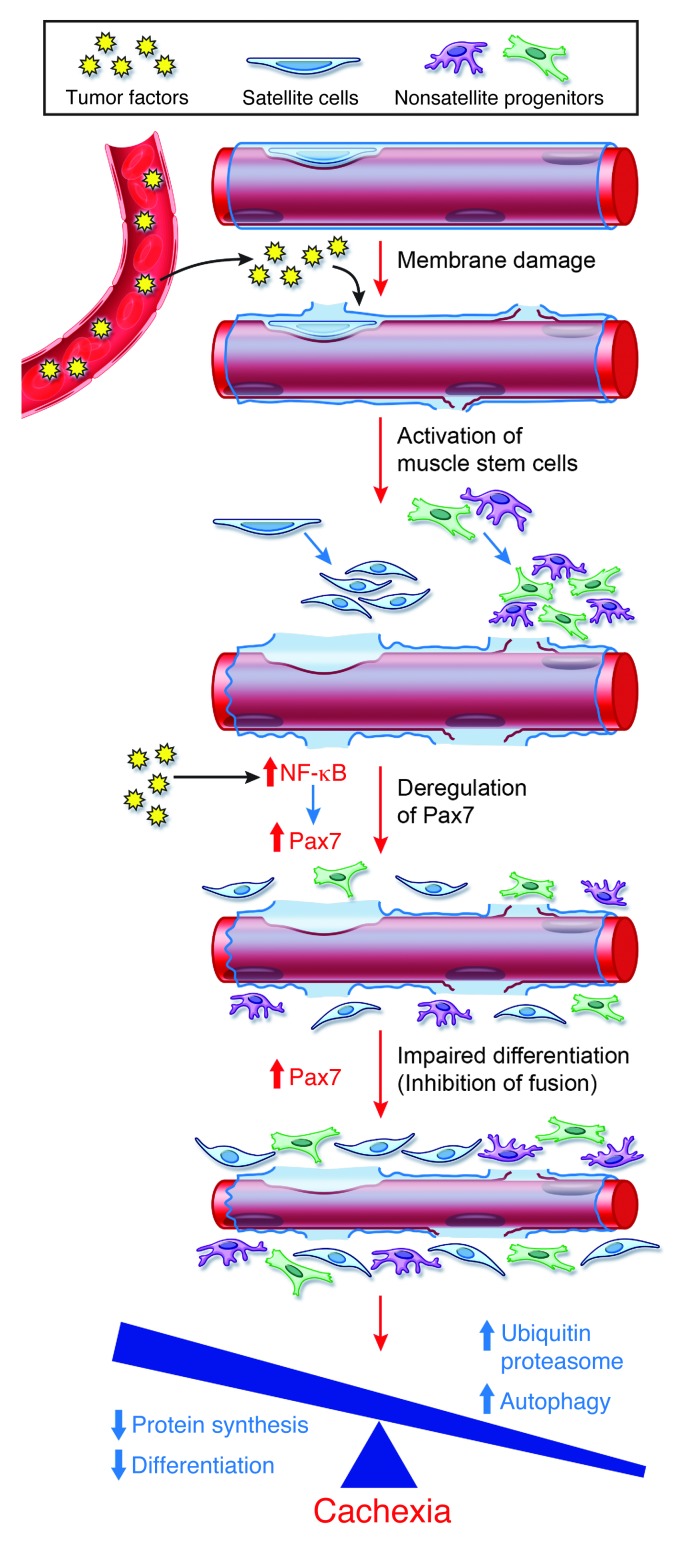
Cachexia is a debilitating condition characterized by extreme skeletal muscle wasting that contributes significantly to morbidity and mortality. Efforts to elucidate the underlying mechanisms of muscle loss have predominantly focused on events intrinsic to the myofiber. In contrast, less regard has been given to potential contributory factors outside the fiber within the muscle microenvironment. In tumor-bearing mice and patients with pancreatic cancer, we found that cachexia was associated with a type of muscle damage resulting in activation of both satellite and nonsatellite muscle progenitor cells. These muscle progenitors committed to a myogenic program, but were inhibited from completing differentiation by an event linked with persistent expression of the self-renewing factor Pax7. Overexpression of Pax7 was sufficient to induce atrophy in normal muscle, while under tumor conditions, the reduction of Pax7 or exogenous addition of its downstream target, MyoD, reversed wasting by restoring cell differentiation and fusion with injured fibers. Furthermore, Pax7 was induced by serum factors from cachectic mice and patients, in an NF-κB-dependent manner, both in vitro and in vivo. Together, these results suggest that Pax7 responds to NF-κB by impairing the regenerative capacity of myogenic cells in the muscle microenvironment to drive muscle wasting in cancer.


Figures

Figure 1
Figure 1. Cancer cachexia promotes myofiber damage and interstitial cell alterations.
(A) Top: EM taken from ultrathin longitudinal sections of skeletal muscle biopsies from NC or PC patients. Middle: Human IgG immunofluorescence staining on muscle cross-sections from noncancer individuals (NC) and pancreatic cancer (PC) patients. Bottom: α-laminin and DAPI staining on the same patient samples. Percentages in parentheses represent body weight loss compared with pre-illness status. (B) EM analysis of ultrathin muscle longitudinal sections from control and C-26 mice. (C) Sections similar to those in A were analyzed for cellular alterations. Higher-magnification views of bracketed regions in B and C are shown at right (enlarged ×4). Scale bars: 0.5 μm (A, top); 100 μm (A, middle); 10 μm (A, bottom; B; and C).
Figure 2
Figure 2. Cancer cachexia is associated with activation and myogenic commitment of satellite cells.
(A) GAST from control and C-26 and LLC tumor–bearing mice blotted for Pax7. Numbers above lanes represent Pax7 quantitation by ImageJ, normalized to α-tubulin. (B) Top: GAST sections probed for Pax7 and α-laminin. Similar results were observed in TA and QUAD. DAPI was used to counterstain nuclei (blue). Insets show 1 Pax7+ cell each at higher magnification (enlarged ×9). Bottom: Isolated myofibers probed for Pax7 and DAPI (blue). Arrows denote Pax7+ cells. (C) Quantitation of Pax7+ cells from single fibers from control and C-26 mice. (D) Cross-sections of muscle biopsies from NC and PC patients, with weight loss as indicated, scored for Pax7+ cells. Quantitation was performed from 20 random fields, counting a minimum of 1,000 fibers per sample. Dashed red line denotes the baseline value of Pax7+ cells in NC patients. (E) Muscles were harvested in duplicate experiments after C-26 administration. Western blots were performed, probing for markers of satellite cells (Pax7), myoblasts (phospho- and total p38, desmin, and MyoD), and differentiation (MyoD and myogenin). Blot was reprobed for α-tubulin. (F) Single fibers were double stained for Pax7 and BrdU. Nuclei were counterstained with DAPI. (G) Quantitation of Pax7+BrdU+ cells in F. Scale bars: 10 μm (B, top); 50 μm (B, bottom); 20 μm (F). ***P < 0.001.
Figure 3
Figure 3. Cancer cachexia is associated with myogenic commitment of multiple muscle progenitor cells.
(A) FACS analysis of nonsatellite progenitor cells in control and C-26 mice by gating for Sca1+CD34+ cells (shown as a percentage). (B) Cells as in A were sorted and probed for Pax7 by Western blot. (C) Single fibers from C-26 mice were stained with Pax7 and Sca1. Nuclei were counterstained with DAPI. Arrows denote Pax7+Sca1 and Pax7+Sca1+ cells. (D) Single fibers from control and C-26 mice were immunostained with Pax7, Sca1, NG2 or PDGFRα, and DAPI as indicated. Dotted white lines indicate myofiber border. (E) Myofibers were isolated from Pax7-CreER;Rosa26-Tomato reporter control and C-26 mice that were induced with tamoxifen before tumor implantation, and myofibers were subsequently immunostained for Pax7 and Sca1. For each condition, at least 300 fibers were counted. Quantitation of 4 cell populations is shown. The major increase of Pax7+Sca1+ cells came from a nonsatellite cell lineage. (F) Distribution of Pax7 expression in the TomatoSca1+ population of control and C-26 mice. Scale bars: 20 μm (C); 25 μm (D). *P < 0.05, **P < 0.01, ***P < 0.001.
Figure 4
Figure 4. Muscle regeneration is compromised in cancer cachexia.
(A and B) 6-week-old mdx mice were injected with C-26 tumors or saline. (A) After 2 weeks, GAST were analyzed by H&E or immunostained with e-MyHC and collagen IV (Col IV). (B) Quantitation of centrally located nuclei (CLN) and e-MyHC+ fibers. (C) TA from control and C-26 mice were injured by freezing and analyzed after 8 days by H&E. (D) TA were injured, and mice were subsequently injected with BrdU. TA was immunostained with BrdU and α-laminin. (E) Athymic nude control and C-26 mice were subjected to TA injury and transplanted with muscle mononuclear cells from desmin-nLacZ transgenic mice. 1 week after injury, TA was stained for LacZ. Arrows denote desmin+ nuclei within myofibers. Inset shows interstitial LacZ+ cells (enlarged ×5). (FH) TA from control (n = 5) and C-26 (n = 7) mice were injured and transplanted with FACS-sorted Tomato+ cells. (F) After transplantation, muscle sections were immunostained for α-laminin and DAPI (blue). Tomato+ myofiber number (G) and size (H) were also quantified. (I and J) As in F, except that the donor population was sorted for CD34+Sca1+ cells from nontumor GFP reporter mice. (I) Immunostaining of GFP, α-laminin, and DAPI (blue). (J) GFP+ myofibers. (K and L) C-26 mice and surgically tumor-resected C-26 mice (TR) were injected with BrdU (n = 5 per group). (K) Sections were stained for BrdU, α-laminin, and DAPI (blue). (L) Sublaminar BrdU+ nuclei from tumor-resected animals. Scale bars: 25 μm (A, H&E); 100 μm (A, immunostaining); 20 μm (C; E; F; I; and K); 10 μm (D). **P < 0.01, ***P < 0.001.
Figure 5
Figure 5. Muscle wasting in cancer cachexia is mediated through deregulation of Pax7.
(A) GAST mass, recorded in young mice (P15) after injection of pBRIT retroviruses expressing empty vector (Vec) or the Pax7 transgene. (B) GAST mass from LLC tumor–bearing Pax7CE/+ and Pax7CE/f mice (n = 10 per group) was measured and normalized to that of Pax7CE/+ heterozygous mice without tumors (set at 100%). (C) Cross-sectional area measurements in LLC tumor–bearing Pax7CE/+ and Pax7CE/f mice from B. (D) Cross-sectional area measurements of muscles from C-26 mice treated with systemic delivery of control siRNA or siRNA targeting Pax7 (n = 5 per group). (E) TA, GAST, and QUAD mass, measured from tumor-bearing mice lacking (n = 7) or expressing (n = 10) DTA under the control of the Pax7-CreER locus. (F) Western blots performed on GAST from mice in C and D, probing for MyoD and myogenin. Blots were reprobed for α-tubulin. (G) GAST mass from C-26 MyoD+/+ and MyoD–/– mice (n = 10 per group) was measured and normalized to that from age- and genotype-matched nontumor control mice (set at 100%). (H) Muscles of C-26 mice were injected with control or MyoD-expressing AAV (n = 5 per group); after 3 weeks of virus expression, muscle mass was measured and normalized to that from nontumor mice. *P < 0.05, **P < 0.01.
Figure 6
Figure 6. Deregulation of Pax7 promotes wasting in cancer by inhibiting regeneration through impairment of myogenic cell fusion.
(A) Cross-sections prepared from muscles of tumor-bearing Pax7+/+ and Pax7+/– mice, mice treated with control or siRNA against Pax7, or mice with conditional Pax7 deletion were immunostained with α-laminin and DAPI. Arrows denote sublaminar nuclei. (B) Quantitation of sublaminar nuclei from A. At least 3,000 fibers were counted per condition. (CF) Sublaminar nuclei per fiber from neonatal muscles injected with pHIT4 vector or Pax7 retroviruses (C); muscles in which Pax7+ progenitors were eliminated by DTA (D); MyoD+/+ and MyoD–/– muscles (E); and muscles injected with control or MyoD-expressing AAV (F). At least 3,000 fibers were counted per condition. (G) Tumor-bearing Pax7+/+ and Pax7+/– mice were injected with BrdU, and muscle fibers were immunostained for Pax7, BrdU, and DAPI. Merged images are shown to denote specificity. (H) Quantitation of Pax7BrdU+ myonuclei per fiber from muscles in G. Scale bars: 10 μm (A); 25 μm (G). **P < 0.01, ***P < 0.001.
Figure 7
Figure 7. Pax7-mediated muscle wasting in cancer is independent of proteasome and autophagy pathways.
Muscles were isolated from tumor-bearing mice either conditionally deleted for Pax7 in Pax7+ cells (A) or systemically treated with siRNA targeting Pax7 (B), and RNA was analyzed by qRT-PCR probing for genes regulated in the ubiquitin proteasome (Atrogin-1 and MuRF1) and autophagy (Atg5, Atg12, Beclin1, and Bnip3) pathways.
Figure 8
Figure 8. Circulating cachectic factors deregulate Pax7 through NF-κB activation.
(A) Pax7 Western blot from C2C12 control cells and IκBα-SR cells (SR) that were treated with 5% serum from control or LLC tumor–bearing mice. Quantitation of Pax7 normalized to α-tubulin is also shown. (B) Similar to A, except serum was collected from NC and PC patients with weight loss as indicated (expressed as a percentage of the premorbid state). n.d., not determined. Asterisks denote samples from Figure 2D. Quantitation of Pax7 normalized to α-tubulin is also shown. (C) Cachectic serum was heat inactivated (HI), delipidated (DL), or size fractionated <10 kDa (SF) and used to treat C2C12 cells. Pax7 was examined by Western blot. (D) Fibers from control and LLC tumor–bearing mice were immunostained for Pax7 and GFP. (E) Quantitation of Pax7+GFP+ cells in D. Scale bars: 20 μm (D). *P < 0.05.
Figure 9
Figure 9. Pax7 deregulation and muscle wasting in cancer cachexia is dependent on NF-κB.
(A) GAST mass from tumor-bearing control Rosa26-IKKβ (n = 15) or Pax7-CreER;Rosa26-IKKβ (n = 20) mice injected with tamoxifen. (B) Western blot for Pax7 from muscles in A. (C) Tomato+ cells from Rosa26-IKKβ and Pax7-CreER;Rosa26-IKKβ muscles probed for α-laminin and DAPI (blue). Higher-magnification views are shown below. (D) Quantitation of Tomato+ cells from C. At least 15 sections and 9 × 10⁵ fibers from 5 mice were counted for each group. (E) GAST mass from tumor-bearing control IKKβf/f (n = 12) or Pax7-CreER;IKKβf/f (n = 18) mice injected with tamoxifen. (F) Western blot of Pax7 from muscles in E. (G) Tomato+ cells from IKKβf/f and Pax7-CreER;IKKβf/f muscle sections immunostained with α-laminin and DAPI (blue). Higher-magnification views are shown below. (H) Quantitation of Tomato+ fibers from G. At least 15 sections and 9 × 10⁵ fibers from 3 mice were counted for each group. Scale bars: 0.5 mm (C and G, top); 100 μm (C and G, bottom). *P < 0.05, **P < 0.01.
Figure 10
Figure 10. Involvement of the muscle microenvironment in cancer cachexia.
Circulating tumor factors induce muscle membrane damage; in response, satellite cells and other myogenic progenitor populations are activated and commit to a myogenic program. Upon additional activation of classical NF-κB, Pax7 is deregulated, which impairs myogenic cell differentiation. Because of this stalled regeneration, committed myoblasts are unable to fuse and rescue damaged fibers. As a result, the loss of muscle repair in conjunction with reduced protein synthesis are unable to compensate against the procachectic activities of the ubiquitin proteasome and autophagy pathways, thus tipping the balance toward a cachectic state.

Similar articles

Cited by

References

    1. Tisdale MJ. Cachexia in cancer patients. Nat Rev Cancer. 2002;2(11):862–871. doi: 10.1038/nrc927. - DOI - PubMed
    1. Andreyev HJ, Norman AR, Oates J, Cunningham D. Why do patients with weight loss have a worse outcome when undergoing chemotherapy for gastrointestinal malignancies? Eur J Cancer. 1998;34(4):503–509. doi: 10.1016/S0959-8049(97)10090-9. - DOI - PubMed
    1. Fearon KC, Glass DJ, Guttridge DC. Cancer cachexia: mediators, signaling, and metabolic pathways. Cell Metab. 2012;16(2):153–166. doi: 10.1016/j.cmet.2012.06.011. - DOI - PubMed
    1. Tisdale MJ. Tumor-host interactions. J Cell Biochem. 2004;93(5):871–877. doi: 10.1002/jcb.20246. - DOI - PubMed
    1. Siegel R, Naishadham D, Jemal A. Cancer statistics, 2012. CA Cancer J Clin. 2012;62(1):10–29. doi: 10.3322/caac.20138. - DOI - PubMed

Publication types

MeSH terms