. 2013;9(9):e1003774.
doi: 10.1371/journal.pgen.1003774. Epub 2013 Sep 5.

An alteration in ELMOD3, an Arl2 GTPase-activating protein, is associated with hearing impairment in humans

Affiliations

An alteration in ELMOD3, an Arl2 GTPase-activating protein, is associated with hearing impairment in humans

Thomas J Jaworek et al. PLoS Genet. 2013.

Abstract

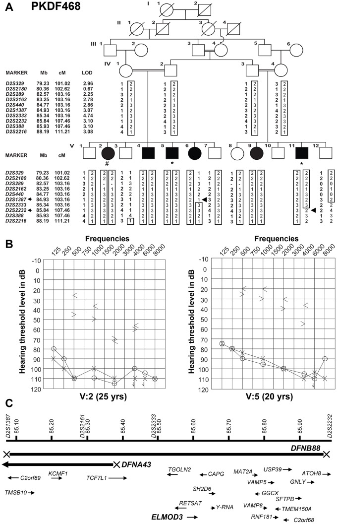
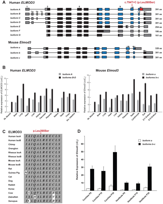
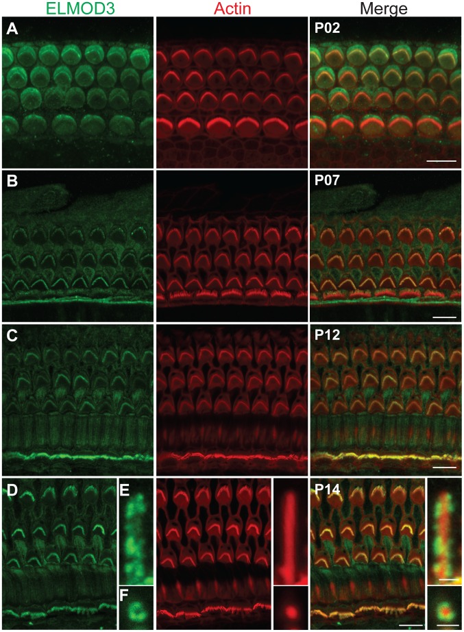
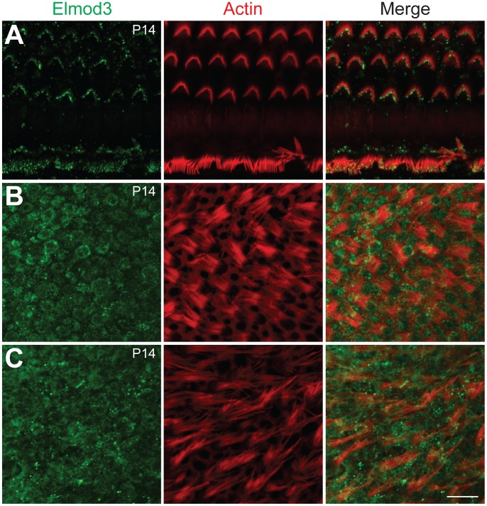
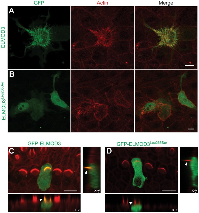
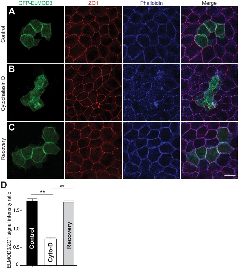
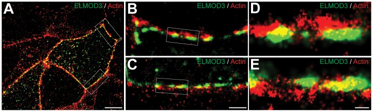
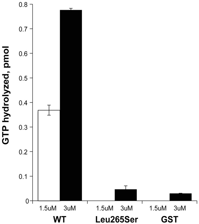
Exome sequencing coupled with homozygosity mapping was used to identify a transition mutation (c.794T>C; p.Leu265Ser) in ELMOD3 at the DFNB88 locus that is associated with nonsyndromic deafness in a large Pakistani family, PKDF468. The affected individuals of this family exhibited pre-lingual, severe-to-profound degrees of mixed hearing loss. ELMOD3 belongs to the engulfment and cell motility (ELMO) family, which consists of six paralogs in mammals. Several members of the ELMO family have been shown to regulate a subset of GTPases within the Ras superfamily. However, ELMOD3 is a largely uncharacterized protein that has no previously known biochemical activities. We found that in rodents, within the sensory epithelia of the inner ear, ELMOD3 appears most pronounced in the stereocilia of cochlear hair cells. Fluorescently tagged ELMOD3 co-localized with the actin cytoskeleton in MDCK cells and actin-based microvilli of LLC-PK1-CL4 epithelial cells. The p.Leu265Ser mutation in the ELMO domain impaired each of these activities. Super-resolution imaging revealed instances of close association of ELMOD3 with actin at the plasma membrane of MDCK cells. Furthermore, recombinant human GST-ELMOD3 exhibited GTPase activating protein (GAP) activity against the Arl2 GTPase, which was completely abolished by the p.Leu265Ser mutation. Collectively, our data provide the first insights into the expression and biochemical properties of ELMOD3 and highlight its functional links to sound perception and actin cytoskeleton.


Conflict of interest statement

The authors have declared that no competing interests exist.

Figures

Figure 1
Figure 1. Hearing loss segregating in family PKDF468 is associated with a missense ELMOD3 allele.
(A) Pedigree of the family in which the DFNB88 locus was mapped. The filled symbols represent affected individuals, and a double horizontal line connecting parents represents a consanguineous marriage. The alleles forming the risk haplotypes are boxed. The short tandem repeat (STR) markers, their relative map positions (Mb) according to UCSC Genome Bioinformatics Build GRCh37 (hg19), and their genetic positions (cM) based on the Marshfield genetic map are shown next to the pedigree. Haplotype analysis revealed a linkage region of 4.3 cM (0.91 Mb) that was delimited by proximal meiotic recombination in individual V:6 (arrowhead) at marker D2S1387 (103.16 cM; arrow) and distal recombination at marker D2S2232 (107.46 cM; arrow) in individual V:11 (arrowhead). (B) Pure-tone audiograms for family PKDF468 V:2 (25-year-old female) and V:5 (20-year-old male). The symbols ‘o’ and ‘x’ denote air conduction pure-tone thresholds, and the ‘<’ and ‘>’ symbols denote bone conduction thresholds in the right and the left ears, respectively. (C) The linkage interval that defines the DFNB88 locus for family PKDF468 partially overlaps with DFNA43. The relative locations and orientations of the genes and mRNAs are indicated with arrows. Individual V:2 DNA sample was used for exome sequencing, while individuals V:5 and V:11 samples were used for Sanger sequencing of coding, non-coding, and flanking sequences of the exon-intron boundaries of the known candidate genes in DFNB88 linkage interval.
Figure 2
Figure 2. The transcripts and expression profiles of the genes that encode ELMOD3 in humans and mice.
(A) Alternative splicing leads to seven isoforms of human ELMOD3. Non-coding segments, sequences encoding ELMO domain and other coding regions of exons are denoted by gray, blue and black boxes, respectively. Also depicted is the mutation that was identified in the DFNB88 family (red arrow). The numbering of the position of mutation c.794T>C (p.Leu265Ser) is based on accession number NM_032213.4. Mice have only three Elmod3 alternative transcripts. Primers used for real-time quantitative PCR analyses are represented with black arrows. (B) Real-time quantitative PCR analysis of human and mouse ELMOD3/Elmod3 isoforms A/a and B–E/b–c. (C) The leucine residue at amino acid position 265 (accession number NP_115589) is completely conserved across a wide variety of species. Identical residues are boxed in gray. (D) Real-time quantitative RT-PCR analysis of Elmod3 isoforms a and b–c in C57BL/6J mouse cochlear and vestibular tissues at three different ages (P0, P10 and P30). CT indicates the observed threshold number of PCR cycles that was required for the detection of the amplification product; the relative expression level is the calculated difference in CT between the Elmod3 and that of an internal control standard (Gapdh), which was measured in the same sample.
Figure 3
Figure 3. ELMOD3 is expressed in sensory cells of rat organ of Corti.
(A–F) The spatio-temporal expression pattern of ELMOD3 in the rat organ of Corti. (A) At P02, ELMOD3 immunoreactivity (green) was found in the stereocilia of outer hair cells and in the kinocilia of both outer and inner hair cells. ELMOD3 immunostaining was also concentrated at the cuticular plate level of auditory hair cells. Rhodamine-phalloidin (red) staining was used to label stereocilia and actin cytoskeleton of cochlear inner hair cells and three rows of outer hair cells. (B–D) Later in development, ELMOD3 immunostaining was detected in the stereocilia bundles and microvilli of sensory cells and also in the supporting cells bodies. Longitudinal (E) and top view (F) cross-sections of stereocilia of inner hair cells, at higher magnification, showed that ELMOD3 immunostaining is distributed in patches all along the length of the stereocilia but is absent from the very tip of each stereocilium. Scale bars in panels A, B, C and D are 10 µm. Scale bar in panels E–F is 1 µm. Images in panels A to D are projections of confocal optical sections, while images in panels E and F are single plane confocal sections.
Figure 4
Figure 4. Immunolocalization of ELMOD3 in the mouse organ of Corti and vestibular sensory epithelia.
(A–C) Double staining for F-actin (red) and ELMOD3 (green) in the mouse inner ear epithelia. (A) In the organ of Corti, at P14, ELMOD3 immunoreactivity (green) was detected along the length of stereocilia of hair cells, with prominent staining in the upper half of the actin-filled structures. Staining was also observed in the microvilli on the apical surface of the hair cells. (B–C) In the vestibular end organs, ELMOD3 immunostaining was localized within the hair cell and supporting cell bodies in the saccule (B) and utricule (C) at P14, but no immunoreactivity was observed in the vestibular hair bundles. All images are projections of confocal optical sections stack. Scale bar applies to all panels and is 10 µm.
Figure 5
Figure 5. ELMOD3 accumulates at actin-based structures.
(A–B) GFP-ELMOD3 localizes on actin structures (elongated microvilli due to co-expression of espin) but the deafness-causing ELMOD3 mutant does not. (A) CL4 epithelial cells were co-transfected with GFP-ELMOD3 and un-tagged Espin expression vectors. Rhodamine phalloidin (red) was used to reveal F-actin and highlight the microvilli on the CL4 cell surface. GFP-ELMOD3 was efficiently targeted to the microvillar actin bundles and the cell membrane. (B) GFP-ELMOD3 harboring the p.Leu265Ser human deafness-associated mutation retained weak microvillar targeting and cell membrane localization; the protein remained in the cytoplasm and accumulated in the nucleus. (C–D) Representative results of the transfection of P2 C57BL/6J mouse inner ear sensory epithelial explants with expression vectors that encoded either GFP-ELMOD3 or p.Leu265Ser ELMOD3. The x–z and y–z plane projections are also presented (lower panels). (C) An inner hair cell (IHC) in the mouse organ of Corti that was transfected with GFP-ELMOD3 reveals localization along the length of stereocilia (arrowhead) and throughout the hair cell body. (D) Mouse organ of Corti hair cell that was transfected with GFP-ELMOD3 (p.Leu265Ser). No concentration and only negligible fluorescence is observed in the stereocilia (arrowhead), and mutated ELMOD3 remains in the cytosol. Scale bar applies to all panels and is 10 µm.
Figure 6
Figure 6. ELMOD3 is linked to the F-actin cytoskeleton.
(A) In transfected MDCK cells, GFP-ELMOD3 (green) localized with F-actin at the cell membrane. Alexa647-phalloidin (blue) was used to stain F-actin, and a ZO1 antibody was used to label the tight junctions (red). (B) When the cells were treated with cytochalasin D (Cyto-D), GFP-ELMOD3 was internalized, and no or negligible localization at the cell membrane was observed. (C) Four hrs post-Cyto-D treatment, GFP-ELMOD3 re-localized with the actin cytoskeleton at the plasma membrane. Scale bar applies to all panels and is 10 µm. (D) Quantification of membranous GFP-ELMOD3 localization. The normalized ratio of GFP-ELMOD3 and ZO1 signal intensities at the cell membrane (n = 30, ** p<0.01) confirmed the internalization of ELMOD3 following Cyto-D treatment.
Figure 7
Figure 7. ELMOD3 is localized at the plasma membrane.
STORM imaging revealed close proximity between actin filaments and over-expressed GFP-ELMOD3 at the plasma membrane in MDCK cells. (A) A representative two-color STORM image of transfected MDCK cells is shown. Alexa647 phalloidin was used to label F-actin and a Cy3B-conjugated GFP antibody was used to detect GFP-ELMOD3. In most places the GFP-ELMOD3 (green) signal did not co-localize with actin (red) at the plasma membrane. However, at few spots we found partial overlap between ELMOD3 and actin. (B–C) Magnification of the regions inside the boxes in A. (D–E) Zoomed-in views of the regions inside the boxes in B and C, respectively. Scale bars: 5 µm for A, 1 µm for B and C, 200 nm for D and E.
Figure 8
Figure 8. ELMOD3 has Arl2 GAP activity.
Human GST-ELMOD3 (WT), the point mutant (Leu265Ser), and GST alone (GST) were expressed and purified from HEK293T cells. The white and black bars represent the Arl2 GAP activities, with 1.5 µM or 3 µM of purified protein, respectively, assayed using 200 nM Arl2 as substrate. The activities shown are the averages of duplicate experiments. The bars indicate the range. This experiment was repeated with a different preparation, and essentially identical results were obtained.

Similar articles

Cited by

References

    1. Brown SD, Hardisty-Hughes RE, Mburu P (2008) Quiet as a mouse: dissecting the molecular and genetic basis of hearing. Nature reviews Genetics 9: 277–290. - PubMed
    1. Frolenkov GI, Belyantseva IA, Friedman TB, Griffith AJ (2004) Genetic insights into the morphogenesis of inner ear hair cells. Nature reviews Genetics 5: 489–498. - PubMed
    1. Petit C, Richardson GP (2009) Linking genes underlying deafness to hair-bundle development and function. Nature neuroscience 12: 703–710. - PMC - PubMed
    1. Richardson GP, de Monvel JB, Petit C (2011) How the genetics of deafness illuminates auditory physiology. Annual review of physiology 73: 311–334. - PubMed
    1. Kitajiri S, Sakamoto T, Belyantseva IA, Goodyear RJ, Stepanyan R, et al. (2010) Actin-bundling protein TRIOBP forms resilient rootlets of hair cell stereocilia essential for hearing. Cell 141: 786–798. - PMC - PubMed

Publication types