Case Reports
. 2013 Aug 26;210(9):1743-59.
doi: 10.1084/jem.20130592. Epub 2013 Jul 29.

Inherited human OX40 deficiency underlying classic Kaposi sarcoma of childhood

Affiliations
Case Reports

Inherited human OX40 deficiency underlying classic Kaposi sarcoma of childhood

Minji Byun et al. J Exp Med. .

Abstract

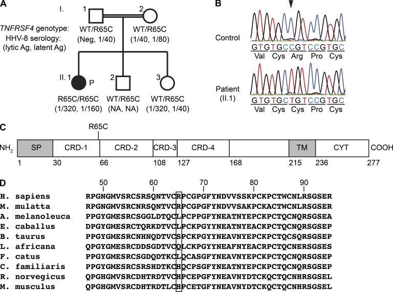
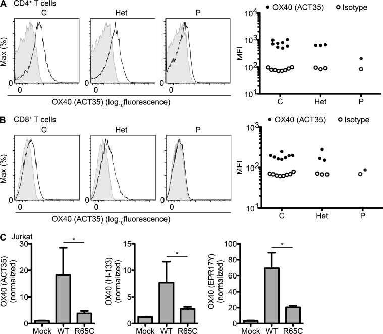
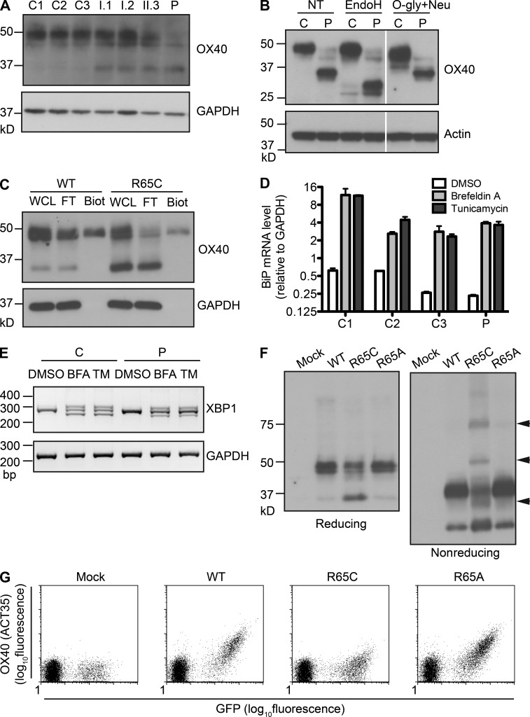
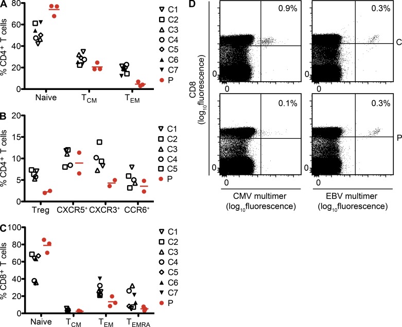
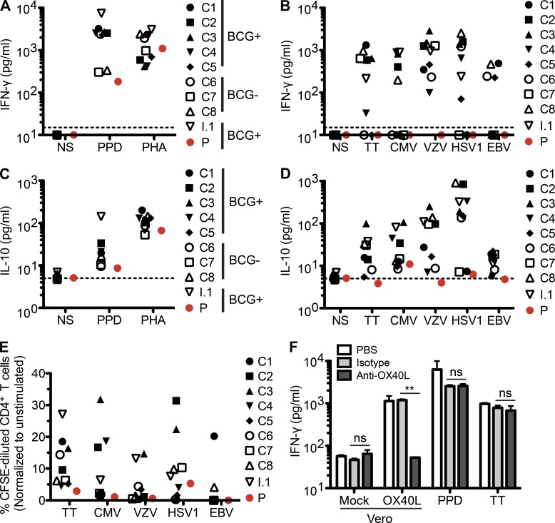
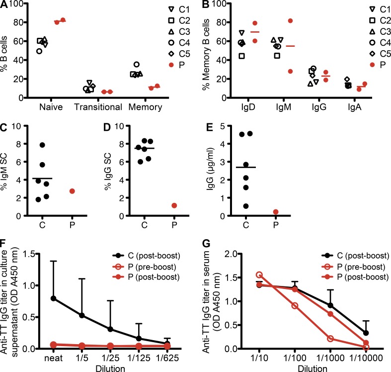
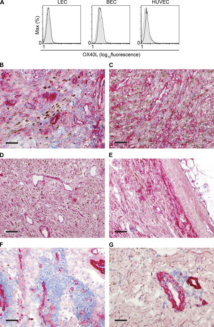
Kaposi sarcoma (KS), a human herpes virus 8 (HHV-8; also called KSHV)-induced endothelial tumor, develops only in a small fraction of individuals infected with HHV-8. We hypothesized that inborn errors of immunity to HHV-8 might underlie the exceedingly rare development of classic KS in childhood. We report here autosomal recessive OX40 deficiency in an otherwise healthy adult with childhood-onset classic KS. OX40 is a co-stimulatory receptor expressed on activated T cells. Its ligand, OX40L, is expressed on various cell types, including endothelial cells. We found OX40L was abundantly expressed in KS lesions. The mutant OX40 protein was poorly expressed on the cell surface and failed to bind OX40L, resulting in complete functional OX40 deficiency. The patient had a low proportion of effector memory CD4(+) T cells in the peripheral blood, consistent with impaired CD4(+) T cell responses to recall antigens in vitro. The proportion of effector memory CD8(+) T cells was less diminished. The proportion of circulating memory B cells was low, but the antibody response in vivo was intact, including the response to a vaccine boost. Together, these findings suggest that human OX40 is necessary for robust CD4(+) T cell memory and confers apparently selective protective immunity against HHV-8 infection in endothelial cells.


Figures

Figure 1.
Figure 1.
Homozygous TNFRSF4 mutation in a patient with classic KS. (A) Family pedigree with TNFRSF4 allele segregation. The black-filled symbol indicates the patient (P). Anti–HHV-8 antibody titers, assessed in serum samples collected 3 yr after the initial HHV-8 serology (case 3 in Sahin et al. [2010]), are indicated in parentheses. Ag, antigen; NA, data not available; Neg, negative. (B) Confirmation of the single nucleotide substitution c.193C>T (indicated by an arrowhead) by Sanger sequencing. TNFRSF4 exon 2 was amplified by PCR from genomic DNA from a healthy control and the patient. Representative chromatograms for three independent experiments are shown. (C) Schematic representation of OX40 protein structure. Numbers shown below the scheme indicate the amino acid residue number. CRD, cysteine-rich domain; CYT, cytosolic domain; SP, signal peptide; TM, transmembrane domain. (D) Multiple sequence alignment of human TNFRSF4 and its orthologues. The Arg65 residue of human OX40 (top row) and the corresponding residues in the other species are boxed.
Figure 2.
Figure 2.
Low levels of mutant OX40 on the cell surface. (A and B) PBMCs from healthy controls (C), R65C heterozygous family members (I.1, I.2, and II.3; Het), and the patient (P) were activated by incubation with PHA for 4 d. Cell surface OX40 levels were determined by flow cytometry with the ACT35 monoclonal antibody. Representative histograms and MFI (mean fluorescence intensity) values for three independent experiments are shown. (A) Gated on the CD3+CD4+ population (CD4+ T cells). (B) Gated on the CD3+CD8+ population (CD8+ T cells). (C) Jurkat cells were transduced with bicistronic retroviruses with an empty vector (Mock) or encoding OX40-WT or OX40-R65C, together with IRES-GFP. The MFI of OX40 on GFP+ cells is shown, as assessed with the antibodies indicated and normalized with respect to isotype controls. The mean values of three independent experiments are plotted. The error bars indicate the SEM. *, P < 0.05.
Figure 3.
Figure 3.
Intracellular accumulation of the mutant OX40 proteins. (A) Immunoblotting of PHA-activated T cell blasts from three healthy controls (C1–C3), R65C heterozygous family members (I.1, I.2, and II.3), and the patient (P) was performed with the anti-OX40 monoclonal antibody EPR17Y. GAPDH served as a loading control. One experiment representative of three performed is shown. (B) Whole-cell lysates of PHA-activated T cell blasts from a healthy control (C) or the patient (P) were treated with the enzymes indicated before immunoblotting. White lines indicate the intervening lanes have been spliced out. One experiment representative of two performed is shown. EndoH, endoglycosidase H; NT, nontreated; O-gly+Neu, O-glycosidase and neuraminidase. (C) HEK-293 cells transduced with retroviral vectors encoding OX40-WT or OX40-R65C were subjected to cell surface biotinylation. Biotinylated proteins were isolated with avidin-coated beads. OX40 immunoblotting was performed on whole-cell lysate (WCL), the flow-through fraction (FT), and the biotinylated fraction (Biot). A GAPDH blot was used to assess whether the biotinylated fraction was free of intracellular proteins. The results of two independent experiments are shown. (D and E) PHA-activated T cell blasts from healthy controls (C1–C3) or the patient (P) were treated with DMSO alone or treated with either Brefeldin A (BFA) or tunicamycin (TM) for 6 h. The results of two independent experiments are shown. (D) The mRNA levels of BiP/GRP78, relative to GAPDH, were assessed by quantitative RT-PCR. Error bars indicate the SEM. (E) PCR was performed using primer pairs that can differentiate various spliced variants of XBP1 mRNA. (F) Whole-cell lysates of HEK-293 cells transduced with retroviral vectors with an empty vector (Mock) or encoding OX40-WT, OX40-R65C, or OX40-R65A were resolved by SDS-PAGE under reducing or nonreducing conditions. The arrowheads indicate protein bands unique to the OX40-R65C sample. One result representative of three independent experiments is shown. (G) Jurkat cells were transduced with bicistronic retroviruses with an empty vector (Mock) or encoding OX40-WT, OX40-R65C, or OX40-R65A, together with IRES-GFP. Cell surface OX40 levels were assessed with ACT35 by flow cytometry. One result representative of three independent experiments is shown.
Figure 4.
Figure 4.
R65C is a loss-of-function mutation. (A) PHA-activated T cell blasts from healthy controls (C), R65C heterozygous family members (Het), or the patient (P) were incubated with biotinylated recombinant soluble OX40L. Unbound OX40L molecules were washed out, and the levels of cell-bound OX40L were measured by flow cytometry with allophycocyanin-labeled streptavidin (SA-APC). Representative histograms and MFIs (mean fluorescence intensities) of CD3+CD4+ cells (CD4+ T cells) and CD3+CD8+ cells (CD8+ T cells) in three independent experiments are shown. (B) PHA-activated T cell blasts from the patient were transduced with bicistronic lentiviral vectors encoding luciferase (Luc), OX40-WT, or OX40-R65C, together with IRES-RFP. Cell surface OX40 levels measured with ACT35 and binding to OX40L measured with biotinylated recombinant soluble OX40L are shown for CD3+CD4+RFP+ cells. One result representative of two independent experiments is shown. (C) OX40L binding to Jurkat or HEK-293 cells transduced with bicistronic retroviruses with an empty vector (Mock) or encoding OX40-WT or OX40-R65C, together with IRES-GFP, was assessed as in A. The MFIs of OX40L binding for GFP+ cells, normalized with respect to those for isotype controls, are shown. The mean of three independent experiments is shown. Error bars indicate the SEM. *, P < 0.05. (D) CFSE-labeled PBMCs from healthy controls (C1–C8) and the patient (P) were incubated for 3 d with 1 ng/ml of plate-bound anti-CD3 antibody and Vero cells infected with retroviruses either with an empty vector (Vero-Mock) or encoding OX40L (Vero-OX40L). Percentages of CFSE-diluted cells in the CD3+CD4+ population are plotted. One result representative of two independent experiments is shown. (E) CFSE-labeled PBMCs from healthy controls (C1–C8) and the patient (P) were incubated for 3 d with the indicated concentrations of plate-bound anti-CD3 antibody or PHA. Percentages of CFSE-diluted cells in the CD3+CD4+ population are plotted. One result representative of two independent experiments is shown.
Figure 5.
Figure 5.
Lower proportions of circulating nonnaive T cell subsets. (A and B) The proportions of the indicated subsets among CD4+ T cells (CD3+CD4+) were assessed in PBMCs collected from five to seven age-matched healthy controls (C) and the patient (P). Two or three independent measurements (of blood drawn at different time points) were performed for the patient (each indicated by a red circle). Mean values are indicated with red horizontal bars. The various subsets were defined as follows: naive (CD45RA+CCR7+); TCM, T central memory (CD45RACCR7+); TEM, T effector memory (CD45RACCR7); Treg, T regulatory cells (CD25highCD127low); CXCR5+, T follicular helper cells (CD45RACXCR5+); CXCR3+, IFN-γ–producing T cells (CD45RACXCR5CXCR3+CCR6); and CCR6+, IL-17–producing T cells (CD45RACXCR5CXCR3CCR6+). (C) Proportions of CD8+ T cell (CD3+CD8+) subsets, assessed as in A, are shown. The various subsets were defined as follows: naive (CD45RA+CCR7+); TCM (CD45RACCR7+); TEM (CD45RACCR7); and TEMRA, CD45RA+ revertant memory T cells (CD45RA+CCR7). (D) The frequency of antigen-specific CD8+ T cells (CD3+CD8+) in an age-matched control (C) or the patient (P) was assessed with HLA-A*0201 multimers loaded with peptides derived from CMV antigen pp65 or EBV antigen BMLF-1.
Figure 6.
Figure 6.
Impaired CD4+ T cell recall antigen response. (A and C) PBMCs from eight healthy controls (C1–C8), an R65C heterozygous family member (I.1), and the patient (P) were stimulated with PPD or PHA for 5 d. IFN-γ (A) or IL-10 (C) levels in the supernatant were measured by ELISA. The dotted lines indicate the limit of detection. One result representative of three independent experiments is shown. BCG+, BCG vaccinated; BCG−, no prior BCG vaccination; NS, nonstimulated. (B and D) IFN-γ (B) or IL-10 (D) production in response to various recall antigens was assessed as in A and C. Except for TT, which was provided as purified protein, the recall antigens were provided as virus-infected crude cell lysate. A lysate of uninfected cells tested in the same experiment did not trigger IFN-γ production (not depicted). The dotted lines indicate the limit of detection. One result representative of three independent experiments is shown. (E) CFSE-labeled PBMCs were incubated with the indicated recall antigens for 6 d. T cell proliferation was assessed by determining the proportion of cells with CFSE levels lower than the undivided peak. Results are shown for CD4+ T cells (CD3+CD4+). One result representative of three independent experiments is shown. (F) PBMCs from two healthy controls were incubated with 1 ng/ml of plate-bound anti-CD3 and Vero cells infected with retroviruses with an empty vector (Vero-Mock) or encoding OX40L (Vero-OX40L) for 3 d, or PPD or TT for 6 d. PBS, isotype antibody, or anti-OX40L antibody was added every other day to a concentration of 1 µg/ml. IFN-γ levels in the culture supernatant were measured by ELISA. Means and SEM from two experiments for one of the healthy controls are shown. **, P < 0.01; ns, not significant.
Figure 7.
Figure 7.
Low frequency of memory B cells but intact antibody response in vivo. (A) The proportions of B cell (CD20+) subsets in PBMCs are shown. The various subsets were defined as follows: naive (CD10CD27), transitional (CD10+CD27), and memory (CD10CD27+). (B) The proportions of memory B cells from the patient and healthy controls expressing the indicated surface markers are shown. (C–F) Purified B cells from six age-matched healthy controls (C) and the patient (P) were cultured in the presence of CD40L, IL-10, IL-21, and CpG for 5 d. The frequencies of total IgM (C)- or IgG-secreting B cells (D) were assessed by ELISPOT. The levels of total IgG (E) or TT-specific secreted IgG (F) in the culture supernatant were quantified by ELISA. (A–E) Horizontal bars indicate the mean. (G) Anti-TT IgG titer in serum was assessed by ELISA. (F and G) Error bars indicate the SD from 6 (F) or 10 (G) healthy controls.
Figure 8.
Figure 8.
OX40L is abundantly expressed in KS lesions. (A) Expression of OX40L on the cell surface was assessed with flow cytometry in three types of primary endothelial cells: human dermal microvascular lymphatic endothelial cells (LEC), human dermal microvascular blood endothelial cells (BEC), and HUVECs. One result representative of two independent experiments is shown. (B–G) OX40L (red) and HHV-8 LANA (brown) expression was assessed by immunohistochemistry in frozen tissue sections. Bars, 5 mm. (B–E) AIDS-related KS lesions in lymph node (B), presenting as a submental mass (C), and in skin (D and E) show HHV-8–positive cells (punctate nuclear brown staining) as well as numerous OX40L-positive cells. (F and G) Non-KS tissues used as controls include normal lymph node (F) and skin with mycosis fungoides (G), showing OX40L in cells lining vascular spaces.

Similar articles

Cited by

References

    1. Abecasis G.R., Cherny S.S., Cookson W.O., Cardon L.R. 2002. Merlin—rapid analysis of dense genetic maps using sparse gene flow trees. Nat. Genet. 30:97–101 10.1038/ng786 - DOI - PubMed
    1. Adzhubei I.A., Schmidt S., Peshkin L., Ramensky V.E., Gerasimova A., Bork P., Kondrashov A.S., Sunyaev S.R. 2010. A method and server for predicting damaging missense mutations. Nat. Methods. 7:248–249 10.1038/nmeth0410-248 - DOI - PMC - PubMed
    1. Akman E.S., Ertem U., Tankal V., Pamir A., Tuncer A.M., Uluoğlu O. 1989. Aggressive Kaposi’s sarcoma in children: a case report. Turk. J. Pediatr. 31:297–303 - PubMed
    1. Alcaïs A., Quintana-Murci L., Thaler D.S., Schurr E., Abel L., Casanova J.L. 2010. Life-threatening infectious diseases of childhood: single-gene inborn errors of immunity? Ann. N. Y. Acad. Sci. 1214:18–33 10.1111/j.1749-6632.2010.05834.x - DOI - PubMed
    1. Andreoni M., El-Sawaf G., Rezza G., Ensoli B., Nicastri E., Ventura L., Ercoli L., Sarmati L., Rocchi G. 1999. High seroprevalence of antibodies to human herpesvirus-8 in Egyptian children: evidence of nonsexual transmission. J. Natl. Cancer Inst. 91:465–469 10.1093/jnci/91.5.465 - DOI - PubMed

Publication types

MeSH terms