. 2013 Oct;33(19):3749-61.
doi: 10.1128/MCB.01387-12. Epub 2013 Jul 22.

Phosphorylation regulates FOXC2-mediated transcription in lymphatic endothelial cells

Affiliations

Phosphorylation regulates FOXC2-mediated transcription in lymphatic endothelial cells

Konstantin I Ivanov et al. Mol Cell Biol. 2013 Oct.

Abstract

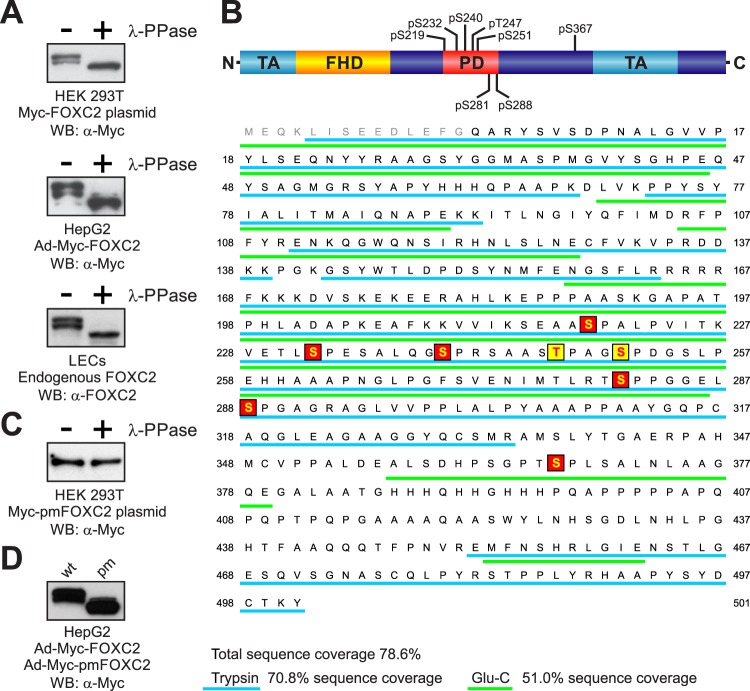
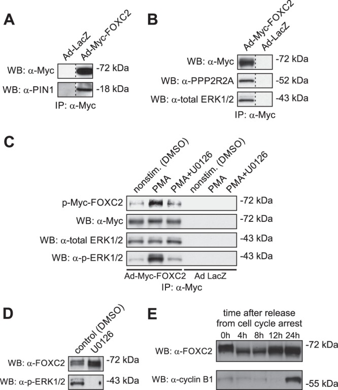
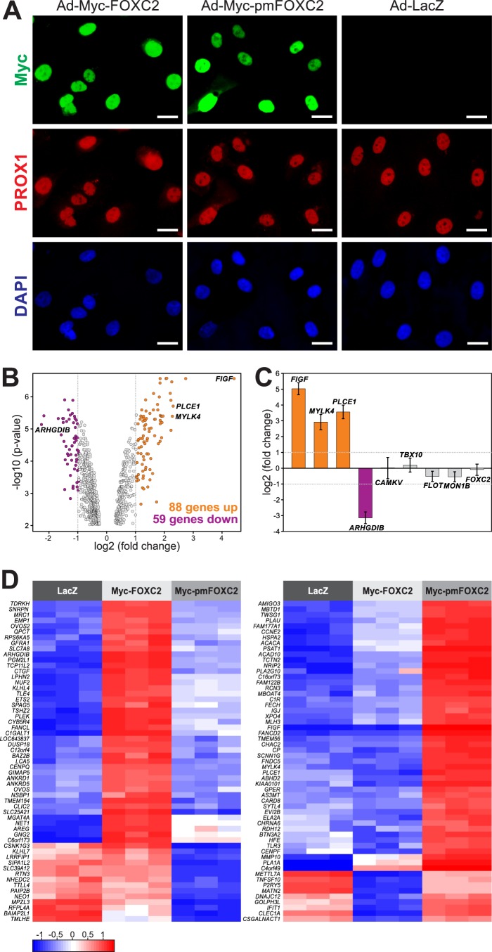
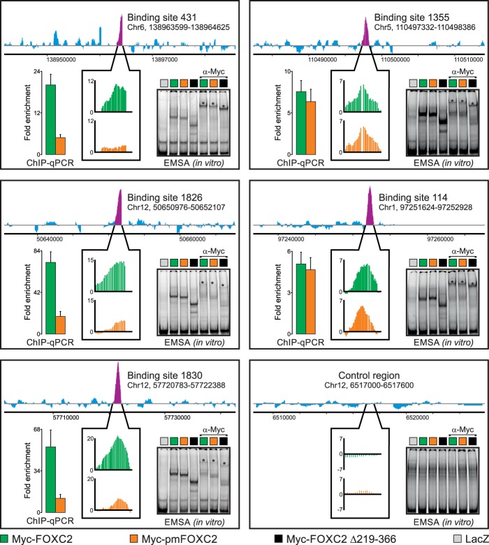
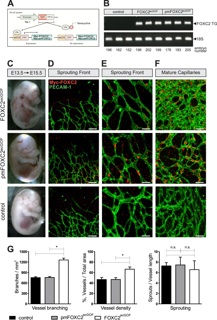
One of the key mechanisms linking cell signaling and control of gene expression is reversible phosphorylation of transcription factors. FOXC2 is a forkhead transcription factor that is mutated in the human vascular disease lymphedema-distichiasis and plays an essential role in lymphatic vascular development. However, the mechanisms regulating FOXC2 transcriptional activity are not well understood. We report here that FOXC2 is phosphorylated on eight evolutionarily conserved proline-directed serine/threonine residues. Loss of phosphorylation at these sites triggers substantial changes in the FOXC2 transcriptional program. Through genome-wide location analysis in lymphatic endothelial cells, we demonstrate that the changes are due to selective inhibition of FOXC2 recruitment to chromatin. The extent of the inhibition varied between individual binding sites, suggesting a novel rheostat-like mechanism by which expression of specific genes can be differentially regulated by FOXC2 phosphorylation. Furthermore, unlike the wild-type protein, the phosphorylation-deficient mutant of FOXC2 failed to induce vascular remodeling in vivo. Collectively, our results point to the pivotal role of phosphorylation in the regulation of FOXC2-mediated transcription in lymphatic endothelial cells and underscore the importance of FOXC2 phosphorylation in vascular development.


Figures

Fig 1
Fig 1
Analysis of FOXC2 phosphorylation. (A) Endogenous and recombinant human FOXC2 are similarly phosphorylated in primary LECs and immortalized cell lines. Cell lysates were treated (+) or not treated (−) with lambda protein phosphatase (λ-PPase) and analyzed by Western blotting with anti-FOXC2 or anti-Myc antibodies. (B) Schematic representation of FOXC2 phosphorylation sites. FHD, forkhead domain; TA, transactivation domains (5, 34); PD, phosphorylated domain. Phosphorylation sites identified by LC-MS/MS are shaded in red; phosphorylation sites identified by mutagenesis are shaded in yellow. Peptides detected by MS in tryptic and Glu-C digests are underlined in cyan and green, respectively. Amino acid numbering is the same as in the endogenous protein (NP_005242). (C) Substitution of eight phosphorylation sites in Myc-FOXC2 with alanine abolishes the phosphorylation-dependent electrophoretic mobility shift. Lysates of cells transfected with a plasmid expressing the phosphorylation-deficient mutant Myc-pmFOXC2 were treated (+) or not treated (−) with λ-PPase and analyzed by Western blotting with anti-Myc antibody. (D) FOXC2 phosphorylation-deficient mutant (pm) has increased electrophoretic mobility compared to the wild-type (wt) protein. Shown is Western blot analysis of lysates from HepG2 cells transduced with adenoviruses expressing Myc-FOXC2 or Myc-pmFOXC2.
Fig 2
Fig 2
FOXC2 interacts with peptidyl-prolyl cis/trans isomerase PIN1, alpha isoform of the regulatory subunit B of the protein phosphatase PP2A (PPP2R2A), and ERK1/2 protein kinases. (A) Coimmunoprecipitation assays with anti-Myc antibody demonstrate the association of Myc-FOXC2 with endogenous PIN1 in HeLa cells transduced with recombinant Ad-Myc-FOXC2. Shown is Western blot (WB) of anti-Myc immunoprecipitates consecutively probed with anti-Myc and anti-PIN1 antibodies. Control immunoprecipitation was performed from extracts of HeLa cells transduced with recombinant adenovirus expressing bacterial β-galactosidase (Ad-LacZ). (B) Myc-FOXC2 binds to endogenous PPP2R2A and ERK1/2 in HepG2 cells transduced with recombinant Ad-Myc-FOXC2. Coimmunoprecipitation assays were performed and analyzed as in A, except that anti-PPP2R2A and anti-total ERK1/2 antibodies were used for immunoblotting. (C) Immunocomplex kinase assays demonstrate that Myc-FOXC2 is phosphorylated in vitro by the coprecipitating endogenous ERK1/2 kinases. Shown is Western blot (WB) of anti-Myc immunoprecipitates incubated in the presence of [γ-32P]ATP and phosphorimage of the corresponding membrane. The blot was consecutively probed with anti-Myc, anti-total ERK1/2 and anti-active ERK1/2 (p-ERK1/2) antibodies. The identity of ERK1/2 was confirmed by immunocomplex kinase assays with anti-Myc antibody using lysates of HepG2 cells stimulated with PMA in the presence or absence of 10 mM U0126, a selective inhibitor of upstream MEK. (D) Inhibition of ERK1/2 does not modify the electrophoretic mobility of endogenous FOXC2 in LECs. Shown is a Western blot of total LEC lysates consecutively probed with anti-FOXC2 and anti-active ERK1/2 antibodies. (E) The electrophoretic mobility of endogenous FOXC2 changes after release from serum starvation-induced cell cycle arrest in LECs, suggesting CDK involvement in FOXC2 phosphorylation.
Fig 3
Fig 3
Phosphorylation regulates FOXC2-mediated transcription in primary LECs. (A) Immunofluorescent staining for Myc (green), lymphatic marker PROX1 (red), and DNA (blue) of LECs transduced with adenoviruses expressing wild-type Myc-FOXC2, phosphorylation-deficient mutant Myc-pmFOXC2, or control bacterial β-galactosidase (LacZ). Note that wild-type and mutant FOXC2 have similar expression levels and subcellular localization. Bars, 20 μm. (B) Phosphorylation regulates FOXC2 transcriptional activity. Gene expression profiling was performed on the adenovirus-transduced LECs shown in panel A. Genes whose expression changed >2-fold in response to the loss of FOXC2 phosphorylation (FDR < 0.01) are shown in orange (upregulated) and purple (downregulated) in the Volcano plot of significance against the fold change in gene expression. Vertical dotted lines mark the 2-fold change limits. (C) RT-PCR validation of the gene expression profiling results. Genes upregulated or downregulated >2-fold in response to the loss of FOXC2 phosphorylation are shown in orange and purple, respectively; genes affected <2-fold are shown in gray. No change in FOXC2 expression reflects equally efficient cell transduction with Ad-Myc-FOXC2 and Ad-Myc-pmFOXC2. The data are presented as log2-transformed fold change in gene expression normalized to a housekeeping gene (GAPDH). Horizontal dotted lines mark the 2-fold change limits. Shown are the means and standard deviations of triplicate determinations in a single experiment representative of two independent experiments. (D) Heat map representation of the differences in gene expression in response to the loss of FOXC2 phosphorylation. The left heat map shows expression levels of 57 of 59 genes downregulated >2-fold (FDR < 0.01) in Ad-Myc-pmFOXC2-transduced LECs compared to Ad-Myc-FOXC2-transduced LECs. The right heat map shows expression levels of 57 out of 88 genes upregulated >2-fold (FDR < 0.01) in the same cells. Three biological replicates are shown for each condition. The color key at the lower left corresponds to the mean-centered, arctan-transformed log2-fold change in gene expression falling within the range from −π/2 to π/2. Blue denotes genes with relative decreased expression; red denotes genes with relative increased expression.
Fig 4
Fig 4
Phosphorylation differentially regulates FOXC2 binding to genomic target sites in the context of native chromatin but not in vitro. We used genome-wide ChIP-chip to compare the binding of adenovirus-expressed wild-type Myc-FOXC2 and the phosphorylation-deficient mutant Myc-pmFOXC2 to physiological binding sites occupied by endogenous FOXC2 in primary LECs. Endogenous FOXC2 enrichment profiles are shown at the top of each panel. Purple peaks indicate FOXC2-enriched regions; their relative occupancies by Myc-FOXC2 and Myc-pmFOXC2 are shown in callout boxes in green and orange, respectively. Vertical axes represent MAT score. Binding sites are numbered as in Norrmén et al. (8); genomic coordinates refer to the hg18 human genome assembly. An unbound control region is shown in the lower right panel. The ChIP-chip results were validated by ChIP-qPCR with primers flanking ∼100-bp sequences within the FOXC2-enriched regions. The results are presented as the fold enrichment over the unbound control region. Green and orange bars correspond to wild-type Myc-FOXC2 and Myc-pmFOXC2, respectively. Shown are the means and standard deviations of triplicate determinations. An EMSA was used to compare the in vitro binding of adenovirus-expressed wild-type Myc-FOXC2, Myc-pmFOXC2, and deletion mutant Myc-FOXC2 D219-366 to naked dsDNA from the ChIP-enriched regions or the unbound control region. Binding specificity was controlled with adenovirus-expressed bacterial β-galactosidase (LacZ) and anti-Myc antibody. Asterisks indicate the positions of the antibody-supershifted complexes.
Fig 5
Fig 5
Phosphorylation regulates FOXC2 function in vivo. (A) Endothelial cell-specific gain-of-function models for the analysis of FOXC2 phosphorylation. (B) Both models express comparable levels of the transgene, as evidenced by RT-PCR analysis of the indicated mRNAs from E15.5 lungs. Transgene expression was initiated at E13.5. (C) Macroscopic appearances of FOXC2ecGOF, pmFOXC2ecGOF, and control E15.5 embryos. (D and E) FOXC2 overexpression does not affect capillary sprouting. (F) Overexpression of FOXC2 but not pmFOXC2 promotes vascular remodeling in maturing capillaries. Note the increased capillary branching and density in FOXC2ecGOF embryos. Whole-mount staining of E15.5 head skin for pan-endothelial marker CD31 (green) and the transgene (red). The transgene expression was detected using anti-Myc antibody. Scale bars: 100 μm (D), 38 μm (E), 35 μm (F). (G) Quantification of vascular branching, density, and sprouting at the vascular front in the control, pmFOXC2ecGOF, and FOXC2ecGOF embryos. n = 3 per genotype. *, P < 0.05. n.s., nonsignificant.

Similar articles

Cited by

References

    1. Wijchers PJ, Burbach JP, Smidt MP. 2006. In control of biology: of mice, men, and Foxes. Biochem. J. 397:233–246 - PMC - PubMed
    1. Iida K, Koseki H, Kakinuma H, Kato N, Mizutani-Koseki Y, Ohuchi H, Yoshioka H, Noji S, Kawamura K, Kataoka Y, Ueno F, Taniguchi M, Yoshida N, Sugiyama T, Miura N. 1997. Essential roles of the winged helix transcription factor MFH-1 in aortic arch patterning and skeletogenesis. Development 124:4627–4638 - PubMed
    1. Winnier GE, Kume T, Deng K, Rogers R, Bundy J, Raines C, Walter MA, Hogan BL, Conway SJ. 1999. Roles for the winged helix transcription factors MF1 and MFH1 in cardiovascular development revealed by nonallelic noncomplementation of null alleles. Dev. Biol. 213:418–431 - PubMed
    1. Fang J, Dagenais SL, Erickson RP, Arlt MF, Glynn MW, Gorski JL, Seaver LH, Glover TW. 2000. Mutations in FOXC2 (MFH-1), a forkhead family transcription factor, are responsible for the hereditary lymphedema-distichiasis syndrome. Am. J. Hum. Genet. 67:1382–1388 - PMC - PubMed
    1. Petrova TV, Karpanen T, Norrmén C, Mellor R, Tamakoshi T, Finegold D, Ferrell R, Kerjaschki D, Mortimer P, Yla-Herttuala S, Miura N, Alitalo K. 2004. Defective valves and abnormal mural cell recruitment underlie lymphatic vascular failure in lymphedema distichiasis. Nat. Med. 10:974–981 - PubMed

Publication types

MeSH terms