Review
. 2013 Sep 13;288(37):26512-20.
doi: 10.1074/jbc.R113.464131. Epub 2013 Jul 16.

The redox proteome

Affiliations
Review

The redox proteome

Young-Mi Go et al. J Biol Chem. .

Abstract

The redox proteome consists of reversible and irreversible covalent modifications that link redox metabolism to biologic structure and function. These modifications, especially of Cys, function at the molecular level in protein folding and maturation, catalytic activity, signaling, and macromolecular interactions and at the macroscopic level in control of secretion and cell shape. Interaction of the redox proteome with redox-active chemicals is central to macromolecular structure, regulation, and signaling during the life cycle and has a central role in the tolerance and adaptability to diet and environmental challenges.

Keywords: Glutathione; Redox Regulation; Systems Biology; Thiol; Thioredoxin.


Figures
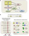
FIGURE 1.
FIGURE 1.
Redox proteome as the interface of the epiproteome, diet, and environment. A, the redox proteome is a subset of post-translational modifications of protein considered here as the epiproteome (indicated by the dashed arrow). This is considered in analogy to the epigenome and genome as a system to provide function and regulation beyond that of the translated sequence of amino acids. The redox proteome includes amino acids that undergo reversible redox reactions (Cys, Met, and Sec) and others that are irreversibly modified by reactive species in oxidative stress (Lys, Trp, and others). The redox proteome impacts the genome and epigenome through altered DNA and RNA binding and altered trafficking, activities, and structures of associated proteins. Metabolic activities, especially in cell signaling, impact the metabolome. The redox metabolome is a subset of the metabolome that alters structure and function of the redox proteome. NADPH/NADP, GSH/GSSG and Cys/cystine are central components of the redox metabolome. The metabolome and redox proteome respond to diet and environmental influences (blue arrows) to protect against oxidant stress and other environmental challenges. B, covalent modifications of Cys provide a versatile structure-function switching system. Cys commonly exists on the surface of proteins either alone (monothiol) or in close proximity to another Cys residues (vicinal dithiols) (top). The reactivities are impacted by vicinal cationic amino acids, which enhance ionization and make Cys more reactive. Vicinal dithiols tend to form disulfides upon oxidation, whereas monothiols undergo reversible oxidation to sulfenic acid and mixed disulfides with low molecular mass thiol chemicals, GSH, Cys, and homocysteine (HCy; left, inside box). Under highly oxidizing conditions, sulfenic acid is further oxidized to sulfinic and sulfonic acids (left, below box). Other modifications also occur, such as S-nitrosylation, S-sulfhydration, and thiohemiacetal formation. These changes, as well as Zn2+ binding and acylation, can result in altered protein-protein interactions, DNA or RNA binding, or membrane interaction (right).
FIGURE 2.
FIGURE 2.
Non-equilibrium steady states of redox couples direct metabolism, structure, and macromolecular trafficking. Central thiol/disulfide couples (left) serve as redox hubs and include small molecules of the redox metabolome and redox-active proteins, such as Trx1 and Trx2. These exist with a range of steady-state redox potentials (Eh, calculated with the Nernst equation (see text)) spanning nearly 400 mV from mitochondrial NADPH/NADP to plasma Cys/cystine (CySS). The difference between redox couples (ΔEh) is proportional to −ΔG, describing the energetics of electron transfer. The ratio of (SH)2 to SS for a dithiol/disulfide couple with Eo = −210 mV illustrates the relative abundance of the forms if the protein is equilibrated at the respective Eh value. Upper right, kinetic limitation is shown for proteins in the pathway from NADPH to NF-κB (p50) under both control and energy-limiting conditions in cultured cells. Lower right, the Eh of cytosolic GSH/GSSG becomes progressively oxidized in the life cycle of cells from proliferation to differentiation to apoptosis. Such change could, in principle, impact proteins via glutaredoxin-dependent S-glutathionylation (30) or reflect changes in H2O2 generation and GSH peroxidase activity. Mito, mitochondria; nuc, nuclear; cyto, cytosolic; ER, endoplasmic reticulum; TR1, Trx reductase 1. Cell images are from Invitrogen and the Genetics Home Reference (http://ghr.nlm.nih.gov).
FIGURE 3.
FIGURE 3.
Types of sulfur switches. Sulfur switches can turn systems on and off, provide allosteric regulation, change the binding interactions, and/or have a chameleon-like effect on the character of a protein (upper). Crystal structures are provided to illustrate each. In PTP1B (PTP), oxidation at the active site Cys215 residue turns off activity (middle left) (45). Oxidation of Cys38 in NF-κB (p65) or Cys62 in NF-κB (p50) results in loss of DNA binding (middle right). Formation of a disulfide (Cys62–Cys69) in a surface α-helix in Trx1 (20) results in an allosteric-like decrease in interaction with Trx reductase (lower left). A switch in cysteinylation of Cys60 (Protein Data Bank) to glutathionylation results in a change from glycosylation-inhibiting factor (GIF) activity to macrophage migration inhibitory factor (MIF) activity (lower right).
FIGURE 4.
FIGURE 4.
Integrated function of the redox proteome. A, redox-sensing Cys residues provide an orthogonal control system to regulate and integrate biologic systems without impacting mechanisms. Redox-signaling mechanisms are presented as pathways (diagonal right arrows) that serve to signal cell stress and other responses. These include controlled H2O2 production by regulated NADPH oxidases and redox-signaling Cys. They function in parallel with other signaling pathways, such as kinase signaling and ion-gated signaling (diagonal right arrows). These are viewed as providing relatively rapid and transient signals. Redox-sensing Cys residues integrate these signaling pathways without impacting their mechanisms (diagonal left arrows). Although poorly defined experimentally, the latter can be conceived as a background poise of the cellular H2O2 generation by mitochondria, Nox4, and other oxidases (lower left) and opposed by reductant systems dependent upon Trx and GSH (upper right). These have slow kinetics relative to signaling mechanisms and thereby provide a more long-term phenotypic control. This allows a single signaling mechanism to be used effectively in different cell types and states of cell division, growth, differentiation, or apoptosis. These signaling and sensing Cys switches operate within the non-equilibrium states maintained by the central redox hubs (Fig. 2) and together provide a versatile system for integrated spatial and temporal control of cell structure and function. ROS/RNS, reactive oxygen and nitrogen species. B, broad ranges of protein thiol reactivity and abundance are known to occur in the redox proteome. These provide a redox proteomic structure to support localized redox signaling within an inherently stable redox system. Upper left panel, protein abundance (C) is given on the y axis from femtomolar to 10 μm, expressed as a function of each respective protein, listed in order of abundance on the x axis. Middle left panel, a hypothetical condition is shown in which the second-order rate constant (k) for protein thiol reaction with H2O2, from 1 to 107 m−1 s−1 (73), is varied in opposition to abundance for the same proteins. Lower left panel, the product of the rate constant and concentration (k·C) for each protein under this condition shows that all proteins contribute equivalently to the rate of H2O2 metabolism. Right, the rate constant (middle panel) is varied in proportion to abundance (upper panel). The product (k·C) for this condition shows that the most abundant protein thiols contribute 14 orders of magnitude more to the rate of H2O2 metabolism than the least abundant protein thiols (lower panel). This comparison shows that opposing co-evolution of abundance and reactivity of specific Cys residues within the proteome can account for an inherent stability of the redox proteome while also having specialized subsets of peptidyl-Cys for redox sensing and redox signaling.

Similar articles

Cited by

References

    1. Keilin D. (1966) The History of Cell Respiration and Cytochrome, Cambridge University Press, Cambridge
    1. Mitchell P. (1979) Keilin's respiratory chain concept and its chemiosmotic consequences. Science 206, 1148–1159 - PubMed
    1. Mason H. S., Fowlks W. L., Peterson J. (1955) Oxygen transfer and electron transport by the phenolase complex. J. Am. Chem. Soc. 77, 2914–2915
    1. Hayaishi O., Katagiri M., Rothberg S. (1955) Mechanism of the pyrocatechase reaction. J. Am. Chem. Soc. 77, 5450–5451
    1. Kovaleva E. G., Lipscomb J. D. (2008) Versatility of biological non-heme Fe(II) centers in oxygen activation reactions. Nat. Chem. Biol. 4, 186–193 - PMC - PubMed

Publication types

LinkOut - more resources