. 2013 Jun 21;8(6):e66464.
doi: 10.1371/journal.pone.0066464. Print 2013.

PUMA Cooperates with p21 to Regulate Mammary Epithelial Morphogenesis and Epithelial-To-Mesenchymal Transition

Affiliations

PUMA Cooperates with p21 to Regulate Mammary Epithelial Morphogenesis and Epithelial-To-Mesenchymal Transition

Yanhong Zhang et al. PLoS One. .

Erratum in

Abstract

Lumen formation is essential for mammary morphogenesis and requires proliferative suppression and apoptotic clearance of the inner cells within developing acini. Previously, we showed that knockdown of p53 or p73 leads to aberrant mammary acinus formation accompanied with decreased expression of p53 family targets PUMA and p21, suggesting that PUMA, an inducer of apoptosis, and p21, an inducer of cell cycle arrest, directly regulate mammary morphogenesis. To address this, we generated multiple MCF10A cell lines in which PUMA, p21, or both were stably knocked down. We found that morphogenesis of MCF10A cells was altered modestly by knockdown of either PUMA or p21 alone but markedly by knockdown of both PUMA and p21. Moreover, we found that knockdown of PUMA and p21 leads to loss of E-cadherin expression along with increased expression of epithelial-to-mesenchymal transition (EMT) markers. Interestingly, we found that knockdown of ΔNp73, which antagonizes the ability of wide-type p53 and TA isoform of p73 to regulate PUMA and p21, mitigates the abnormal morphogenesis and EMT induced by knockdown of PUMA or p21. Together, our data suggest that PUMA cooperates with p21 to regulate normal acinus formation and EMT.


Conflict of interest statement

Competing Interests: The authors have declared that no competing interests exist.

Figures

Figure 1
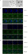
Figure 1. Mammary epithelial cells cultured on ECM form functional acini.
A, Representative phase-contrast microscopic images of MCF10A cells in 2-D culture (a, 200×) and 3-D culture (b, 40×; c, 100×). B, Serial confocal images of cross-sections through the middle of acini stained with To-Pro-3 and antibody against E-cadherin in MCF10A cells. C, Serial confocal images of cross-sections through the middle of acini stained with To-Pro-3 and antibody against β-catenin in MCF10A cells. D, Serial confocal images of cross-sections through the middle of acini stained with To-Pro-3 and antibody against laminin V in MCF10A cells. Scale bar, 20 µm.
Figure 2
Figure 2. PUMA is necessary for morphogenesis of MCF10A cells.
A, Generation of MCF10A cells in which PUMA (clones #2 and 3) was stably knocked down. Western blots were performed with extracts from MCF10A cells untreated or treated with 0.2 µM doxorubicin for 24 h and then probed with antibodies against PUMA, ΔNp73 and actin, respectively. B, Representative images of MCF10A cells or MCF10A cells with PUMA-KD in 2-D culture (a and d, 200×) and 3-D culture (b and e, 40×; c and f, 100×). Black arrow indicates elongated spindle–liked MCF10A cells. C, Representative confocal images of cross-sections through the middle of acini stained with To-Pro-3 and antibody against E-cadherin in MCF10A cells with PUMA-KD. D, Representative confocal images of cross-sections through the middle of acini stained with To-Pro-3 and antibody against β-catenin in MCF10A cells with PUMA-KD. White arrows indicate the accumulation and translocation of β-catenin in acinus structure. E, Representative confocal images of cross-sections through the middle of acini stained with To-Pro-3 and antibody against laminin V in MCF10A cells with PUMA-KD. Scale bar, 20 µm.
Figure 3
Figure 3. p21 is necessary for morphogenesis of MCF10A cells.
A, Generation of MCF10A cells in which p21 was stably knocked down (clones #2 and #4). Western blot were performed with extracts from MCF10A cells untreated or treated with 0.2 µM doxorubicin for 24 h and then probed with antibodies against p21, ΔNp73 and actin, respectively. B, Representative phase-contrast microscopic images of MCF10A cells with p21-KD in 2-D culture (a, 200×,) and 3-D culture (b, 40×, and c, 100×). Black arrow indicates elongated spindle–liked MCF10A cells. C, Representative confocal images of cross-sections through the middle of an acinus stained with To-Pro-3 and antibody against E-cadherin. D, Representative confocal images of cross-sections through the middle of acini stained with To-Pro-3 and antibody against β-catenin. White arrows indicate the accumulation and translocation of β-catenin in an acinus structure. E, Representative confocal images of cross-sections through the middle of an acinus stained with To-Pro-3 and antibody against laminin V. Scale bar, 20 µm.
Figure 4
Figure 4. PUMA cooperates with p21 to regulate morphogeneis of MCF10A cells.
A, Generation of MCF10A cells in which both PUMA and p21 were stably knocked down (clone #1 and #3). Western blots were prepared with extracts from MCF10A cells untreated or treated with 0.2 µM doxorubicin for 24 h and then probed with antibodies against PUMA, p21, ΔNp73, and actin, respectively. B, Representative images of MCF10A cells with PUMA&p21-KD in 2-D culture (a, 200×) and 3-D culture (b, 40×; c, 100×). Black arrow indicates elongated spindle-liked MCF10A cells. C, Representative confocal images of cross-sections through the middle of an acinus stained with To-Pro-3 and antibody against E-cadherin. D, Representative confocal images of cross-sections through the middle of acini stained with To-Pro-3 and antibody against β-catenin. White arrows indicate the accumulation and translocation of β-catenin in an acinus structure. E, Representative confocal images of cross-sections through the middle of an acinus stained with To-Pro-3 and antibody against laminin V. Scale bar, 20 µm.
Figure 5
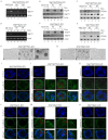
Figure 5. Knockdown of PUMA and p21 enhances EMT.
A-B, Western blots were prepared with extracts from MCF10A cells (lane 1), and MCF10A cells with p21-KD (lane 2), PUMA-KD (lane 3), or PUMA&p21-KD (lane 4). MCF10A cells were grown in Matrigel for 20 days. The blots were probed with antibodies against E-cadherin (A), β-catenin (A), laminin V (A), Snail-1 (B), Slug (B), Twist (B), and actin (A–B), respectively. C, Top panel: Colony formation assay was performed with MCF10A cells, or MCF10A cells with p21-KD, PUMA-KD or PUMA&p21-KD. Cells were cultured for a period of 12 days, then fixed and stained with crystal violet. Bottom panel: The number of colonies was counted and presented as Mean ± SD from three separate experiments. D, Top panel: Wound healing assay was performed with MCF10A cells and MCF10A cells with p21-KD, PUMA-KD or PUMA&p21 -KD. Cell migration was determined by visual assessment of cells migrating into the wound for a period of 24 h using a phase-contrast microscopy. Bottom panel: The time required for wound closure was measured and presented as Mean ± SD from three separate experiments.
Figure 6
Figure 6. Knockdown ofΔNp73 counters the effect of PUMA-KD or p21-KD on MCF10A cell morphogenesis.
A-F, Generation of MCF10A cells in which both ΔNp73 and PUMA were stably knocked down (A-C, clones #2 and #3) or ΔNp73 and p21 were stably knocked down (D-F, clones #2 and #3). The levels of ΔNp73 mRNA were measured by RT-PCR (A and D). The protein levels of TAp73α (B and E), ΔNp73α (B and E), PUMA (C and F), and p21 (C and F) were measured by Western blotting with antibodies against TAp73, ΔNp73, p21, and PUMA, respectively. MCF10A cells were untreated or treated with 0.2 µM doxorubicin for 24 h and total RNAs and cell extracts were collected for RT-PCR and Western blotting, respectively. G-H, Representative images of MCF10A cells with ΔNp73& PUMA -KD (G) or with ΔNp73&p21-KD (H) in 2-D culture (a, 200×) and 3-D culture (b, 40×; c, 100×). I and L, Representative confocal images of cross-sections through the middle of acini stained with To-Pro-3 and antibody against E-cadherin in MCF10A cells with ΔNp73&PUMA -KD (I) or with ΔNp73&p21 -KD (L). J and M, Representative confocal images of cross-sections through the middle of acini stained with To-Pro-3 and antibody against β-catenin in MCF10A cells with ΔNp73&PUMA-KD (J) or with ΔNp73&p21-KD (M). K and N, Representative confocal images of cross-sections through the middle of acini stained with To-Pro-3 and antibody against laminin V in MCF10A cells with ΔNp73&PUMA-KD (K) or with ΔNp73&p21-KD (N). Scale bar, 20 µm.
Figure 7
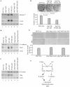
Figure 7. Knockdown ofΔNp73 mitigates EMT induced by PUMA-KD or p21-KD.
A–C,MCF10A cells were grown in Matrigel for 20 days. Western blots were prepared using extracts from MCF10A cells (lane 1), MCF10A cells with ΔNp73-KD (lane 2), with p21-KD (lane 3), with PUMA-KD (lane 4), with ΔNp73&p21-KD (lane 5) or with ΔNp73&PUMA-KD (lane 6). The blots were probed with antibodies against laminin V (A), Twist (A), E-cadherin (B), Snail-1 (B), β-catenin (C), Slug (C), and actin (A-C), respectively. D, Top panel: Colony formation assay was performed with MCF10A cells and MCF10A cells with ΔNp73&p21-KD or with ΔNp73&PUMA-KD. Bottom panel: the number of colonies was counted and presented as Mean ± SD from three separate experiments. E, Top panel: Wound healing assay was performed with MCF10A cells and MCF10A cells with ΔNp73&p21-KD or with ΔNp73&PUMA-KD. Cell migration was determined by visual assessment of cells migrating into the wound for a period of 24 h using a phase-contrast microscopy. Bottom panel: The time required for wound closure was measured and presented as Mean ± SD from three separate experiments. F, A model of PUMA, p21 and ΔNp73 in cell polarity.

Similar articles

Cited by

References

    1. Debnath J, Mills KR, Collins NL, Reginato MJ, Muthuswamy SK, et al. (2002) The role of apoptosis in creating and maintaining luminal space within normal and oncogene-expressing mammary acini. Cell 111: 29–40. - PubMed
    1. Schmeichel KL, Bissell MJ (2003) Modeling tissue-specific signaling and organ function in three dimensions. J Cell Sci 116: 2377–2388. - PMC - PubMed
    1. Strange R, Metcalfe T, Thackray L, Dang M (2001) Apoptosis in normal and neoplastic mammary gland development. Microsc Res Tech 52: 171–181. - PubMed
    1. Bissell MJ, Radisky D (2001) Putting tumours in context. Nat Rev Cancer 1: 46–54. - PMC - PubMed
    1. Reginato MJ, Mills KR, Becker EB, Lynch DK, Bonni A, et al. (2005) Bim regulation of lumen formation in cultured mammary epithelial acini is targeted by oncogenes. Mol Cell Biol 25: 4591–4601. - PMC - PubMed

MeSH terms