

Wnt activation in nail epithelium couples nail growth to digit regeneration
- PMID: 23760480
- PMCID: PMC3936678
- DOI: 10.1038/nature12214
Wnt activation in nail epithelium couples nail growth to digit regeneration
Abstract
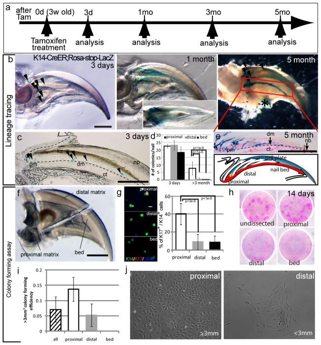
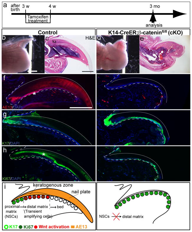
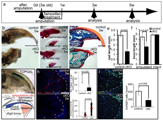
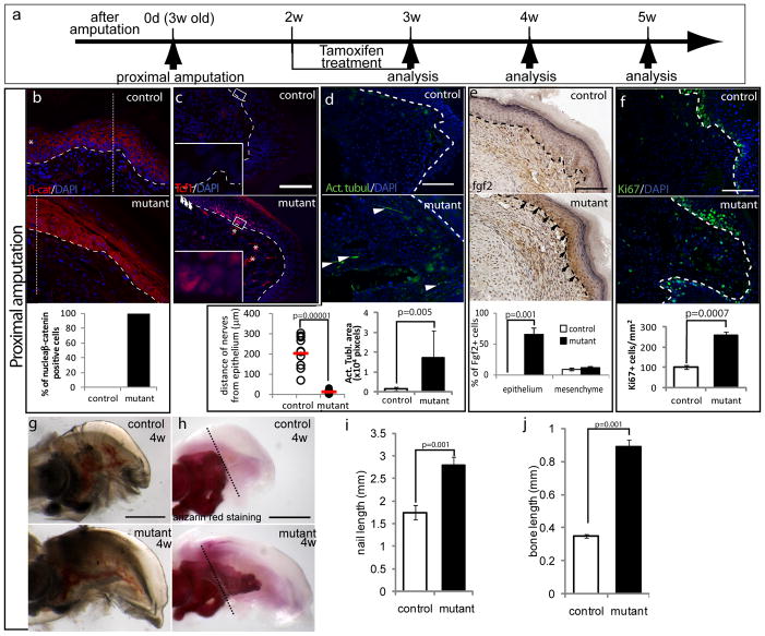
The tips of mammalian digits can regenerate after amputation, like those of amphibians. It is unknown why this capacity is limited to the area associated with the nail. Here we show that nail stem cells (NSCs) reside in the proximal nail matrix and that the mechanisms governing NSC differentiation are coupled directly with their ability to orchestrate digit regeneration. Early nail progenitors undergo Wnt-dependent differentiation into the nail. After amputation, this Wnt activation is required for nail regeneration and also for attracting nerves that promote mesenchymal blastema growth, leading to the regeneration of the digit. Amputations proximal to the Wnt-active nail progenitors result in failure to regenerate the nail or digit. Nevertheless, β-catenin stabilization in the NSC region induced their regeneration. These results establish a link between NSC differentiation and digit regeneration, and suggest that NSCs may have the potential to contribute to the development of novel treatments for amputees.
Conflict of interest statement
The authors declare no competing financial interests.
Figures
Similar articles
-
Lgr6 marks nail stem cells and is required for digit tip regeneration.Proc Natl Acad Sci U S A. 2015 Oct 27;112(43):13249-54. doi: 10.1073/pnas.1518874112. Epub 2015 Oct 12. Proc Natl Acad Sci U S A. 2015. PMID: 26460010 Free PMC article.
-
Are fingernails a key to unlocking the puzzle of mammalian limb regeneration?Exp Dermatol. 2017 Jun;26(6):478-482. doi: 10.1111/exd.13246. Epub 2017 Feb 19. Exp Dermatol. 2017. PMID: 27761955
-
Epithelium-Derived Wnt Ligands Are Essential for Maintenance of Underlying Digit Bone.J Invest Dermatol. 2016 Jul;136(7):1355-1363. doi: 10.1016/j.jid.2016.03.018. Epub 2016 Mar 25. J Invest Dermatol. 2016. PMID: 27021406 Free PMC article.
-
The Potential of Nail Mini-Organ Stem Cells in Skin, Nail and Digit Tips Regeneration.Int J Mol Sci. 2021 Mar 11;22(6):2864. doi: 10.3390/ijms22062864. Int J Mol Sci. 2021. PMID: 33799809 Free PMC article. Review.
-
The roles of Wnt/β-catenin pathway in tissue development and regenerative medicine.J Cell Physiol. 2018 Aug;233(8):5598-5612. doi: 10.1002/jcp.26265. Epub 2018 Mar 7. J Cell Physiol. 2018. PMID: 29150936 Review.
Cited by
-
Regeneration of amputated mice digit tips by including Wnt signaling pathway with CHIR99021 and IWP-2 chemicals in limb organ culture system.Iran J Basic Med Sci. 2024;27(10):1251-1259. doi: 10.22038/ijbms.2024.76957.16643. Iran J Basic Med Sci. 2024. PMID: 39229572 Free PMC article.
-
Hallmarks of regeneration.Cell Stem Cell. 2024 Sep 5;31(9):1244-1261. doi: 10.1016/j.stem.2024.07.007. Epub 2024 Aug 19. Cell Stem Cell. 2024. PMID: 39163854 Review.
-
CRISPR-mediated genomic deletion of Sox2 in the axolotl shows a requirement in spinal cord neural stem cell amplification during tail regeneration.Stem Cell Reports. 2014 Sep 9;3(3):444-59. doi: 10.1016/j.stemcr.2014.06.018. Epub 2014 Aug 7. Stem Cell Reports. 2014. PMID: 25241743 Free PMC article.
-
Bifunctional ectodermal stem cells around the nail display dual fate homeostasis and adaptive wounding response toward nail regeneration.Proc Natl Acad Sci U S A. 2014 Oct 21;111(42):15114-9. doi: 10.1073/pnas.1318848111. Epub 2014 Oct 2. Proc Natl Acad Sci U S A. 2014. PMID: 25277970 Free PMC article.
-
Lgr6-expressing functional nail stem-like cells differentiated from human-induced pluripotent stem cells.PLoS One. 2024 May 14;19(5):e0303260. doi: 10.1371/journal.pone.0303260. eCollection 2024. PLoS One. 2024. PMID: 38743670 Free PMC article.
References
-
- Douglas BS. Conservative management of guillotine amputation of the finger in children. Aust Paediatr J. 1972;8:86–89. - PubMed
-
- Borgens RB. Mice regrow the tips of their foretoes. Science. 1982;217:747–750. - PubMed
-
- Mohammad KS, Day FA, Neufeld DA. Bone growth is induced by nail transplantation in amputated proximal phalanges. Calcif Tissue Int. 1999;65:408–410. - PubMed
Publication types
MeSH terms
Substances
Associated data
- Actions
- Actions
- Actions
- Actions
- Actions
Grants and funding
LinkOut - more resources
Full Text Sources
Other Literature Sources
Molecular Biology Databases
