. 2013 Aug;87(15):8606-23.
doi: 10.1128/JVI.00805-13. Epub 2013 May 29.

Constitutive interferon-inducible protein 16-inflammasome activation during Epstein-Barr virus latency I, II, and III in B and epithelial cells

Affiliations

Constitutive interferon-inducible protein 16-inflammasome activation during Epstein-Barr virus latency I, II, and III in B and epithelial cells

Mairaj Ahmed Ansari et al. J Virol. 2013 Aug.

Erratum in

Abstract

Epstein-Barr virus (EBV), etiologically linked with human B-cell malignancies and nasopharyngeal carcinoma (NPC), establishes three types of latency that facilitate its episomal genome persistence and evasion of host immune responses. The innate inflammasome responses recognize the pathogen-associated molecular patterns which lead into the association of a cytoplasmic sensor such as NLRP3 and AIM2 proteins or nuclear interferon-inducible protein 16 (IFI16) with adaptor ASC protein (apoptosis-associated speck-like protein with a caspase recruitment domain) and effector procaspase-1, resulting in active caspase-1 formation which cleaves the proforms of inflammatory interleukin-1β (IL-1β), IL-18, and IL-33 cytokines. Whether inflammasome responses recognize and respond to EBV genome in the nuclei was not known. We observed evidence of inflammasome activation, such as the activation of caspase-1 and cleavage of pro-IL-1β, -IL-18, and -IL-33, in EBV latency I Raji cells, latency II NPC C666-1 cells, and latency III lymphoblastoid cell lines (LCL). Interaction between ASC with IFI16 but not with AIM2 or NLRP3 was detected in all three latencies and during EBV infection of primary human B cells. IFI16 and cleaved caspase-1, IL-1β, IL-18, and IL-33 were detected in the exosomes from Raji cells and LCL. Though EBV nuclear antigen 1 (EBNA1) and EBV-encoded small RNAs (EBERs) are common to all forms of EBV latency, caspase-1 cleavage was not detected in cells expressing EBNA1 alone, and blocking EBER transcription did not inhibit caspase-1 cleavage. In fluorescence in situ hybridization (FISH) analysis, IFI16 colocalized with the EBV genome in LCL and Raji cell nuclei. These studies demonstrated that constant sensing of latent EBV genome by IFI16 in all types of latency results in the constitutive induction of the inflammasome and IL-1β, IL-18, and IL-33 maturation.


Figures

Fig 1
Fig 1
Activation of the inflammasome in cells latently infected with EBV. (A to E) Equal protein concentrations of whole-cell lysates were analyzed by Western blotting (WB) for the following: procaspase-1 and activated caspase-1 (p20) in Ramos cells (EBV-negative Burkitt's lymphoma), a lymphoblastoid cell line (EBV+ latency III LCL), and Raji cells (EBV+ latency I Burkitt's lymphoma) (A); procaspase-1 and activated caspase-1 in 184B5 (EBV negative-control normal human mammary epithelial cell line) and in C666-1 (EBV+ latency II epithelial cell line from undifferentiated nasopharyngeal carcinoma [NPC] xenograft) (B); pro-IL-1β and cleaved mature IL-1β (p17.5) in Ramos, LCL, Raji, 184B5 and C666-1 cells (C); pro-IL-18, cleaved mature IL-18 (p18), pro-IL-33, and cleaved mature IL-33 (p18) in Ramos, LCL, and Raji cells (D) and in 184B5 and NPC cells (E). β-Actin was used as loading control. (F) Equal protein concentrations of nuclear and cytoplasmic (Cyto) proteins of LCL, Raji, and C666-1 cells were analyzed by Western blotting for distribution of cleaved caspase-1. The purity of cytoplasmic and nuclear fractions was confirmed by tubulin and TATA-binding protein (TBP), respectively.
Fig 2
Fig 2
Expression of inflammasome proteins, identification of IFI16-mediated inflammasome induction, cytoplasmic and nuclear distribution of inflammasome proteins, and transcript expression levels in EBV latency III LCL cells. (A) Equal protein concentrations of whole-cell lysates from Ramos (EBV BL) and LCL (EBV+ latency III) cells were analyzed by Western blotting for inflammasome sensors IFI16, AIM2, and NLRP3 proteins and for the adaptor ASC molecule. Equal loading was confirmed by β-actin. (B) Equal protein concentrations of whole-cell lysates Ramos and LCL cells were immunoprecipitated (IP) with anti-ASC antibodies and subjected to Western blotting for IFI16, NLRP3, and AIM2. Immunoprecipitations were confirmed by blotting for ASC. (C) Equal protein concentrations of Ramos and LCL cells were immunoprecipitated for caspase-1 and subjected to Western blotting for IFI16, NLRP3, and ASC. Immunoprecipitations were confirmed by blotting for caspase-1. (D) Equal protein concentrations of nuclear and cytoplasmic proteins from Ramos and LCL cells were analyzed by Western blotting for the distribution of inflammasome-associated proteins. The purity of cytoplasmic and nuclear fractions was confirmed by tubulin and TATA-binding protein (TBP), respectively. (E, F, and G) Equal protein concentrations of cytoplasmic and nuclear proteins of LCL cells were immunoprecipitated with anti-ASC (E), anti-caspase-1 (F), or anti-IFI16 (G) antibodies and subjected to Western blotting for IFI16 (E and F) and ASC (G). Immunoprecipitations were confirmed by blotting for ASC, caspase-1, and IFI16. (H) Equal quantities of LCL whole-cell lysates were immunoprecipitated with control rabbit IgG antibodies (lane 1) and rabbit anti-caspase-1 antibodies (lane 2) and subjected to Western blotting for caspase-1. Heavy and light chains of IgG are indicated. (I) Equal quantities of LCL whole-cell lysates were immunoprecipitated with control mouse IgG antibodies (lane 1) and mouse anti-IFI16 antibodies (lane 2) and subjected to Western blotting for IFI16. IgG heavy chain is indicated.
Fig 3
Fig 3
Expression of inflammasome proteins, identification of IFI16-mediated inflammasome induction, and cytoplasmic and nuclear distribution of inflammasome proteins in EBV latency I Raji cells. (A) Equal protein concentrations of whole-cell lysates from Ramos (EBV BL) and Raji (EBV+ latency I) cells were analyzed by Western blotting for NLRP3, IFI16, AIM2, and ASC proteins. Equal loading was confirmed by β-actin. (B) Equal protein concentrations of whole-cell lysates of Ramos and Raji cells were immunoprecipitated with anti-caspase-1 antibodies and subjected to Western blotting for NLRP3, IFI16, AIM2, and ASC. Immunoprecipitations were confirmed by blotting for caspase-1. (C) Equal protein concentrations of nuclear and cytoplasmic proteins of Ramos and Raji cells were analyzed by Western blotting for the distribution of IFI16, AIM2, and ASC. The purity of cytoplasmic and nuclear fractions was confirmed by tubulin and TBP, respectively.
Fig 4
Fig 4
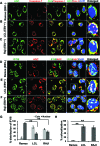
Immunofluorescence microscopic analysis of ASC–caspase-1 and ASC-IFI16 association in EBV-negative Ramos and EBV+ LCL (latency III) and Raji (latency I) cells. Cells were washed, fixed in acetone, permeabilized by Triton X-100, blocked with Image-iT FX signal enhancer, and reacted with anti-ASC and -caspase-1 antibodies (A, B, and C) and anti-IFI16 and -ASC antibodies (D, E and F); samples were then washed and incubated with Alexa Fluor-488 (green) and Alexa Fluor-594 (red) secondary antibodies. The images were merged with DAPI-stained nuclei (blue). The boxed areas are enlarged and shown in the rightmost panels. Colocalization in the cytoplasm (yellow spots) is indicated by red arrows, and yellow arrows indicate nuclear colocalization (white spots). Magnification, ×60. Mean pixel intensities from panels D to F were used to determine IFI16 cytoplasmic and nuclear percent distribution (G) and percent colocalization of IFI16 with ASC (H). **, P ≤ 0.01.
Fig 5
Fig 5
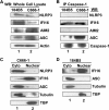
Expression of inflammasome proteins, identification of IFI16-mediated inflammasome induction, and cytoplasmic and nuclear distribution of inflammasome proteins in EBV latency II C666-1 cells. (A) Equal protein concentrations of whole-cell lysates from 184B5 (EBV-negative mammary epithelial cell line) and C666-1 (EBV+ latency II NPC epithelial cell line) were analyzed by Western blotting for NLRP3, IFI16, AIM2, and ASC proteins. Equal loading was confirmed by β-actin. (B) Equal protein concentrations of whole-cell lysates of 184B5 and C666-1 cells were immunoprecipitated with anti-caspase-1 antibodies and subjected to Western blotting for NLRP3, IFI16, AIM2, and ASC. Immunoprecipitations were confirmed by blotting for caspase-1. (C and D) Equal protein concentrations of nuclear and cytoplasmic proteins of C666-1 (C) and 184B5 (D) were analyzed by Western blotting for the distribution of IFI16, NLRP3, and ASC. The purity of cytoplasmic and nuclear fractions was confirmed by tubulin and TBP, respectively.
Fig 6
Fig 6
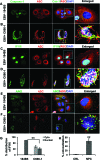
Immunofluorescence microscopic analysis of ASC–caspase-1, ASC-IFI16, and ASC-AIM2 association in EBV-negative 184B5 epithelial and EBV+ NPC epithelial C666-1 (latency II) cells. Cells cultured for 48 h in fibronectin and layered in eight-well chambered glass slides were fixed by 4% paraformaldehyde, permeabilized by 0.1% Triton X-100, blocked with signal enhancer, and reacted with anti-ASC and -caspase-1 antibodies (A and B), anti-ASC and -IFI16 antibodies (C and D), and anti-ASC and -AIM2 antibodies (E and F); samples were then washed and incubated with Alexa Fluor-488 (green) and Alexa Fluor-594 (red) secondary antibodies. The images were merged with DAPI-stained nuclei (blue). The boxed areas are enlarged and shown in the rightmost panels. Colocalization in the cytoplasm (yellow spots) is indicated by red arrows, and yellow arrows indicate nuclear colocalization (white spots). Magnification, ×60. Mean pixel intensities from panels C and D were used to determine IFI16 cytoplasmic and nuclear percent distribution (G) and percent colocalization of IFI16 with ASC in 184B5 (CRL, control) and C666-1 (NPC) cells (H). **, P ≤ 0.01.
Fig 7
Fig 7
Immunofluorescence microscopy analysis of ASC-IFI16 association in EBV-infected primary B cells (latency III). Human B cells purified from PBMCs were infected with EBV for 4 h, washed to remove the virus, and incubated further at 37°C for the indicated time points (A to F). These cells were washed, fixed, permeabilized, blocked with signal enhancer, and reacted with anti-ASC and -IFI16 antibodies; samples were then washed and incubated with Alexa Fluor-488 (green) and Alexa Fluor-594 (red) secondary antibodies. The boxed areas are enlarged and shown in the right panels. Far-right panels show the enlarged boxed areas with DAPI staining to depict the nucleus. Colocalizations in the nucleus (white spots) and in the cytoplasm (yellow spots) are indicated by red arrows. Magnification, ×60.
Fig 8
Fig 8
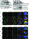
Detection of IFI16, caspase-1, IL-1β, IL-18, and IL-33 in the exosomes released from LCL and Raji cells latently infected with EBV and detection of the Rab27a interaction with caspase-1 and IL-1β. Culture supernatants from Ramos, LCL, and Raji cells cultured in exosome-depleted medium for 72 h were used to harvest the exosomes. (A) The purity of exosome fractions was determined by the presence of multivesicular body-derived Alix and Tsg101 proteins and the absence of the ER protein calnexin. Proteins were loaded at 10 μg per lane for Western blot analysis. (B) Exosomes were analyzed by Western blotting for the presence of IFI16 and cleaved forms of caspase-1, IL-1β, IL-18, and IL-33. (C to H) Cells were immunostained for colocalization of exolysosome/exosome marker Rab27a (green) and caspase-1 or IL-1β (red). Cells were fixed with acetone, permeabilized with 0.1% Triton X-100, blocked with signal enhancer, and reacted with anti-Rab27, -caspase-1, and -IL-1β antibodies; samples were then washed and incubated with Alexa Fluor-488 (green) and Alexa Fluor-594 (red) secondary antibodies. The images were merged with DAPI-stained nuclei (blue). The boxed areas are enlarged and shown in the rightmost panels. Colocalization in the cytoplasm (yellow spots) is indicated by red arrows. Magnification, ×60.
Fig 9
Fig 9
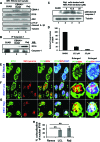
IFI16 recognition of the EBV genome is the sensor trigger for inflammasome induction in EBV latently infected LCL and Raji cells. (A and B) EBV latency (I, II, and III)-associated EBNA1 is not involved in IFI16-inflammasome induction. Equal protein concentrations of whole-cell lysates from BJAB (EBV BL), EBNA1-expressing BJAB, and Raji (EBV+ latency I) cells were analyzed by Western blotting for EBNA1, IFI16, ASC, and cleaved IL-1β proteins. Equal loading was confirmed by tubulin. (B) Equal protein concentrations of the above cell lysates were immunoprecipitated with anti-caspase-1 antibodies and subjected to Western blotting for IFI16. Immunoprecipitation was confirmed by blotting for caspase-1 in the immunoprecipitates. (C) Equal protein concentrations of whole-cell lysates were analyzed by Western blotting for activated caspase-1 (p20) in BJAB (lane 1) and in Raji cells treated with RNA Pol III inhibitor for 24 h (lanes 2 to 4) at the indicated concentrations (0, DMSO). Equal loading was confirmed by tubulin. (D) The gene expression levels of EBERs were analyzed in Raji cells treated with RNA Pol III inhibitor (12.5 μM and 25 μM) by real-time RT-PCR. Relative percent expression of EBER was calculated considering levels in Raji cells treated with DMSO as 100 after expression was normalized to that of the GAPDH gene as a control. Each bar represents the relative percent expression ± standard deviation of three independent experiments. (E, F, and G) Colocalization of IFI16 with the EBV genome present in the nuclei of EBV+ LCL and Raji cells. Ramos, LCL, and Raji cells were immunostained with mouse anti-IFI16 antibody followed by anti-mouse Alexa Fluor-488 (green) secondary antibody. These cells were then subjected to in situ hybridization with a biotinylated EBV FISH probe and analyzed by immunofluorescence. Cell nuclei were visualized by DAPI (blue). Red and yellow arrows point to the yellow and white colocalization spots, respectively, of IFI16 with EBV genome in the nucleus. Magnification, ×60. (H) The mean pixel intensities from panels E to G were analyzed for percent colocalization of nuclear IFI16 with EBV genome-specific FISH probe in Ramos, LCL, and Raji cells. **, P ≤ 0.01.
Fig 10
Fig 10
Schematic model depicting the constitutive activation of IFI16-mediated inflammasome during EBV latency in B and epithelial cells with latency I, II, and III. During primary infection of B or epithelial cells (step 1), EBV enters the target cells either via the fusion of its envelope with the plasma membrane or via endocytosis; released nucleocapsid travels to the nuclear pore, and linear DNA enters the nucleus, circularizes, and establishes latent infection (step 2). Multiple copies of the EBV genome are maintained in the cells with latency I, II, and III (steps 3 to 5). Studies presented here demonstrate that EBV episomal DNA is sensed in the infected cell during latency by innate DNA sensor IFI16 in the nucleus, leading to recruitment of the adaptor protein ASC and procaspase-1 (Pro-Casp-1) to form an inflammasome complex, which is followed by its translocation to the cytoplasm, activation of caspase-1, and cleavage of pro-IL-1β, pro-IL-18, and pro-IL-33 into their mature forms (steps 6 to 8). IFI16 and cleaved IL-1β, IL-18, and IL-33 are sorted and released to the exterior of the cells via exosomes, which could be an EBV-mediated strategy to subvert their inflammatory functions. Colocalization of IFI16 with most of the EBV genome in FISH analyses suggest that IFI16 must be sensing the EBV genome continuously in the latently infected cells and thereby contributing to constitutive inflammasome activation. IFI16 interaction with the EBV genome probably triggers changes that allow it to interact with ASC, leading to the formation of the inflammasome complex with ASC and procaspase-1 and rapid translocation to the cytoplasm, while a new IFI16 molecule probably comes into contact with the EBV genome, resulting in a continuum of the above-described processes.

Similar articles

Cited by

References

    1. Maeda E, Akahane M, Kiryu S, Kato N, Yoshikawa T, Hayashi N, Aoki S, Minami M, Uozaki H, Fukayama M, Ohtomo K. 2009. Spectrum of Epstein-Barr virus-related diseases: a pictorial review. Jpn. J. Radiol. 27:4–19 - PubMed
    1. Savard M, Belanger C, Tardif M, Gourde P, Flamand L, Gosselin J. 2000. Infection of primary human monocytes by Epstein-Barr virus. J. Virol. 74:2612–2619 - PMC - PubMed
    1. Gulley ML, Tang W. 2008. Laboratory assays for Epstein-Barr virus-related disease. J. Mol. Diagn. 10:279–292 - PMC - PubMed
    1. Amon W, Farrell PJ. 2005. Reactivation of Epstein-Barr virus from latency. Rev. Med. Virol. 15:149–156 - PubMed
    1. Young LS, Arrand JR, Murray PG. 2007. EBV gene expression and regulation, p 461–489 Arvin A, Campadelli-Fiume G, Mocarski E, Moore PS, Roizman B, Whitley R, Yamanishi K. (ed), Human herpesviruses biology, therapy, and immunoprophylaxis. Cambridge University Press, Cambridge, United Kingdom - PubMed

Publication types

LinkOut - more resources