Ribosome profiling: a Hi-Def monitor for protein synthesis at the genome-wide scale
- PMID: 23696005
- PMCID: PMC3823065
- DOI: 10.1002/wrna.1172
Ribosome profiling: a Hi-Def monitor for protein synthesis at the genome-wide scale
Erratum in
-
Ribosome profiling: a Hi-Def monitor for protein synthesis at the genome-wide scale.Wiley Interdiscip Rev RNA. 2017 Sep;8(5):e1434. doi: 10.1002/wrna.1434. Wiley Interdiscip Rev RNA. 2017. PMID: 28815950 Free PMC article. No abstract available.
Abstract
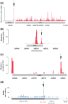
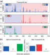
Ribosome profiling or ribo-seq is a new technique that provides genome-wide information on protein synthesis (GWIPS) in vivo. It is based on the deep sequencing of ribosome protected mRNA fragments allowing the measurement of ribosome density along all RNA molecules present in the cell. At the same time, the high resolution of this technique allows detailed analysis of ribosome density on individual RNAs. Since its invention, the ribosome profiling technique has been utilized in a range of studies in both prokaryotic and eukaryotic organisms. Several studies have adapted and refined the original ribosome profiling protocol for studying specific aspects of translation. Ribosome profiling of initiating ribosomes has been used to map sites of translation initiation. These studies revealed the surprisingly complex organization of translation initiation sites in eukaryotes. Multiple initiation sites are responsible for the generation of N-terminally extended and truncated isoforms of known proteins as well as for the translation of numerous open reading frames (ORFs), upstream of protein coding ORFs. Ribosome profiling of elongating ribosomes has been used for measuring differential gene expression at the level of translation, the identification of novel protein coding genes and ribosome pausing. It has also provided data for developing quantitative models of translation. Although only a dozen or so ribosome profiling datasets have been published so far, they have already dramatically changed our understanding of translational control and have led to new hypotheses regarding the origin of protein coding genes.
Copyright © 2013 John Wiley & Sons, Ltd.
Figures


Similar articles
-
GWIPS-viz: development of a ribo-seq genome browser.Nucleic Acids Res. 2014 Jan;42(Database issue):D859-64. doi: 10.1093/nar/gkt1035. Epub 2013 Oct 31. Nucleic Acids Res. 2014. PMID: 24185699 Free PMC article.
-
Genome-wide annotation and quantitation of translation by ribosome profiling.Curr Protoc Mol Biol. 2013 Jul;Chapter 4:4.18.1-4.18.19. doi: 10.1002/0471142727.mb0418s103. Curr Protoc Mol Biol. 2013. PMID: 23821443 Free PMC article.
-
Studying Selenoprotein mRNA Translation Using RNA-Seq and Ribosome Profiling.Methods Mol Biol. 2018;1661:103-123. doi: 10.1007/978-1-4939-7258-6_8. Methods Mol Biol. 2018. PMID: 28917040
-
Translation Analysis at the Genome Scale by Ribosome Profiling.Methods Mol Biol. 2016;1361:105-24. doi: 10.1007/978-1-4939-3079-1_7. Methods Mol Biol. 2016. PMID: 26483019 Review.
-
Ribosomal profiling adds new coding sequences to the proteome.Biochem Soc Trans. 2015 Dec;43(6):1271-6. doi: 10.1042/BST20150170. Biochem Soc Trans. 2015. PMID: 26614672 Review.
Cited by
-
LncRNA-encoded peptides in cancer.J Hematol Oncol. 2024 Aug 12;17(1):66. doi: 10.1186/s13045-024-01591-0. J Hematol Oncol. 2024. PMID: 39135098 Free PMC article. Review.
-
Ribosome profiling reveals downregulation of UMP biosynthesis as the major early response to phage infection.Microbiol Spectr. 2024 Apr 2;12(4):e0398923. doi: 10.1128/spectrum.03989-23. Epub 2024 Mar 7. Microbiol Spectr. 2024. PMID: 38451091 Free PMC article.
-
Biosynthesis and function of 7-deazaguanine derivatives in bacteria and phages.Microbiol Mol Biol Rev. 2024 Mar 27;88(1):e0019923. doi: 10.1128/mmbr.00199-23. Epub 2024 Feb 29. Microbiol Mol Biol Rev. 2024. PMID: 38421302 Review.
-
No country for old methods: New tools for studying microproteins.iScience. 2024 Jan 20;27(2):108972. doi: 10.1016/j.isci.2024.108972. eCollection 2024 Feb 16. iScience. 2024. PMID: 38333695 Free PMC article. Review.
-
Ribosome profiling: a powerful tool in oncological research.Biomark Res. 2024 Jan 25;12(1):11. doi: 10.1186/s40364-024-00562-4. Biomark Res. 2024. PMID: 38273337 Free PMC article. Review.
References
-
- Weiss RB, Atkins JF. Molecular biology. Translation goes global. Science. 2011;334:1509–1510. - PubMed
-
- Steitz JA. Polypeptide chain initiation: nucleotide sequences of the three ribosomal binding sites in bacteriophage R17 RNA. Nature. 1969;224:957–964. - PubMed
-
- Ingolia NT. Genome-wide translational profiling by ribosome footprinting. Methods Enzymol. 2010;470:119–142. - PubMed
FURTHER READING
-
- GWIPS-viz browser for visualization of Genome Wide Information on Protein Synthesis. http://gwips.ucc.ie.
Publication types
MeSH terms
Substances
Grants and funding
LinkOut - more resources
Full Text Sources
Other Literature Sources

