. 2013 Jul;87(14):8004-16.
doi: 10.1128/JVI.00506-13. Epub 2013 May 15.

Activation of the B cell antigen receptor triggers reactivation of latent Kaposi's sarcoma-associated herpesvirus in B cells

Affiliations

Activation of the B cell antigen receptor triggers reactivation of latent Kaposi's sarcoma-associated herpesvirus in B cells

Semra Kati et al. J Virol. 2013 Jul.

Abstract

Kaposi's sarcoma-associated herpesvirus (KSHV) is an oncogenic herpesvirus and the cause of Kaposi's sarcoma, primary effusion lymphoma (PEL) and multicentric Castleman's disease. Latently infected B cells are the main reservoir of this virus in vivo, but the nature of the stimuli that lead to its reactivation in B cells is only partially understood. We established stable BJAB cell lines harboring latent KSHV by cell-free infection with recombinant virus carrying a puromycin resistance marker. Our latently infected B cell lines, termed BrK.219, can be reactivated by triggering the B cell receptor (BCR) with antibodies to surface IgM, a stimulus imitating antigen recognition. Using this B cell model system we studied the mechanisms that mediate the reactivation of KSHV in B cells following the stimulation of the BCR and could identify phosphatidylinositol 3-kinase (PI3K) and X-box binding protein 1 (XBP-1) as proteins that play an important role in the BCR-mediated reactivation of latent KSHV.


Figures
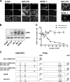
Fig 1
Fig 1
Establishment of BrK.219 cells, LANA expression, and persistence of viral genomes. (A) Immunofluorescence staining of the latent viral nuclear antigen LANA was carried out in BrK.219 cells, the uninfected parental BJAB cell line, and the PEL cell line BCBL-1. Cells infected with rKSHV.219 express GFP constitutively. Samples were fixed, blocked, and incubated either with primary antibody against LANA, or without primary antibody, followed by a Cy5-conjugated secondary antibody. Nuclei were stained with DAPI (4′,6′-diamidino-2-phenylindole). Magnified images of the boxed regions B1 and B2 are shown on the right. (B) Expression of LANA shown by immunoblotting. (C) BrK.219 cells are BJAB-derived. BrK.219 cells established by coculture with rKSHV.219-infected Vero cells as described in Materials and Methods were analyzed using STR analysis for two gene loci, CSF1PO and FGA. The result indicates that BrK.219 cells and BJAB cells share the same genetic profile but differ from Vero cells. (D) Persistence of KSHV genomes in the presence or absence of puromycin. Samples were taken twice a week, and the number of KSHV genomes was measured by qPCR and normalized to CRP (a cellular gene) copy numbers.
Fig 2
Fig 2
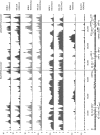
Comparison of global histone modification patterns of KSHV genomes in BrK.219 and PEL cells. Global patterns of histone H3 trimethylated at lysine 4 (H3K4-me3), lysine 9 (H3K9-me3), or lysine 27 (H3K27-me3) or acetylated at lysine 9 and/or lysine 14 (H3K27-me3 and H3K9/14-ac). Modification patterns in BrK.219 cells were analyzed by ChIP-on-chip analysis using high-resolution KSHV tiling microarrays. Values shown on the y axis represent relative enrichment of normalized signals from the immunoprecipitated over input material. Histone modification patterns in the PEL line BCBL-1 (47) are shown for comparison. Regions highlighted in gray indicate the position of the ORF50/Rta and the ORF73/LANA promoters, both of which carry activating H3K4-me3 and H3K9/14-ac marks. In addition, an H3K19/14-ac peak, located upstream of the vIRF-3 coding region and marked by arrows, is more prominent in BCBL-1 compared to BrK.219 cells, which may reflect the lower basal expression levels of vIRF-3 during latency in BrK.219 cells. The ORF50 promoter additionally acquires repressive H3K27-me3 marks and thus exhibits the hallmarks of bivalent chromatin, which is transcriptionally silent, but “poised” for reactivation (47).
Fig 3
Fig 3
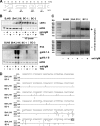
Latent KSHV in BrK.219 cells is reactivated following triggering of the BCR. BrK.219 cells and their uninfected counterparts were treated with increasing amounts of antibodies against IgM or IgG for 3 days. (A) Expression of latent (LANA), immediate-early (RTA), early (KbZIP), and late (gpK8.1) proteins shown by immunoblotting. (B) Infectious KSHV titers released from antibody-stimulated BrK.219 cells, as measured on HEK 293 cells. (C) Expression of viral proteins in BrK.219 cells treated with anti-IgM and F(ab′)2 anti-IgM. (D) Infectious KSHV titers released from BrK.219 cells treated with anti-IgM or F(ab′)2 anti-IgM, as measured on HEK 293 cells.
Fig 4
Fig 4
Comparison of KSHV reactivation induced by antibody against IgM with other chemical stimuli in BrK.219 and in PEL cells. (A) BrK.219 cells and the uninfected parental cell line BJAB were treated with different inducers of the KSHV lytic cycle (2.5 μg of anti-IgM/ml, 50 ng of TPA/ml, 1.25 mM sodium butyrate, 1.25 mM valproic acid [VPA]) for 3 days, and the expression of viral proteins was detected by immunoblotting as described in the legend of Fig. 2. (B) The PEL cell line BC-3, BrK.219 cells, and parental BJAB cells were treated with anti-IgM or sodium butyrate, and the expression of viral proteins was monitored by immunoblotting. (C) KSHV genome copy number in induced BC-3 and BrK.219 cells, as measured by qPCR. The data shown are means of triplicates with the standard deviations.
Fig 5
Fig 5
vIRF-3 is an inducible gene with two 5′ start sites in BrK.219 cells. (A) Gene arrangement in the vIRF region shown between ORFs 57 and 58. (B) BJAB cells, their rKSHV.219-infected counterparts (BrK.219), and PEL cell lines (BC-3 and BC-1) were treated with either 2.5 μg of anti-IgM/ml or 25 ng of TPA/ml for 3 days or left untreated as a control. Viral protein expression was investigated by immunoblotting. For analysis of vIRF-3 expression in PEL cells, only 1/5 of the cell lysates were used. (C) 5′-RACE of vIRF-3 transcripts. BrK.219 cells, uninfected parental BJAB cells, and the PEL cell line BC-3 were treated for 3 days with anti-IgM or left untreated. Reverse transcription and PCR was performed with total RNAs. Agarose gels show the products of the 5′-RACE, which was also performed on RNA samples without reverse transcription to control for the potential amplification of contaminating DNA. (D) Location of 5′ start sites of vIRF-3 mRNAs determined by 5′-RACE. The 5′-RACE products as shown in panel C of two independent experiments were cloned, and multiple clones were sequenced to determine their 5′ ends. Numbers (ranging between 1 and 5) reflect the frequency and position of individual 5′ start sites. Whereas in BC-3 cells most transcripts originate within the first 100 bp upstream of the vIRF-3 ATG, in anti-IgM-stimulated BrK.219 cells, additional peaks of initiation are located up to further 150 bp upstream at position −101 to position −250.
Fig 6
Fig 6
PI3K is important for the reactivation of latent KSHV following triggering of the BCR. (A) LY294002 inhibits the reactivation of latent KSHV following triggering of the BCR. BJAB and BrK.219 were treated for 1 h with increasing amounts of inhibitor or vehicle control (DMSO). Next, reactivation was induced by the addition of antibodies against IgM (2.5 μg/ml). After incubation for 3 days, supernatants were titered on HEK 293 cells. The results are represented as the means of duplicate samples ± the standard deviations. (B) Cell lysates of the same experiments (left panel) and their nonreactivated control samples (right panel) were analyzed by SDS-PAGE and immunoblotting with the indicated antibodies. (C) PI-103 inhibits KSHV reactivation induced by treatment with anti-IgM but not with TPA (2 ng/ml). (D) The metabolic activity of BrK.219 cells treated for 3 days with the inhibitor LY294002 (5 or 7.5 μM), PI-103 (0.5 or 0.75 μM) or vehicle control (DMSO) was analyzed by MTT assay. The results are presented as the means of triplicates ± the standard deviations.
Fig 7
Fig 7
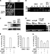
Lytic KSHV induction following BCR activation requires the splicing of XBP-1 mRNA. (A) Confocal images of BrK.219 cells transduced with lentiviral vectors expressing either GFP (“control”) only or the spliced form of XBP-1 (“active XBP-1”). Expression of RFP marks RTA-expressing BrK.219 cells. (B) RT-PCR analysis of the DTT-induced splicing of XBP-1 transcript in BrK.219 cells with or without treatment with IRE1α inhibitor (IRE1i; 25 μM). cDNA products derived from the spliced XBP-1 mRNA are resistant to PstI digestion (upper band, 223 bp), whereas the products for the unspliced XBP-1 mRNA are cleaved (the two lower bands, 104 and 145 bp). WB denotes water blank control. The band sizes (in bp) of the DNA ladder are as indicated. (C) Western blot analysis of KSHV RTA induction by anti-IgM antibodies (1 μg/ml) with or without IRE1i (25 μM) treatment. (D) Semiquantitative RT-PCR analysis of a KSHV late gene (ORF29) expression at 48 h posttreatment (upper panel), using serially 10-fold-diluted cDNA obtained from BrK.219 cells treated with either DMSO, anti-IgM antibodies (1 μg/ml), or anti-IgM antibodies plus IRE1i (25 μM). Input cDNA was normalized to a cellular gene (β2M lower panel). Primers for both virus and cellular genes span across intron(s), and no amplification of genomic DNA was observed under the PCR conditions used. The data from one representative experiment out of two independent experiments are shown. (E) Relative KSHV titer in supernatants from BrK.219 cells 3 days after BCR cross-linking, with or without the presence of IRE1i. Cell supernatants from BrK.219 cells treated with either DMSO, anti-IgM, or anti-IgM plus IRE1i were titrated on TE671 cells. The data are shown as the relative infectious titer where all samples were normalized to the positive control (anti-IgM) as 100% and plotted as means ± the standard deviations from three independent experiments. (F) Effect of IRE1i on the relative KSHV titer of anti-IgM induced supernatant from BrK.219 cells. The cell supernatant from BrK.219 cells induced with anti-IgM was split into two, and each half mixed with either DMSO or IRE1i; the resulting supernatants were titrated on TE671 cells. The data are shown as relative infectious titer where all samples were normalized to the positive control (anti-IgM plus DMSO) as 100% and plotted as means ± the standard deviations from three independent experiments. (G) The relative cell viability of BrK.219 cells treated with either DMSO, IRE1i, anti-IgM plus DMSO, or anti-IgM plus IRE1i is plotted as the percent concentration of ATP (proportional to the number of live cells) normalized to the DMSO control as 100%. The data (means ± the standard deviations) from one representative experiment out of two independent experiments are shown.

Similar articles

Cited by

References

    1. Cesarman E. 2011. Gammaherpesvirus and lymphoproliferative disorders in immunocompromised patients. Cancer Lett. 305:163–174 - PMC - PubMed
    1. Taylor GS, Blackbourn DJ. 2011. Infectious agents in human cancers: lessons in immunity and immunomodulation from gammaherpesviruses EBV and KSHV. Cancer Lett. 305:263–278 - PubMed
    1. Saha A, Robertson ES. 2011. Epstein-Barr virus-associated B-cell lymphomas: pathogenesis and clinical outcomes. Clin. Cancer Res. 17:3056–3063 - PMC - PubMed
    1. Cesarman E, Chang Y, Moore PS, Said JW, Knowles DM. 1995. Kaposi's sarcoma-associated herpesvirus-like DNA sequences in AIDS-related body-cavity-based lymphomas. N. Engl. J. Med. 332:1186–1191 - PubMed
    1. Soulier J, Grollet L, Oksenhendler E, Cacoub P, Cazals-Hatem D, Babinet P, d'Agay MF, Clauvel JP, Raphael M, Degos L. 1995. Kaposi's sarcoma-associated herpesvirus-like DNA sequences in multicentric Castleman's disease. Blood 86:1276–1280 - PubMed

Publication types

MeSH terms