. 2013 May 13;8(5):e62516.
doi: 10.1371/journal.pone.0062516. Print 2013.

Stromal Claudin14-heterozygosity, but not deletion, increases tumour blood leakage without affecting tumour growth

Affiliations

Stromal Claudin14-heterozygosity, but not deletion, increases tumour blood leakage without affecting tumour growth

Marianne Baker et al. PLoS One. .

Abstract

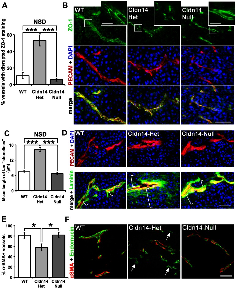
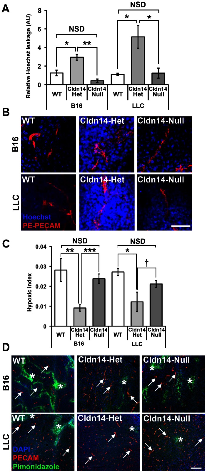
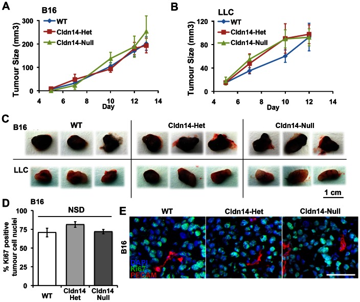
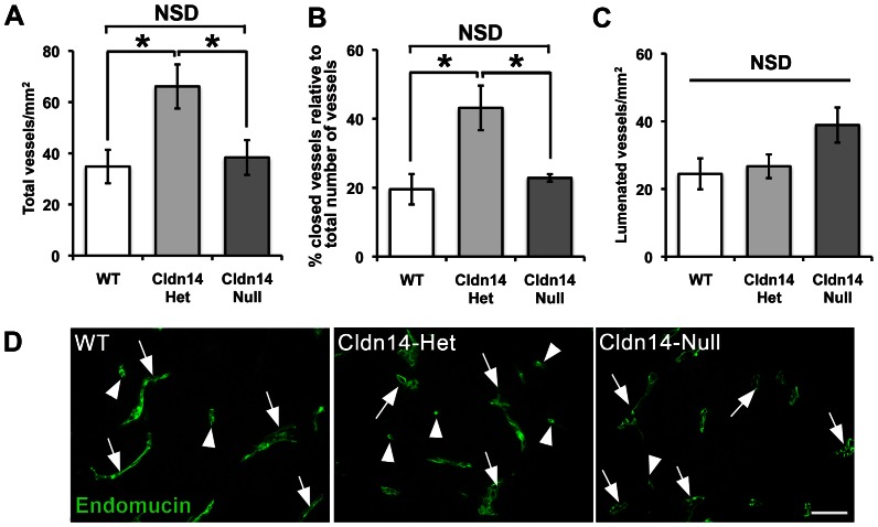
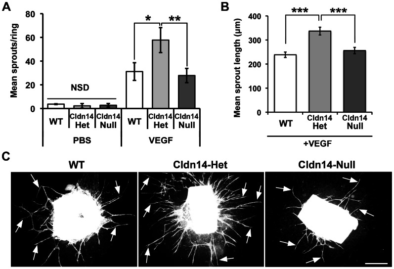
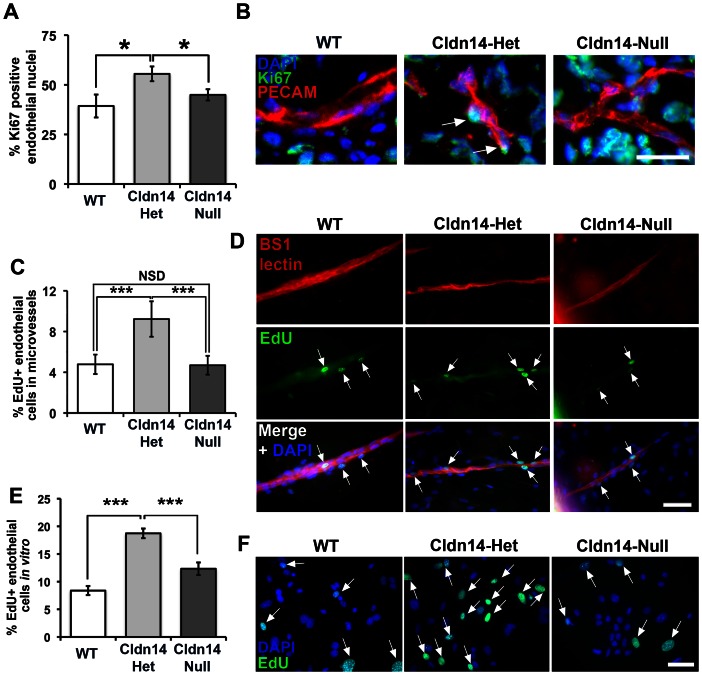
The maintenance of endothelial cell-cell junctions is vital for the control of blood vessel leakage and is known to be important in the growth and maturation of new blood vessels during angiogenesis. Here we have investigated the role of a tight junction molecule, Claudin 14, in tumour blood vessel leakage, angiogenesis and tumour growth. Using syngeneic tumour models our results showed that genetic ablation of Claudin 14 was not sufficient to affect tumour blood vessel morphology or function. However, and surprisingly, Claudin 14-heterozygous mice displayed several blood vessel-related phenotypes including: disruption of ZO-1-positive cell-cell junctions in tumour blood vessels; abnormal distribution of basement membrane laminin around tumour blood vessels; increased intratumoural leakage and decreased intratumoural hypoxia. Additionally, although total numbers of tumour blood vessels were increased in Claudin 14-heterozygous mice, and in VEGF-stimulated angiogenesis ex vivo, the number of lumenated vessels was not changed between genotypes and this correlated with no difference in syngeneic tumour growth between wild-type, Claudin 14-heterozygous and Claudin 14-null mice. Lastly, Claudin 14-heterozygosity, but not complete deficiency, also enhanced endothelial cell proliferation significantly. These data establish a new role for Claudin 14 in the regulation of tumour blood vessel integrity and angiogenesis that is evident only after the partial loss of this molecule in Claudin 14-heterozyous mice but not in Claudin 14-null mice.


Conflict of interest statement

Competing Interests: The authors have declared that no competing interests exist.

Figures

Figure 1
Figure 1. Cldn14 heterozygosity destabilises tumour blood vessels.
B16F10 tumours were grown for 10 days in WT, Cldn14-het and Cldn14-null mice and midline sections from size-matched tumours were analysed for blood vessel stabilisation by immunostaining for the tight junction component ZO-1, basement membrane laminin, and pericyte coverage using an anti-αSMA antibody. (A) The tight junction adapter protein ZO-1 staining pattern was observed at cell-cell borders in PECAM-positive tumour blood vessels. A higher proportion of blood vessels exhibited a disrupted ZO-1 staining pattern in Cldn14-het tumour sections, compared to tumours from WT and Cldn14-null mice. (B) Representative images of ZO-1 staining and the endothelial cell marker PECAM, with nuclei DAPI-counterstained. Inserts show higher magnification of ZO-1 at cell-cell junctions. Scale bars: main panels  = 50 µm, insets  = 10 µm. (C) The AxioVision software linear measuring tool was used to analyse the spread (in µm) of laminin surrounding PECAM-positive blood vessels in immunostained tumour sections. Laminin expression was close to blood vessel walls in tumours from WT and Cldn14-null mice but disorganised around tumour blood vessels in Cldn14-het mice; a “shorelining” effect of laminin deposition was evident in these sections. (D) Representative images of tumour cryosections immunostained for basement membrane laminin and the endothelial cell marker PECAM, with nuclei DAPI-counterstained. White brackets indicate the spread of laminin staining radiating from PECAM-positive vessels and demonstrates the quantification method. (E) α-Smooth Muscle Actin (αSMA) antibody conjugated to Cy3 fluorescent dye was used to label supporting cells (pericytes) around endomucin-labelled blood vessels in midline tumour sections. The percentage of αSMA-positive vessels was quantified. (F) Representative images of endomucin and αSMA double-stained tumour sections. Arrows, αSMA-negative vessels. Scale bar 50 µm. N = 4–6 tumours per genotype. Bar charts represent means ± SEM. * P<0.05, *** P<0.001.
Figure 2
Figure 2. Heterozygosity for Cldn14 increases tumour blood vessel leakage and decreases intratumoural hypoxia.
Wild-type, Cldn14-heterozygous and Cldn14-null mice were injected subcutaneously in the flank with 0.5×10⁶ B16F10 melanoma or Lewis Lung Carcinoma (LLC) cells. (A) At 10 days post inoculation, PE-conjugated anti-PECAM antibody and Hoechst dye were injected via the tail vein prior to sacrifice. Midline sections (100 µm) of snap-frozen tumours were fixed, mounted and imaged using a Zeiss LSM 510 confocal microscope. The extent of Hoechst leakage was measured in z-stacks using ImageJ. Bars show mean Hoechst leakage relative to PECAM signal ± SEM. Blood vessel leakage is increased significantly in Cldn14-het mice when compared with WT and Cldn14-null mice. (B) Representative images of Hoechst (blue) and PECAM (red) detection. (C) Tumour-bearing mice from each genotype were injected with pimonidazole prior to sacrifice to measure hypoxic areas within the tumour. 8 µm tumour cryosections were then double stained with anti-pimonidazole antibody (green) to highlight hypoxic areas and anti-PECAM antibody to identify blood vessels. The hypoxic index was quantified relative to PECAM staining using image J software. Bars represent mean relative hypoxic index ± SEM. (D) Representative images of pimonidazole detection and PECAM-positive blood vessels in tumour sections. Arrows, blood vessels; Asterisks, pimonidazole-positive staining. Scale bars: A 50 µm; D 200 µm. N = 4 tumours per genotype. NSD: not statistically different, * P<0.05, ** P<0.01, *** P<0.001, † P = 0.09.
Figure 3
Figure 3. Stromal Cldn14 heterozygosity does not affect tumour size or tumour cell proliferation.
Wild-type and Cldn14-het and Cldn14-null mice were injected subcutaneously with 0.5×10⁶ B16F10 melanoma or Lewis Lung Carcinoma (LLC) cells. (A and B) Tumour size was measured every two days for up to 13 days. No difference in B16 or LLC tumour growth rate was observed between the genotypes. (C) Representative images of B16 and LLC endpoint tumours from each genotype. N = 12–17 mice per tumour type per genotype. (D) Tumour cryosections were immunostained for the proliferation marker Ki67 and the endothelial marker PECAM, with a DAPI nuclear stain. The percentage of Ki67-positive/PECAM-negative tumour cells was counted. Bars show mean ± SEM. (E) Representative images of Ki67-stained tumour sections. Scale bar 50 µm. NSD: no significant difference.
Figure 4
Figure 4. Cldn14-heterozygous mice have increased tumour blood vessel density, but show no difference in the number of lumenated tumour blood vessels.
Wild-type and Cldn14-het and Cldn14-null mice were injected subcutaneously with 0.5×10⁶ B16F10 melanoma cells. Whole midline sections of frozen 13 day old tumours were fixed and stained with anti-endomucin antibody. (A) The total number of blood vessels was counted across entire tumour sections and divided by the section area to give total mean blood vessel density for each genotype. (B) Graph showing the percentage of total blood vessels that are closed in tumour sections. (C) Graph showing mean numbers of lumenated vessels per mm2 of midline tumour section. (D) Representative images of endomucin-positive vessels in all genotypes Arrows, lumenated vessels; arrowheads, non-lumenated vessels. Scale bar 50 µm. N = 6 mice per genotype. For all graphs, bars show means ± SEM. NSD: no significant difference. * P<0.05.
Figure 5
Figure 5. Cldn14 heterozygosity increases VEGF-stimulated aortic ring microvessel sprouting and sprout length.
A. Quantitation of wild-type, Cldn14-heterozygous and Cldn14-null VEGF-stimulated aortic ring microvessel sprouting at 9 days in culture. PBS was used as a negative control. VEGF-stimulated microvessel numbers were increased significantly in Cldn14-het samples when compared with similarly treated WT and Cldn14 –null samples. B. Quantification of microvessel sprout length in using the ImageJ line tool on scaled images. N = 25–91 rings per genotype. VEGF-stimulated microvessel length was increased significantly in Cldn14-het samples when compared with similarly treated WT and Cldn14-null samples. C. Representative images of VEGF-treated BS1 lectin-stained aortic rings fixed and stained after 9 days in culture. Arrows, endothelial microvessel sprouts. Scale bar 500 µm. * P<0.05, ** P<0.01, *** P<0.001.
Figure 6
Figure 6. Cldn14 gene copy number affects endothelial cell proliferation in vivo, ex vivo and in vitro.
(A) Percentages of Ki67-positive endothelial cells were counted in cryosections of 13-day B16F10 tumours from WT, Cldn14-het and Cldn14-null mice co-stained with PECAM. Endothelial cell proliferation was enhanced significantly in Cldn14-het mice. (B) Representative images of tumour sections in each genotype. Arrows, Ki67-positive endothelial cell nuclei. Scale bar 25 µm. (C) Proliferating cells in VEGF-stimulated wild-type, Cldn14-het and Cldn14-null collagen-embedded aortic explants were detected by EdU incorporation. The number of proliferating (EdU-positive) nuclei, counterstained with DAPI, was divided by the total number of cell nuclei also BS1-lectin positive to give % proliferating endothelial cells in VEGF-treated aortic rings. Bars show mean % of proliferating cells ± SEM. n = 6–8 rings per genotype, 513–717 nuclei per genotype. (D) Representative images of VEGF-stimulated WT, Cldn14-het and Cldn14-null microvessels from aortic ring explants stained for EdU and BS1 lectin. Scale bar 50 µm. (E) WT, Cldn14-het and Cldn14-null primary endothelial cells were examined for EdU incorporation in the presence of 30 ng/ml VEGF. Cells were counterstained with DAPI and the number of EdU-positive cells recorded for each genotype. Bars show mean % EdU-positive cells ± SEM. N = 1217–3464 nuclei per genotype, 3 mice per genotype. (F) Representative images of primary endothelial cells in culture. Scale bar 50 µm. Arrows, EdU-positive nuclei. NSD: no significant difference. * P<0.05, *** P<0.001.

Similar articles

Cited by

  • Antagonizing Integrin β3 Increases Immunosuppression in Cancer.
    Su X, Esser AK, Amend SR, Xiang J, Xu Y, Ross MH, Fox GC, Kobayashi T, Steri V, Roomp K, Fontana F, Hurchla MA, Knolhoff BL, Meyer MA, Morgan EA, Tomasson JC, Novack JS, Zou W, Faccio R, Novack DV, Robinson SD, Teitelbaum SL, DeNardo DG, Schneider JG, Weilbaecher KN. Su X, et al. Cancer Res. 2016 Jun 15;76(12):3484-95. doi: 10.1158/0008-5472.CAN-15-2663. Epub 2016 May 23. Cancer Res. 2016. PMID: 27216180 Free PMC article.
  • Claudin-based barrier differentiation in the colonic epithelial crypt niche involves Hopx/Klf4 and Tcf7l2/Hnf4-α cascades.
    Lili LN, Farkas AE, Gerner-Smidt C, Overgaard CE, Moreno CS, Parkos CA, Capaldo CT, Nusrat A. Lili LN, et al. Tissue Barriers. 2016 Jul 19;4(3):e1214038. doi: 10.1080/21688370.2016.1214038. eCollection 2016 Jul-Sep. Tissue Barriers. 2016. PMID: 27583195 Free PMC article.
  • Mutations in Chromatin Modifier and Ephrin Signaling Genes in Vein of Galen Malformation.
    Duran D, Zeng X, Jin SC, Choi J, Nelson-Williams C, Yatsula B, Gaillard J, Furey CG, Lu Q, Timberlake AT, Dong W, Sorscher MA, Loring E, Klein J, Allocco A, Hunt A, Conine S, Karimy JK, Youngblood MW, Zhang J, DiLuna ML, Matouk CC, Mane S, Tikhonova IR, Castaldi C, López-Giráldez F, Knight J, Haider S, Soban M, Alper SL, Komiyama M, Ducruet AF, Zabramski JM, Dardik A, Walcott BP, Stapleton CJ, Aagaard-Kienitz B, Rodesch G, Jackson E, Smith ER, Orbach DB, Berenstein A, Bilguvar K, Vikkula M, Gunel M, Lifton RP, Kahle KT. Duran D, et al. Neuron. 2019 Feb 6;101(3):429-443.e4. doi: 10.1016/j.neuron.2018.11.041. Epub 2018 Dec 18. Neuron. 2019. PMID: 30578106 Free PMC article.
  • Emerging multifunctional roles of Claudin tight junction proteins in bone.
    Alshbool FZ, Mohan S. Alshbool FZ, et al. Endocrinology. 2014 Jul;155(7):2363-76. doi: 10.1210/en.2014-1173. Epub 2014 Apr 23. Endocrinology. 2014. PMID: 24758302 Free PMC article. Review.
  • Platelets and cancer: a casual or causal relationship: revisited.
    Menter DG, Tucker SC, Kopetz S, Sood AK, Crissman JD, Honn KV. Menter DG, et al. Cancer Metastasis Rev. 2014 Mar;33(1):231-69. doi: 10.1007/s10555-014-9498-0. Cancer Metastasis Rev. 2014. PMID: 24696047 Free PMC article. Review.

References

    1. Carmeliet P, Jain RK (2000) Angiogenesis in cancer and other diseases. Nature 407: 249–257. - PubMed
    1. Carmeliet P (2005) VEGF as a key mediator of angiogenesis in cancer. Oncology 69: 4–10. - PubMed
    1. Lamalice L, Le Boeuf F, Huot J (2007) Endothelial Cell Migration During Angiogenesis. Circ Res 100: 782–794. - PubMed
    1. Dudley AC (2012) Tumor endothelial cells. Cold Spring Harb Perspect Med 2: 1–18. - PMC - PubMed
    1. Tlsty TD, Coussens LM (2006) Tumor stroma and regulation of cancer development. Annu Rev Pathol 1: 119–150. - PubMed

Publication types

MeSH terms

LinkOut - more resources