Review
. 2013:42:217-39.
doi: 10.1146/annurev-biophys-083012-130404.

Molecular mechanisms of RNA interference

Affiliations
Review

Molecular mechanisms of RNA interference

Ross C Wilson et al. Annu Rev Biophys. 2013.

Abstract

Small RNA molecules regulate eukaryotic gene expression during development and in response to stresses including viral infection. Specialized ribonucleases and RNA-binding proteins govern the production and action of small regulatory RNAs. After initial processing in the nucleus by Drosha, precursor microRNAs (pre-miRNAs) are transported to the cytoplasm, where Dicer cleavage generates mature microRNAs (miRNAs) and short interfering RNAs (siRNAs). These double-stranded products assemble with Argonaute proteins such that one strand is preferentially selected and used to guide sequence-specific silencing of complementary target mRNAs by endonucleolytic cleavage or translational repression. Molecular structures of Dicer and Argonaute proteins, and of RNA-bound complexes, have offered exciting insights into the mechanisms operating at the heart of RNA-silencing pathways.


Figures
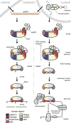
Figure 1
Figure 1
The siRNA (left) and miRNA (right) pathways of RNA interference. Protein domain architecture is approximated in the cartoons here, and domain coloring is maintained in subsequent figures. For clarity, one dsRBD has been omitted from each of the Dicer and microprocessor cartoons.
Figure 2
Figure 2
The microprocessor complex. (a) Domain structures of the human microprocessor constituents Drosha and DGCR8. Regions rich in proline, arginine and serine, or tryptophan are labeled. (b) The crystal structure of the DGCR8 core (protein data bank (PDB) ID: 2YT4). Models of dsRNA are oriented based on typical dsRBD binding and show that the DGCR8 core cannot use both dsRBDs to bind a single pre-miRNA unless extensive deformation of the helix occurs.
Figure 3
Figure 3
The Dicer family’s diverse architecture. (a) Domain structures of Dicer enzymes from humans, Giardia intestinalis (Gin), or K. polysporus (Kpo). The domain of unknown function (DUF) and anticipated ruler domain are indicated for the human enzyme. K. polysporus Dicer bears a single RNase III domain and an N-terminal domain (NTD) that mediates dimerization. (b) Dicer’s measurement and cleavage of pre-miRNA as illustrated by the G. intestinalis crystal structure (PDB ID: 2FFL). Purple spheres represent erbium atoms present in the crystal, which reflect the position of the Mg2+ ions critical to RNase III enzyme catalysis. In this model, a dsRNA’s 2 nt 3′-terminal overhang is docked into Dicer’s PAZ domain, orienting it for two cleavage events 65 Å away at the active site, generating a 25 nt duplex. (c) The inside-out mechanism of K. polysporus Dicer (PDB ID: 3RV0). This enzyme lacks a PAZ domain and uses neighboring molecules to measure its product. This model depicts two homodimers of the enzyme, each bearing a single RNase III domain, contacting each other to measure a 23 nt product. The resulting dsRNA will originate from the center of the substrate duplex, in contrast to the end-derived products generated by PAZ-containing Dicers. (d) The global architecture of human Dicer. Homologous domains have been docked into a segmented EM map (PDB IDs: 4A36, 2KOU, 2FFL, and 3C4T). The helicase domain resembles a clamp and is optimally oriented to guide an incoming dsRNA substrate (speculatively modeled here in black) towards the RNase IIIa/b active center and PAZ domain. Relative to the G. intestinalis enzyme, a ruler domain is inserted and the PAZ domain is reoriented with respect to the RNase III catalytic center. These changes likely relate to the fact that human Dicer products are 4 nt shorter than those of G. intestinalis.
Figure 4
Figure 4
The structure of dsRNA-binding proteins. (a) The domain structure of human TRPB, a typical dsRBP of the RNAi pathway. The well-folded dsRBDs are separated by flexible ~70 aa linkers, lending the protein a “beads on a string” quality. The first two domains bind dsRNA while the third instead binds to Dicer. (b) The crystal structure of TRBP’s second dsRBD in complex with dsRNA (PDB ID: 3ADL). The protein uses three interfaces to recognize successive portions of minor, major, and minor groove along one face of a helix.
Figure 5
Figure 5
Snapshots of RISC in action. (a) The domain structure of Argonaute is relatively well conserved in the AGO clade. PIWI clade proteins lack the N and PAZ domains. (b) The crystal structure of human Ago2 bound to an RNA guide strand (PDB ID: 4EI1). The seed region (nt 2–6) is pre-arranged in A-form geometry while the downstream portions of strand cannot be modeled due to disorder, as is typical in the absence of a target strand. Helix 7 is observed to rest against guide strand bases 6 and 7, distorting their geometry and providing an apparent barrier to target binding that is presumably circumvented via a conformational change. (c) The PAZ domain of human Ago2 recognizes the 3′-terminal 2 nt overhang typical of helices involved in RNAi (PDB ID: 1SI3). Conserved residues contacting the 3′-terminus are represented as sticks and labeled. A hydrophobic pocket receives the terminal nucleobase. Nucleotides from the 5′-terminus can be seen in the bottom right, making only slight contact with the PAZ domain. (d) The MID domain is responsible for recognition of a phosphorylated 5′-terminus (PDB ID: 3LUJ). This UMP-bound crystal structure reveals the polar contacts that drive phosphate recognition as well as elucidating the base-specific contacts that grant the MID domain its preference for a 5′-terminal A or U. (e) A pair of T. thermophilus crystal structures illustrate a conformational change that results upon extensive base pairing between guide and target (PDB ID: left, 3DLH; right, 3HM9). RISC binding to a 19 nt target strand allows formation of an A-form helix that induces release of the guide strand’s 3′-terminus from the PAZ domain along with a drastic opening of the two Argonaute lobes. The target-bound model shows that the N domain blocks formation of a longer helix. (f) In the catalytic center of T. thermophilus Argonaute’s PIWI domain, three aspartic acid residues coordinate a pair of Mg2+ ions for cleavage of the target strand, shown as white sticks with nucleotides numbered in gray (PDB ID: 3HVR). (g) In T. thermophilus the target-induced conformation change also involves reorientation of L2, which contains a glutamic acid residue. If target binding is incomplete or absent, the inactive state is sampled (yellow). In the target-bound state (brown), the glutamic acid is deposited adjacent to the aforementioned catalytic triad, completing a tetrad typical of RNase H enzymes (PDB ID: bound to a 12 nt target and inactive, 3HO1; bound to a 19 nt target and active, 3HM9). (h) The pre-ordered catalytic tetrad as observed in the absence of target strand in K. polysporus Argonaute (PDB ID: 4F1N). Residue 1013 here corresponds to T. thermophilus residue 512. (i) The PIWI domain of human Ago2 bears two tryptophan binding sites that complement the side chain geometry expected from a GW protein binding partner (PDB ID: 4EI1). Free tryptophan was present in the crystallization conditions and the pair of bound amino acids is represented as sticks.
Figure 6
Figure 6
Molecular assemblies in RNAi. (a) The interface between the C-terminal portion of PABPC1 and the DUF of TNRC6C, a GW protein (PDB ID: 2X04). Interacting side chains are shown as sticks. (b) The human endonuclease C3PO comprises hetero- and homodimers of Trax (yellow) and/or Translin (green or teal). The teal-colored Translin homodimer in the foreground is partially cut away to reveal the side chains of Trax’s active site, rendered as sticks. In this crystal structure, the complex adopts a hollow, egg-like shape with no obvious means for its passenger strand substrate to access the enclosed active sites (PDB ID: 3PJA).

Similar articles

Cited by

References

    1. Bazzini AA, Lee MT, Giraldez AJ. Ribosome profiling shows that miR-430 reduces translation before causing mRNA decay in zebrafish. Science. 2012;336(6078):233–237. - PMC - PubMed
    1. Bellemer C, Bortolin-Cavaillé M-L, Schmidt U, Jensen SMR, Kjems J, Bertrand E, Cavaillé J. Microprocessor dynamics and interactions at endogenous imprinted C19MC microRNA genes. J. Cell. Sci. 2012;125(Pt 11):2709–2720. - PubMed
    1. Berezikov E. Nat. Rev. Genet. 12. Vol. 12. Nature Publishing Group; 2011. Evolution of microRNA diversity and regulation in animals; pp. 846–860. - PubMed
    1. Bernstein E, Caudy AA, Hammond SM, Hannon GJ. Role for a bidentate ribonuclease in the initiation step of RNA interference. Nature. 2001;409(6818):363–366. - PubMed
    1. Boland A, Huntzinger E, Schmidt S, Izaurralde E, Weichenrieder O. Crystal structure of the MID-PIWI lobe of a eukaryotic Argonaute protein. Proc. Natl. Acad. Sci. U.S.A. 2011;108(26):10466–10471. - PMC - PubMed

Publication types