Review
. 2013 Apr 2;17(4):491-506.
doi: 10.1016/j.cmet.2013.03.002.

Mitochondrial dynamics in the regulation of nutrient utilization and energy expenditure

Affiliations
Review

Mitochondrial dynamics in the regulation of nutrient utilization and energy expenditure

Marc Liesa et al. Cell Metab. .

Abstract

Mitochondrial fusion, fission, and mitophagy form an essential axis of mitochondrial quality control. However, quality control might not be the only task carried out by mitochondrial dynamics. Recent studies link mitochondrial dynamics to the balance between energy demand and nutrient supply, suggesting changes in mitochondrial architecture as a mechanism for bioenergetic adaptation to metabolic demands. By favoring either connected or fragmented architectures, mitochondrial dynamics regulates bioenergetic efficiency and energy expenditure. Placement of bioenergetic adaptation and quality control as competing tasks of mitochondrial dynamics might provide a new mechanism, linking excess nutrient environment to progressive mitochondrial dysfunction, common to age-related diseases.


Figures

Figure 1
Figure 1. Regulation of cellular bioenergetic efficiency under conditions of nutrient excess
In the balanced state fuel/nutrient “supply” is sufficient to sustain energy (ATP) “demand”. Under this condition “waste” or inefficiency in the form of heat is minor. Nutrient excess, characterized by “excessive supply” in the absence of a parallel increase in “demand” represent a situation were the energy required to satisfy ATP demand is lower than the available energy. This is compensated for by adding an energy sink that does not involve ATP synthesis. This component is inefficiency/waste in the form of heat. The major mechanism for inefficiency/waste in the form of heat is mitochondrial proton “leak”. This mechanism can slow down nutrient accumulation and prevent the development of reductive stress (accumulation of NADH), and ROS production.
Figure 2
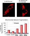
Figure 2. Nutrient excess induces mitochondrial fragmentation in the beta-cell
INS-1 cells treated for 4 hours with different concentrations of glucose and fatty acids (palmitate conjugated to BSA). The upper panel show representative images of INS-1 cells cultured with physiological glucose concentrations (5mM Glucose) and with high glucose and high fatty acids concentrations (20mM Glucose+ 0.4 mM Palmitate BSA at 4:1 ratio) for 4 hours. Mitochondria are shown in red and were stained with DsRed targeted to the mitochondria. Cells exposed to high levels of nutrients (20mM Glucose+ 0.4 mM Palmitate) show fragmentation and the formation of spherical mitochondria (ball-shape), whereas mitochondria with 5mM Glucose appear tubular. The bar graph shows the percentage of cells with fragmented mitochondria after 4 hours incubation with different concentrations of glucose and palmitate (in mM). Note the additive effect of glucose and fatty acids causing fragmentation. See Molina et al., 2009 for more details.
Figure 3
Figure 3. The balance of energy supply/demand is associated with corresponding changes to mitochondrial architecture and to bioenergetic efficiency
Physiological processes associated with increased energy demand and decreased energy supply, such as acute stress, starvation, G1/S phase, are characterized by mitochondrial elongation and by respiration coupled to ATP synthesis. On the other hand, physiological processes associated with decreased energy demand and increased supply (high levels of nutrients, obesity and diabetes…) are associated with mitochondrial fragmentation and decreased coupling (associated with heat generation or decreased mitochondrial function).
Figure 4
Figure 4. Mitochondrial fragmentation induced by nutrient excess in the beta cell is caused by decreased mitochondrial fusion
Mitochondrial fusion activity was quantified using mitochondrial matrix-targeted photoactivatable GFP (mtPAGFP, green). A portion of the mitochondrial population within a cell is labeled by laser photo-conversion and the sharing of the photo-converted molecules across the mitochondrial population through fusion events is monitored. Over 55 minutes the majority of mitochondria acquire photoconverted PA-GFP molecules. As a result of the dilution of the signal across the cell the intensity is diminished. TMRE (red) labeling was used to visualize the entire mitochondrial population. Right panels: INS-1 cells expressing mtPAGFP exposed to nutrient overload (20 mM Glucose + 0.4 mM Palmitate-BSA) for 4 hours show a dramatic reduction in fusion rates, as shown by the lack of mtPAGFP sharing with other mitochondria. Note that due to the lack of fusion the mtPAGFP intensity in the labeled mitochondria remains unchanged. Left Panels: INS-1 control cells (5 mM Glucose) present almost all mitochondria labeled with mtPAGFP 55 min after photoactivation. The images are adapted from Molina et al., 2009 with permission.
Figure 5
Figure 5. The life cycle of mitochondria and its regulation by nutrient availability
A) The life cycle of the mitochondria. The cycle is characterized by fusion and fission events. Fusion generates a network in which components of the two mitochondria are mixed and reorganized (1). Fission that follows within minutes splits fused mitochondria into two daughter mitochondria with disparate membrane potential (2). The daughter with the higher membrane potential is the first to return to the cycle of fusion/fission, while the daughter with more depolarized membrane potential will remain solitary until its membrane potential recovers (3). If membrane potential remains depolarized, this mitochondrion will loose its ability to fuse and become part of the pre-autophagic pool characterized by solitary, depolarized mitochondria (4). With a delay of 1–3 hours, these mitochondria are eliminated by autophagy (5). B) Changes to nutrient availability and energy demand can divert mitochondria from the life cycle and extend their stay in the post fusion state (elongation) or the post fission state (fragmentation). Elongation of mitochondria is a result of increased fusion or decreased fission activity (top section). This is typical for states of increased energy efficiency (starvation, acute stress, senescence). Shortening of mitochondria is a result of decreased fusion activity or increased fission activity (bottom section). This is typical for states of reduced bioenergetic efficiency (increased respiratory leak). Since bioenergetic adaptation high energy supply requires the arrest of the mitochondria life cycle, extended exposure to excess nutrient environment is expected to impact quality control, a condition that will contribute to reduced longevity.

Similar articles

Cited by

References

    1. Affourtit C, Brand MD. Stronger control of ATP/ADP by proton leak in pancreatic beta-cells than skeletal muscle mitochondria. Biochem J. 2006;393:151–159. - PMC - PubMed
    1. Almind K, Manieri M, Sivitz WI, Cinti S, Kahn CR. Ectopic brown adipose tissue in muscle provides a mechanism for differences in risk of metabolic syndrome in mice. Proc Natl Acad Sci U S A. 2007;104:2366–2371. - PMC - PubMed
    1. Aquila H, Link TA, Klingenberg M. The uncoupling protein from brown fat mitochondria is related to the mitochondrial ADP/ATP carrier. Analysis of sequence homologies and of folding of the protein in the membrane. EMBO J. 1985;4:2369–2376. - PMC - PubMed
    1. Asami DK, McDonald RB, Hagopian K, Horwitz BA, Warman D, Hsiao A, Warden C, Ramsey JJ. Effect of aging, caloric restriction, and uncoupling protein 3 (UCP3) on mitochondrial proton leak in mice. Exp Gerontol. 2008;43:1069–1076. - PMC - PubMed
    1. Ashcroft FM, Harrison DE, Ashcroft SJ. Glucose induces closure of single potassium channels in isolated rat pancreatic beta-cells. Nature. 1984;312:446–448. - PubMed

Publication types

Substances