. 2014 Mar 6;33(10):1258-64.
doi: 10.1038/onc.2013.71. Epub 2013 Mar 18.

Epstein-Barr virus maintains lymphomas via its miRNAs

Affiliations

Epstein-Barr virus maintains lymphomas via its miRNAs

D T Vereide et al. Oncogene. .

Abstract

Epstein-Barr virus (EBV) has evolved exquisite controls over its host cells, human B lymphocytes, not only directing these cells during latency to proliferate and thereby expand the pool of infected cells, but also to survive and thereby persist for the lifetime of the infected individual. Although these activities ensure the virus is successful, they also make the virus oncogenic, particularly when infected people are immunosuppressed. Here we show, strikingly, that one set of EBV's microRNAs (miRNAs) both sustain Burkitt's lymphoma (BL) cells in the absence of other viral oncogenes and promote the transformation of primary B lymphocytes. BL cells were engineered to lose EBV and found to die by apoptosis and could be rescued by constitutively expressing viral miRNAs in them. Two of these EBV miRNAs were found to target caspase 3 to inhibit apoptosis at physiological concentrations.


Conflict of interest statement

Conflict of Interest

The authors declare no conflict of interest.

Figures

Figure 1
Figure 1. Ectopic expression of BART miRNAs partially complements the loss of EBV in BL cells
(a) The population growth of S1-1 cells was measured over time as EBV was evicted following induction of dominant negative EBNA1 (dnEBNA1) at day 0 with doxycycline. The average of three independent experiments, each with one technical replicate ± SD is shown. * p=0.05, one-sided Wilcoxon signed rank test, comparing total number of cell doublings. (b) Cells with or without exogenously introduced BART miRNAs plus or minus induction of dnEBNA1 to evict EBV were assayed for global cell death by staining cells with trypan blue and examining cell morphology. For each data point at least 600 cells were analyzed. The average of three independent experiments ± SD (at least 200 cells per experiment) is shown. (c) Cells as treated in B were assayed for the induction of apoptosis by detecting the activation of caspases (Caspase-Glo 3/7, Promega). Signals were normalized to homeostatic levels (cells transduced with an empty vector in which dnEBNA1 is not induced) which were arbitrarily set to one. The average of three independent experiments ± SD is shown. (d) Cells were cultured for 20 days with or without induction of dnEBNA1 and then scored for the presence of viral DNA by FISH analysis. For each experiment, at least 200 cells were counted per condition. The average percentage of EBV-negative cells ± SD from three independent experiments is shown. (e) Real-time PCR measurements of the levels of two BART miRNAs, BART 1-5p (left graph) and BART 7 (right graph), were made 20 days after induction of dnEBNA1 in S1-1 cells. The average number of miRNA molecules per 10 picograms of total RNA ± SD is shown from three independent experiments.
Figure 2
Figure 2. Identification of BART miRNA targets
(a) RNA-seq was performed on total mRNA and RISC immunoprecipitations from S1-1 cells. The top 10 candidate genes (ranked by their expression level in the S1-1 cells) are listed with their detected expression values for each condition. As a control, the expression values of the BART5 target BBC3 (PUMA) are also listed. BART5 is inefficiently expressed from the BART retroviral vectors. (b) Reporter assays were conducted with constructs encoding the 3′-UTRs of CASP3 and IPO7 downstream of luciferase. The predicted target sites and corresponding miRNA seed sequences are shown. (c) 293 cells were transfected with luciferase vectors and mature synthetic miRNA, and the luciferase activity was normalized as described in the Materials and Methods. Data are the average of three independent experiments ± s.d. (*p<0.05, Wilcoxon rank sum test). (d) The vectors were introduced into EBV-positive or EBV-negative Oku-BL cells, and the normalized luciferase activity in the EBV-negative cells was set to 100%. Data are the average of six independent experiments ± s.d. (**p =0.037, ***p=0.006, Wilcoxon rank sum test). (e) Importin-7 and caspase-3 levels from cell lysates of S1-1 cells treated as in Figure 1 were measured by Western blot. Importin-7 and caspase-3 levels were normalized to alpha-tubulin levels, and then compared to the EBV depleted cells (cells transduced with empty vectors, dnEBNA1 turned on) whose normalized level was arbitrarily set to 100%. Data are the average of two independent experiments ± s.d. The blot images are given in Supplementary Figure 5.
Figure 3
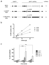
Figure 3. BART miRNAs promote the transformation of primary human B cells
(A) A simplified diagram of the pre-miRNAs of the three recombinant viruses studied is shown. The upper diagram represents 2089, which is the recombinant version of the prototypic EBV strain B95-8. Closed circles represent encoded pre-miRNAs; open circles represent deleted pre-miRNAs. EBV field strains other than the reference strain B95.8 encode up to 25 pre-miRNAs, which result in four mature BHRF1 miRNAs and 40 BART miRNAs. Two reconstituted EBV mutants that ectopically express the full set of 40 BART miRNAs were assembled from sub-genomic fragments as described . To construct the mutant EBV +mirBART (4080), the BART miRNA locus was inserted into the BALF1 gene of EBV, where it is expressed under the control of the CMV immediate early promoter. To construct the mutant EBV +mirBARTbis (4888), the assembled BART miRNA locus was inserted into the prokaryotic plasmid backbone of the prototypic 2089 strain, where it is expressed from the composite CAG promoter . (B) Titration of virus stocks. 1×10⁵ Raji cells were infected with different volumes of virus stocks as shown. Three days post infection, the percentage of GFP-positive Raji cells was determined on a FACS machine, calculated and plotted. The virus stocks 2089, 4080, and 4888 contained 7.1×10⁴±1×10⁴, 1.5×10⁵±4.6×10³, and 1×10⁵±9.3×10³ green Raji units [GRU]/ml, respectively. (C) Primary human B cells isolated from adenoids from five different donors were infected with the three virus stocks at a multiplicity of infection of 1×10⁻³, incubated for 18 hours, and seeded in fresh medium at a density of 1.5×10⁵ cells per ml. At the days indicated the cells were harvested and their proliferation analyzed by FACS. To determine the absolute number of cells counted, a volume standard was added prior to FACS analysis as described , . To correct for variations between donors’ B cells, the raw cell counts were normalized to those infected with 2089 at day 14 (whose value was arbitrarily set to 100%). The average of five different donors ± SD is shown. p-values (p<0.001 ***, p<0.0001 ****) for the time points are indicated (two way ANOVA linked to Bonferroni post-tests; www.graphpad.com, Prism vers. 5.0).

Similar articles

Cited by

References

    1. Nanbo A, Sugden A, Sugden B. The coupling of synthesis and partitioning of EBV’s plasmid replicon is revealed in live cells. EMBO J. 2007;26:4252–62. - PMC - PubMed
    1. Vereide D, Sugden B. Insights into the evolution of lymphomas induced by Epstein-Barr virus. Advances in cancer research. 2010;108:1–19. - PubMed
    1. Vereide DT, Sugden B. Lymphomas differ in their dependence on Epstein-Barr virus. Blood. 2011;117:1977–85. - PMC - PubMed
    1. Kirchmaier AL, Sugden B. Dominant-negative inhibitors of EBNA-1 of Epstein-Barr virus. J Virol. 1997;71:1766–75. - PMC - PubMed
    1. Lee DY, Sugden B. The LMP1 oncogene of EBV activates PERK and the unfolded protein response to drive its own synthesis. Blood. 2008;111:2280–9. - PMC - PubMed

Publication types

MeSH terms