. 2013;8(2):e56265.
doi: 10.1371/journal.pone.0056265. Epub 2013 Feb 18.

Multiple cationic amphiphiles induce a Niemann-Pick C phenotype and inhibit Ebola virus entry and infection

Affiliations

Multiple cationic amphiphiles induce a Niemann-Pick C phenotype and inhibit Ebola virus entry and infection

Charles J Shoemaker et al. PLoS One. 2013.

Erratum in

  • PLoS One. 2013;8(10). doi:10.1371/annotation/76780c06-ac81-48a3-8cce-509da6858fe5

Abstract

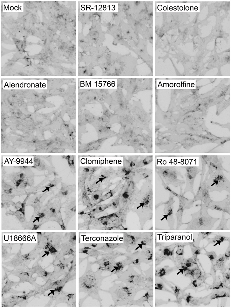
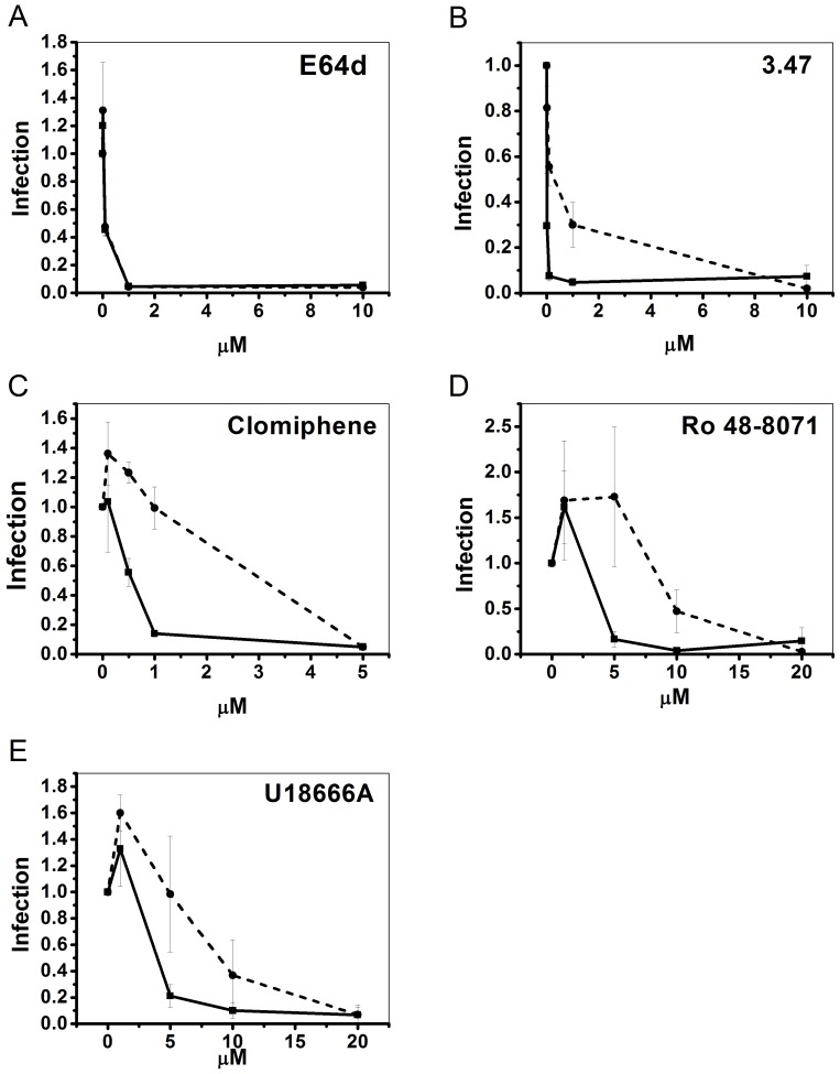
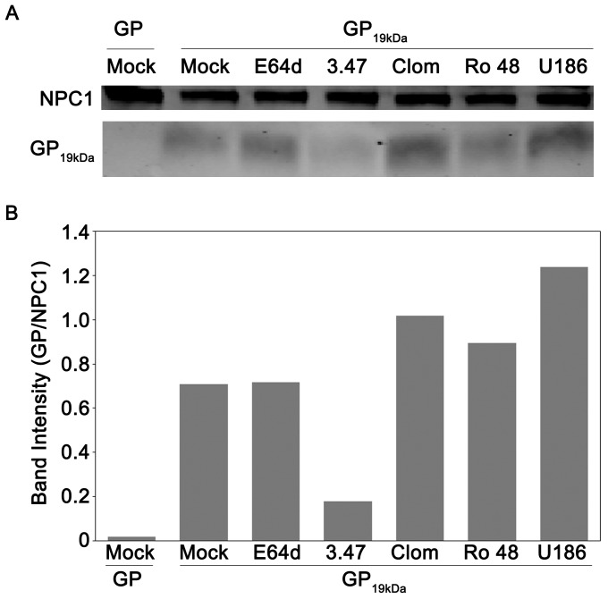
Ebola virus (EBOV) is an enveloped RNA virus that causes hemorrhagic fever in humans and non-human primates. Infection requires internalization from the cell surface and trafficking to a late endocytic compartment, where viral fusion occurs, providing a conduit for the viral genome to enter the cytoplasm and initiate replication. In a concurrent study, we identified clomiphene as a potent inhibitor of EBOV entry. Here, we screened eleven inhibitors that target the same biosynthetic pathway as clomiphene. From this screen we identified six compounds, including U18666A, that block EBOV infection (IC(50) 1.6 to 8.0 µM) at a late stage of entry. Intriguingly, all six are cationic amphiphiles that share additional chemical features. U18666A induces phenotypes, including cholesterol accumulation in endosomes, associated with defects in Niemann-Pick C1 protein (NPC1), a late endosomal and lysosomal protein required for EBOV entry. We tested and found that all six EBOV entry inhibitors from our screen induced cholesterol accumulation. We further showed that higher concentrations of cationic amphiphiles are required to inhibit EBOV entry into cells that overexpress NPC1 than parental cells, supporting the contention that they inhibit EBOV entry in an NPC1-dependent manner. A previously reported inhibitor, compound 3.47, inhibits EBOV entry by blocking binding of the EBOV glycoprotein to NPC1. None of the cationic amphiphiles tested had this effect. Hence, multiple cationic amphiphiles (including several FDA approved agents) inhibit EBOV entry in an NPC1-dependent fashion, but by a mechanism distinct from that of compound 3.47. Our findings suggest that there are minimally two ways of perturbing NPC1-dependent pathways that can block EBOV entry, increasing the attractiveness of NPC1 as an anti-filoviral therapeutic target.


Conflict of interest statement

Competing Interests: Lisa M. Johansen is and Hassan Pajouhesh was an employee of Zalicus, Inc. This does not alter the authors’ adherence to all the PLOS ONE policies on sharing data and materials.

Figures

Figure 1
Figure 1. Effects of Sterol Pathway Inhibitors on EBOV Infection.
Dose response curves for the indicated sterol pathway inhibitors are shown. The compounds were evaluated, in parallel at the indicated concentrations, for their ability to inhibit EBOV infection (black) and for inhibition of cell proliferation (gray) in Vero cells. The maximal % inhibition and the IC50 (µM) for their effects on EBOV infection are indicated. Data for clomiphene are presented in Johansen, et al. (manuscript in preparation).
Figure 2
Figure 2. Effects of sterol pathway inhibitors on EBOV VLP entry.
SNB19 cells were pretreated with inhibitor for 1 hr, and EBOV VLP-GPΔ was then bound by spinfection at 4°C for 1 hr. Cells were then washed in media with inhibitor, incubated at 37°C for 3 hr (in inhibitor), and then processed for VLP entry as described in the Materials and Methods section. Compounds were tested in multiple (n) experiments (each in triplicate) at either the highest concentration under which no toxicity was observed (for compounds that did not inhibit infection), or at the concentration that resulted in maximum inhibition of EBOV infection (Fig. 1) with minimal toxicity: SR 12813 (5 µM), (n = 4), colestolone (10 µM), (n = 5), alendronate (20 µM), (n = 3), BM 15766 (2 µM), (n = 4), amorolfine (6 µM), (n = 4), AY-9944 (5 µM), (n = 3), clomiphene (5 µM), (n = 9), Ro 48-8071 (5 µM, (n = 6), U18666A (5 µM), (n = 7), terconazole (10 µM), (n = 4), and triparanol (5 µM), (n = 3). Error bars represent standard error: * (P<2.96×10⁻³) or ** (P<6.14×10⁻⁵). Dashed line represents the observed threshold for entry inhibition needed to observe corresponding inhibition of live EBOV infection (Fig. 1). As indicated in the key, colors denote classes of molecules.
Figure 3
Figure 3. Ro 48-8071 inhibits EBOV entry at a post internalization step and does not inhibit endosome acidification or cathepsin levels.
Effects of Ro 48-8071 (indicated concentration in A, 5 µM in B-E) on: (A) VLP-GPΔ and VLP-G entry; one representative of two experiments (done in triplicate). (B) VLP-GPΔ and VLP-LCMV entry; one representative of three experiments (done in duplicate). (C) VLP-GPΔ internalization and entry; 50 µM EIPA was used as the positive control for an inhibitor of EBOV internalization ; 10 µM EIPA was used as the control for the entry assay (50 µM EIPA caused high background fluorescence in the entry assay); one representative of two experiments (done in triplicate). (D) Low endosomal pH was detected by incubating cells with Lysotracker Red; 10 mM NH4Cl was used as the control for pH neutralization; representative images from multiple coverslips from a single experiment. (E) Cathepsin B, L, and combined B/L activity; 10 µM E64d was used as the positive control for inhibition of cysteine protease activity; results from a single experiment performed in duplicate. In all assays, SNB19 cells were pre-treated with the indicated concentration of inhibitor for 1 hr at 37°C, and inhibitors were maintained throughout the assays. Error bars represent standard deviation from the mean of mock-treated samples: * (P<.01), ** (P<.001), or *** (P<.0001).
Figure 4
Figure 4. U18666A inhibits EBOV entry at a post internalization step and does not inhibit endosome acidification or cathepsin levels.
Effects of U18666A (indicated concentration in A, 5 µM in B-E) on: (A) VLP-GPΔ and VLP-G entry; one representative of two experiments (done in triplicate). (B) VLP-GPΔ and VLP-LCMV entry; one representative of three experiments (done in duplicate). (C) VLP-GPΔ internalization and entry (controls as in Fig. 3C); one representative of two experiments (done in triplicate). (D) Endosomal pH detected by Lysotracker Red; 10 mM NH4Cl was used as the control for pH neutralization; representative images from multiple coverslips from a single experiment (E) Cathepsin B, L, and combined B/L activity; E64d was used as the positive control as in Fig. 3E; results from a single experiment performed in duplicate. In all assays, SNB19 cells were pre-treated with the indicated concentration of inhibitor for 1 hr at 37°C, and inhibitors were maintained throughout the assays. Error bars represent standard deviation from the mean of mock-treated samples: * (P<.01), ** (P<.001), or *** (P<.0001).
Figure 5
Figure 5. CADs that strongly inhibit EBOV entry and infection cause cholesterol accumulation in LE/Lys.
SNB19 cells were treated for 21 hr with either DMSO or inhibitor (concentrations as in Fig. 2). Cells were then fixed, stained with filipin, and imaged on a fluorescence microscope. Images were inverted and uniformly adjusted for contrast and brightness. Representative images are shown. Arrows indicate sites of cholesterol accumulation. Each compound was tested at least 3 times, and scored (+/−) by a blind observer (Table 1).
Figure 6
Figure 6. CADs inhibit EBOV GP-mediated infection in an NPC1-dependent manner.
Parental CHO cells (−−−−) and stably overexpressing CHO NPC1 cells (− − −) were pre-treated with the indicated concentration of inhibitor for 1 hr at 37°C, and then infected with VSV-GPΔ for 18 hr in the continued presence of inhibitor. Each concentration of inhibitor was tested (in duplicate) in the following number of experiments: E64d (n = 2), compound 3.47 (n = 2), clomiphene (n = 3), Ro 48-8071 (n = 3), and U18666A (n = 4). Infection values were normalized to DMSO treated samples and averaged across experiments. Error bars represent standard error.
Figure 7
Figure 7. CADs do not disrupt the interaction of 19 kDa GP and NPC1.
(A) NPC1-FLAG-enriched LE/Lys membranes from CHO NPC1 cells were disrupted and then incubated with inhibitors for 30 min at RT: mock (4% DMSO), E64d (10 µM), compound 3.47 (13 µM), clomiphene (242 µM), Ro 48-8071 (174 µM), and U18666A (800 µM); each inhibitor was used at a concentration 100 fold over its IC50 for inhibition of infection. The samples were then incubated with 3 µg uncleaved (GP) or cleaved (GP19 kDa) EBOV GP ectodomains for 1 hr at RT. Samples were then lysed, and incubated overnight with anti-FLAG beads. Bound NPC1 and GP were then eluted from beads, and run on an SDS-PAGE gel. The gel was then transferred, blotted for both NPC1 and EBOV GP, and imaged for fluorescent signal. As predicted, uncleaved GP (∼130 kDa) did not co-precipitate with NPC1 , . (B) The intensities of the GP, GP19 kDa, and NPC1 bands from each sample of the blot shown in Fig. 7A were quantified and GP or GP19 kDa was normalized to its respective NPC1 band signal. The experiment was conducted four times with similar results, and a representative experiment is shown.
Figure 8
Figure 8. Models for how CADs May Block EBOV Entry.
(A) In the first model, the CADs (yellow star) interact directly with NPC1, but at a site distinct from the C loop of NPC1 (which binds GP19 kDa). Binding to NPC1 inhibits a second function of NPC1 (i.e. in addition to its role in binding GP19 kDa) that is critical for EBOV entry. (B) In the second model, the CADs intercalate into the LE/Lys membrane, indirectly inhibiting a second function of NPC1 that promotes EBOV entry. (C) In a final model, the CADs disrupt a target distinct from NPC1 that is critical for EBOV fusion with LE/Lys) and is regulated by NPC1. The target may be another LE/Lys protein (e.g. ASM) or a lipid of the LE/Lys membrane system. (See text for details.) Alternatively, the CADs may interfere with NPC1-dependent membrane trafficking , such that the virus is never found in an NPC1-containing compartment. In all of the models, the yellow star denotes a CAD and red in each middle image denotes the target molecule.

Similar articles

Cited by

References

    1. Feldmann H, Geisbert TW (2010) Ebola haemorrhagic fever. Lancet 377: 849–862. - PMC - PubMed
    1. Hoenen T, Groseth A, Falzarano D, Feldmann H (2006) Ebola virus: unravelling pathogenesis to combat a deadly disease. Trends Mol Med 12: 206–215. - PubMed
    1. Sullivan NJ, Martin JE, Graham BS, Nabel GJ (2009) Correlates of protective immunity for Ebola vaccines: implications for regulatory approval by the animal rule. Nat Rev Microbiol 7: 393–400. - PMC - PubMed
    1. Geisbert TW, Jahrling PB (1995) Differentiation of filoviruses by electron microscopy. Virus Res 39: 129–150. - PubMed
    1. Beniac DR, Melito PL, Devarennes SL, Hiebert SL, Rabb MJ, et al. (2012) The organisation of Ebola virus reveals a capacity for extensive, modular polyploidy. PLoS One 7: e29608. - PMC - PubMed

Publication types

MeSH terms