The cancer antigenome
- PMID: 23258224
- PMCID: PMC3553384
- DOI: 10.1038/emboj.2012.333
The cancer antigenome
Abstract
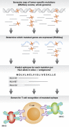
Cancer cells deviate from normal body cells in two immunologically important ways. First, tumour cells carry tens to hundreds of protein-changing mutations that are either responsible for cellular transformation or that have accumulated as mere passengers. Second, as a consequence of genetic and epigenetic alterations, tumour cells express a series of proteins that are normally not present or present at lower levels. These changes lead to the presentation of an altered repertoire of MHC class I-associated peptides. Importantly, while there is now strong clinical evidence that cytotoxic T-cell activity against such tumour-associated antigens can lead to cancer regression, at present we fail to understand which tumour-associated antigens form the prime targets in effective immunotherapies. Here, we describe how recent developments in cancer genomics will make it feasible to establish the repertoire of tumour-associated epitopes on a patient-specific basis. The elucidation of this 'cancer antigenome' will be valuable to reveal how clinically successful immunotherapies mediate their effect. Furthermore, the description of the cancer antigenome should form the basis of novel forms of personalized cancer immunotherapy.
Conflict of interest statement
The authors declare that they have no conflict of interest.
Figures


Similar articles
-
A new era for cancer immunotherapy based on the genes that encode cancer antigens.Immunity. 1999 Mar;10(3):281-7. doi: 10.1016/s1074-7613(00)80028-x. Immunity. 1999. PMID: 10204484 Review. No abstract available.
-
T-cell-receptor-like antibodies - generation, function and applications.Expert Rev Mol Med. 2012 Feb 24;14:e6. doi: 10.1017/erm.2012.2. Expert Rev Mol Med. 2012. PMID: 22361332 Review.
-
The potential of melanoma antigen expression in cancer therapy.Cancer Treat Rev. 1999 Aug;25(4):219-27. doi: 10.1053/ctrv.1999.0126. Cancer Treat Rev. 1999. PMID: 10448130 Review.
-
Personalized vaccines for cancer immunotherapy.Science. 2018 Mar 23;359(6382):1355-1360. doi: 10.1126/science.aar7112. Science. 2018. PMID: 29567706 Review.
-
Antigenic targets for renal cell carcinoma immunotherapy.Expert Opin Biol Ther. 2004 Nov;4(11):1791-801. doi: 10.1517/14712598.4.11.1791. Expert Opin Biol Ther. 2004. PMID: 15500407 Review.
Cited by
-
Characterization of the immunophenotypes and antigenomes of colorectal cancers reveals distinct tumor escape mechanisms and novel targets for immunotherapy.Genome Biol. 2015 Mar 31;16(1):64. doi: 10.1186/s13059-015-0620-6. Genome Biol. 2015. PMID: 25853550 Free PMC article.
-
T-Cell-Based Immunotherapy for Osteosarcoma: Challenges and Opportunities.Front Immunol. 2016 Sep 14;7:353. doi: 10.3389/fimmu.2016.00353. eCollection 2016. Front Immunol. 2016. PMID: 27683579 Free PMC article. Review.
-
Groundwork for AI: Enforcing a benchmark for neoantigen prediction in personalized cancer immunotherapy.Soc Stud Sci. 2023 Oct;53(5):787-810. doi: 10.1177/03063127231192857. Epub 2023 Aug 31. Soc Stud Sci. 2023. PMID: 37650579 Free PMC article.
-
HLA-binding properties of tumor neoepitopes in humans.Cancer Immunol Res. 2014 Jun;2(6):522-9. doi: 10.1158/2326-6066.CIR-13-0227. Epub 2014 Mar 3. Cancer Immunol Res. 2014. PMID: 24894089 Free PMC article. Review.
-
SLC45A2: A Melanoma Antigen with High Tumor Selectivity and Reduced Potential for Autoimmune Toxicity.Cancer Immunol Res. 2017 Aug;5(8):618-629. doi: 10.1158/2326-6066.CIR-17-0051. Epub 2017 Jun 19. Cancer Immunol Res. 2017. PMID: 28630054 Free PMC article.
References
-
- Altman JD, Moss PA, Goulder PJ, Barouch DH, McHeyzer-Williams MG, Bell JI, McMichael AJ, Davis MM (1996) Phenotypic analysis of antigen-specific T lymphocytes. Science 274: 94–96 - PubMed
-
- Andersen RS, Kvistborg P, Frosig TM, Pedersen NW, Lyngaa R, Bakker AH, Shu CJ, Straten P, Schumacher TN, Hadrup SR (2012a) Parallel detection of antigen-specific T cell responses by combinatorial encoding of MHC multimers. Nat Protoc 7: 891–902 - PubMed
-
- Andersen RS, Thrue CA, Junker N, Lyngaa R, Donia M, Ellebaek E, Svane IM, Schumacher TN, thor Straten P, Hadrup SR (2012b) Dissection of T-cell antigen specificity in human melanoma. Cancer Res 72: 1642–1650 - PubMed
-
- Bakker AH, Hoppes R, Linnemann C, Toebes M, Rodenko B, Berkers CR, Hadrup SR, van Esch WJ, Heemskerk MH, Ovaa H, Schumacher TN (2008) Conditional MHC class I ligands and peptide exchange technology for the human MHC gene products HLA-A1, -A3, -A11, and -B7. Proc Natl Acad Sci USA 105: 3825–3830 - PMC - PubMed
Publication types
MeSH terms
Substances
LinkOut - more resources
Full Text Sources
Other Literature Sources
Research Materials

