. 2013 Feb;19(2):230-42.
doi: 10.1261/rna.034926.112. Epub 2012 Dec 18.

MicroRNA-182-5p targets a network of genes involved in DNA repair

Affiliations

MicroRNA-182-5p targets a network of genes involved in DNA repair

Keerthana Krishnan et al. RNA. 2013 Feb.

Abstract

MicroRNAs are noncoding regulators of gene expression, which act by repressing protein translation and/or degrading mRNA. Many have been shown to drive tumorigenesis in cancer, but functional studies to understand their mode of action are typically limited to single-target genes. In this study, we use synthetic biotinylated miRNA to pull down endogenous targets of miR-182-5p. We identified more than 1000 genes as potential targets of miR-182-5p, most of which have a known function in pathways underlying tumor biology. Specifically, functional enrichment analysis identified components of both the DNA damage response pathway and cell cycle to be highly represented in this target cohort. Experimental validation confirmed that miR-182-5p-mediated disruption of the homologous recombination (HR) pathway is a consequence of its ability to target multiple components in that pathway. Although there is a strong enrichment for the cell cycle ontology, we do not see primary proliferative defects as a consequence of miR-182-5p overexpression. We highlight targets that could be responsible for miR-182-5p-mediated disruption of other biological processes attributed in the literature so far. Finally, we show that miR-182-5p is highly expressed in a panel of human breast cancer samples, highlighting its role as a potential oncomir in breast cancer.


Figures
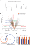
FIGURE 1.
FIGURE 1.
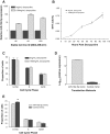
Identifying targets of miR-182-5p via biotin pull-down. (A) Hierarchical clustering of microarray data was performed using the plotSampleRelations function in the lumi package. Total vertical distance between samples indicates similarity. (B) A “volcano plot” showing the log2-transformed fold-change (mock/pull-down) versus the log10-transformed P-value for that fold-change for every gene detected above background in the microarray. (Blue) Genes that are targets validated by previous studies; (orange) genes predicted by TargetScan to be targets of miR-182-5p; (green) genes selected for validation using luciferase assays (D). There is an apparent enrichment of the targets in the pull-downs compared with the controls. (C) Venn diagram showing the overlap of genes between TargetScan-predicted targets of miR-182-5p (also expressed above background in HEK293Ts) and biotinylated miR-182-5p pull-down-predicted targets. This overlap is significantly more than expected by chance. (D) Dual luciferase assay used to validate CHEK2, SMARCD3, CDKN1B, and NFKBIB as targets of miR-182-5p. HEK293T cells were transiently cotransfected with 20 nM miR-182-5p or control mimic with a pmirGlo-luciferase construct containing the predicted binding site from the indicated target gene. Luciferase activity was normalized to Renilla activity; (*) P < 0.05 in a Student’s t-test. The data plotted are the mean and SEM of three independent biological replicates.
FIGURE 2.
FIGURE 2.
Overexpression of miR-182-5p does not induce a proliferative defect. (A) Expression of miR-182-5p as assessed by qRT-PCR in MDA-MB-231 cells (with low endogenous expression of miR-182-5p) stably transfected with miR-182-5p whose expression is induced in response to doxycycline. Shown here are three independent cell lines grown in the presence of 0 or 1000 ng/mL of doxycycline for 48 h. RNU6B was used as an endogenous control for normalization of expression. (B) MTT cell proliferation assays of MDA-MB-231 cells stably expressing miR-182-5p. The graph plots the mean and SEM of the previously studied stable cell lines grown with either 0 or 1000 ng/mL doxycycline. The induction of miR-182-5p does not affect the proliferation rates of MDA-MB-231 cells. (C) DNA profile analysis of MDA-MB-231 cells stably expressing miR-182-5p. The graph shows the mean and SEM of the percentage of cells in different cell cycle phases, as assessed by FACS. There was no significant difference between MDA-MB-231 cells expressing or not expressing miR-182-5p. (D) Expression of miR-182-5p in HeLa cells transiently transfected with miR-182-5p mimic or negative mimic control as assessed by qRT-PCR. RNU6B was used as an endogenous control for normalization of expression. (E) A graph showing the DNA profile analysis of HeLa cells transiently transfected with either miR-182-5p mimic or a control mimic. Shown is the mean and SEM of three independent biological replicates, each performed in technical triplicates. (*) P < 0.05 in a Student’s t-test (n = 3).
FIGURE 3.
FIGURE 3.
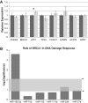
Enrichment of targets in the DNA damage response is specific to miR-182-5p overexpression. (A) Real-time PCR analysis to evaluate the relative mRNA levels of known DNA repair genes was performed in MDA-MB-231 cells transiently transfected with 20 nM miR-182-5p mimic or negative mimic. (*) Significant changes (P < 0.05). Data were normalized using HPRT as the internal control. The results shown are from three independent biological replicates, each performed in technical quadruplicates. (B) Functional enrichment analysis was performed using biotin-enriched targets of several miRNAs including miR-182-5p. As shown, the canonical pathway “Role of BRCA1 in DNA Damage Response” is highly significant in the miR-182-5p pull-down with its −log(significance) at least four standard deviations away (top arrow) from the mean −log(significance) (lower arrow) of the other miRNAs. This confirms that the enrichment seen for targets in the DNA damage response is miR-182-5p specific and not occurring by chance.
FIGURE 4.
FIGURE 4.
Overview of biotinylated miR-182-5p pull-down-predicted targets involved in the DNA damage response pathway genes involved in different canonical pathways underlying the DNA damage response, including the G2M cell cycle checkpoint, BRCA1-dependent HR-mediated pathway, cyclins and role of cell cycle regulators, role of CHK proteins, and ATM signaling. (Dark gray) miR-182-5p targets identified by biotin pull-down. These canonical pathways were found to be significantly enriched compared with 10 random gene lists of similar size using IPA (P-value < 0.02).
FIGURE 5.
FIGURE 5.
The effect of miR-182-5p on sensitivity to PARP inhibition in MDA-MB-231cells. (A) A schematic diagram depicting the principle of the PARP inhibitor assays performed here. Typically, cells with functional PARP repair single-strand breaks via the base excision repair pathway. In PARP inhibition assays, the inhibited PARP leads single-stranded breaks to decay to double-stranded breaks, which are then repaired via the homologous recombination (HR) pathway. However, in cells with a dysfunctional HR pathway (i.e., overexpressing miR-182-5p mimic leading to down-regulation of the DNA damage response [DDR] components), the DNA damage persists, leading to cell death. If a DDR target of an miR-182-5p target (e.g., BRCA1 or CHEK2 open reading frame [ORF]) is reintroduced, this would rescue any effect of miR-182-5p leading to increased cell survival. (B) To assess the level of miR-182-5p overexpression, we performed real-time analysis in MDA-MB-231 cells transiently transfected with 10 nM miR-182-5p or negative control mimic and find an ∼1000-fold increase from the base level expression in these breast cancer cells. (C) BRCA1 and CHEK2 mediate sensitivity to PARP1 inhibitor, induced by overexpression of miR-182-5p. Cells were transiently transfected with miR-182-5p mimic or control mimic (±ORF). Cell viability was assessed using the clonogenic survival assay in the presence of 4-amino-1,8-naphthalimide (ANI; PARP1 inhibitor) at the indicated concentrations on the x-axis. The data plotted are the mean and SEM of at least three independent biological replicates. (*) P < 0.05 in a Student’s t-test.
FIGURE 6.
FIGURE 6.
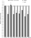
Dual luciferase assay used to validate targets of miR-182-5p. MDA-MB-231 cells were transiently cotransfected with 20 nM miR-182-5p or control mimic with a pmirGlo-luciferase construct containing the predicted binding site from the indicated target gene. Luciferase activity was normalized to Renilla activity; (**) P < 0.05 as indicated in a Student’s t-test. The data plotted are the mean and SEM of three independent biological replicates with three technical replicates.
FIGURE 7.
FIGURE 7.
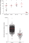
Expression analysis of miR-182-5p across tumor subtypes and normal tissue from human breast cancer patient samples. (Upper panel) Real-time PCR was performed in breast cancer patient samples of various subtypes to assess miR-182-5p expression, where lines and error bars represent the mean and SEM of miR-182-5p expression (normalized to RNU6B) across sample subtypes: Invasive Ductal Carcinoma of Triple Negative (n − 19); Her2+ (n = 4) or ER+/PR+ (n = 9) phenotype; Invasive Lobular Carcinomas (n = 3); and normal breast tissue (n = 6). (▪) Expression levels for individual patients; (*) indicate where the difference in the means subtype was (P < 0.0001) when compared with the normal samples. (Lower panel) Expression of miR-182-5p across several breast cancer patient samples as assessed by miRNA-seq. Shown in the x-axis is reads per million of miR-182-5p in data downloaded from the TCGA web portal, which included tumors (n = 741) and normal tissue (n = 99). (*) Where the difference in the means was (P < 0.0001) when compared with the normal samples.
FIGURE 8.
FIGURE 8.
Model showing the effect of miR-182-5p on the DNA repair pathway. When a cell undergoes double-strand breaks (DSB), “sensor” genes activate a signaling cascade including transducer and effector genes that leads to an efficient repair of the DNA damage. Under this model, miR-182-5p-mediated deregulation of the DNA damage response pathway (orange) typically results in impaired DNA repair with subsequent effects on the cell cycle, apoptosis, or genetic stability leading to tumorigenesis. We have also highlighted previously validated and novel targets that we have mechanistically shown to interact with miR-182-5p, belonging to the DNA damage response pathway.

Similar articles

Cited by

References

    1. Baek D, Villen J, Shin C, Camargo FD, Gygi SP, Bartel DP 2008. The impact of microRNAs on protein output. Nature 455: 64–71 - PMC - PubMed
    1. Bartel DP 2009. MicroRNAs: Target recognition and regulatory functions. Cell 136: 215–233 - PMC - PubMed
    1. Benjamini Y, Hochberg Y 1995. Controlling the false discovery rate: A practical and powerful approach to multiple testing. J R Stat Soc Series B Stat Methodol 57: 289–300
    1. Bentwich I 2005. Prediction and validation of microRNAs and their targets. FEBS Lett 579: 5904–5910 - PubMed
    1. Brennecke J, Stark A, Russell RB, Cohen SM 2005. Principles of microRNA–target recognition. PLoS Biol 3: e85 10.1371/journal.pbio.0030085 - DOI - PMC - PubMed

Publication types

MeSH terms

Associated data