. 2013 Feb 15;288(7):5198-209.
doi: 10.1074/jbc.M112.410274. Epub 2012 Dec 14.

Transforming growth factor-β directly induces p53-up-regulated modulator of apoptosis (PUMA) during the rapid induction of apoptosis in myc-driven B-cell lymphomas

Affiliations

Transforming growth factor-β directly induces p53-up-regulated modulator of apoptosis (PUMA) during the rapid induction of apoptosis in myc-driven B-cell lymphomas

Lindsay C Spender et al. J Biol Chem. .

Abstract

c-Myc transformed human Burkitt's lymphoma (BL) cells are highly sensitive to TGF-β-induced apoptosis. Previously we demonstrated that TGF-β-mediated cell death in BL cells is regulated via the mitochondrial intrinsic apoptosis pathway, which is dependent on the activation of BAX and/or BAK. TGF-β directly induces transcription of the BH3-only protein BIK and represses expression of the pro-survival factor BCL-X(L) but has no effect on the direct BAX/BAK "activators" BIM or BID (tBID). Here we show that TGF-β induces the BH3-only activator PUMA to aid induction of the intrinsic cell death pathway. TGF-β also induced PUMA in normal germinal center CD77-positive centroblasts isolated from human tonsil tissue. PUMA was a direct TGF-β target gene in B-cells, and we identify a putative Smad-binding region within the human PUMA promoter that recruits Smad3 and Smad4 in cells in response to TGF-β signaling. Constitutive activity of the isolated Smad-binding region in luciferase reporter assays was dependent on Smad consensus sequences and was partially dependent on endogenous TGF-β signaling and Smad4. Knockdown of PUMA in BL cells using lentiviral shRNA resulted in slower kinetics of the TGF-β-mediated apoptotic response. Analysis of Eμ-Myc cell lines demonstrated that c-myc-driven murine lymphomas are also sensitive to TGF-β-mediated apoptosis. Moreover, Puma(-/-) Eμ-Myc lines demonstrated significantly delayed kinetics of the apoptotic response when compared with wild type lymphomas. TGF-β therefore induces a polygenic response in Myc-driven lymphomas involving transcription of PUMA, which is necessary for the rapid induction of cell death.


Figures
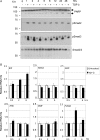
FIGURE 1.
FIGURE 1.
Transcripts of PUMA and BIK are elevated in TGF-β-treated Ramos BL cells. A, time course of TGF-β-induced apoptosis in human BL cells (Ramos). The cells were treated with 1 ng/ml TGF-β for the times indicated. Radioimmune precipitation assay lysates were analyzed by Western blotting for TGF-β signaling (phosphorylation of Smad2 and Smad3) and apoptosis (cleavage of PARP). A Western blot for total Smad2/3 is included as a loading control. B, quantitative real time qRT-PCR analysis of BH3-only members of the BCL-2 family in TGF-β-treated Ramos cells. The mean (± S.D.) fold increases relative to untreated cells (set at 1) are shown.
FIGURE 2.
FIGURE 2.
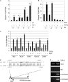
PUMA is induced by TGF-β in human BL cell lines and in normal tonsil CD77-positive B-cells. A, real time qRT-PCR analysis of PUMA in a panel of TGF-β responsive BL cell lines following 2 h of treatment. The mean (± S.D.) fold increase above background (set at 1 in untreated cells) is shown. B, Western blot analysis of radioimmune precipitation assay extracts from BL cells either untreated or treated with TGF-β for 2–8 h. Immunoreactive bands showing both the TGF-β-inducible PUMA-α (23 kDa) and PUMA-β isoforms are indicated. A Western blot for total Smad2/3 is included as a loading control. C and D, real time qRT-PCR analysis of RNA isolated from human primary CD77-positive tonsil B-cells treated with z-VAD.FMK and TGF-β for 2 h (C) and over an extended 24-h time course of TGF-β treatment (D). The mean (± S.D.) fold TGF-β-induced increase in normalized RNA expression compared with untreated cells (set at 1) is shown (C). The mean (± S.D.) relative levels of PUMA RNA normalized to 18 S are shown in D.
FIGURE 3.
FIGURE 3.
TGF-β directly regulates hPUMA transcription and induces recruitment of activated Smad complexes to the hPUMA promoter. A, real time qRT-PCR for PUMA expression in CA46 cells treated with or without TGF-β and the protein synthesis inhibitors cycloheximide and anisomycin (C+A) as indicated. The cells were pretreated with cycloheximide and anisomycin for 2 h, and TGF-β was added at 0 h. The mean (± S.D.) PUMA RNA expression levels relative to the 2-h untreated sample is shown (2-h untreated sample value was set at 1). The fold induction relative to the untreated sample at each time point is shown. B, real time qRT-PCR for PUMA expression in CA46 cells untreated or pretreated for 1 h with the transcription inhibitor actinomycin D (ActD), followed by a time course of TGF-β treatment. RNA expression is expressed relative to the levels detected in the 2-h untreated sample (assigned a value of 1). C, a panel of BL cell lines was pretreated for 30 min with 25 μm of the c-Myc inhibitor (Myc i) 10058-F4 followed by 2 h of TGF-β treatment (1 ng/ml). RNA was extracted and analyzed by qPCR for PUMA expression. Expression levels were normalized to the internal standard 18 S RNA and are shown as the mean (± S.D.) fold expression level relative to untreated controls (set at 1). D, sequence of a known SBR within the human BIK promoter (3) compared with a putative Smad-binding element within the human PUMA promoter. The putative PUMA SBR at position −1923 to −1885 was analyzed by ChIP using specific primers encompassing this region. E, ChIP assay for Smad recruitment to the endogenous PUMA promoter in CA46 cells cultured with and without TGF-β for 1 h. Input lanes are from 10% of samples used in the IPs performed with control IgG, Smad3, and Smad4 antibodies.
FIGURE 4.
FIGURE 4.
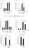
hPUMA promoter SBR luciferase reporter constructs. A and B, transient transfection of CA46 BL cells with control reporter vector pBV-luciferase (pBV-luc) or pBV containing the human PUMA promoter sequence spanning ∼2 kb upstream from the transcription start site (pBV-PUMA FragE (31)). Transfected cells were treated with 5 ng/ml TGF-β (A) or 10 μm SB-431542 (B) as indicated. C, wild type and mutant SBE reporter plasmids were generated by cloning 2× concatemerized oligonucleotides of the sequences shown into the pGL3-promoter vector (Promega). D and E, the wild type SBE (pGL3–2×WT SBE) and the mutant SBE (pGL3–2xMut SBE) reporter constructs were assayed following exogenous TGF-β addition (D) or addition of SB-431542 (E) as indicated. F and G, CAGA12 (F) or pGL3–2×WT SBE (G) constructs were transfected into CA46 cells with either nonsilencing vector (ns) or shRNA vector targeting Smad4. After 48 h, the transfections were left untreated or treated with 5 ng/ml TGF-β. Transfection results throughout are expressed as the means (± S.D.) (n = 3) luciferase reporter activity relative to untreated control samples (set at 1). All data shown are normalized to either co-transfected Renilla luciferase or β-galactosidase activity.
FIGURE 5.
FIGURE 5.
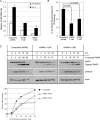
Knockdown of PUMA retards TGF-β-induced apoptosis in BL lines. A, independent stable cell lines expressing nonsilencing (Scrambled) or shRNAs targeted against PUMA (shRNA1 + 2#1 and shRNA1 + 2#2) were generated by lentiviral infection of BL30 cells. Real time qPCR was used to demonstrate the efficacy of knockdown of PUMA in stable BL30 cells in untreated cells and cells treated for 2 h with TGF-β. Mean relative amounts of PUMA RNA are expressed after normalization to 18 S rRNA levels. B, determination of apoptosis by propidium iodide (PI) staining and flow cytometry following 24 h TGF-β treatment of the cell lines described in A. Statistical analysis was performed using Student's t test. C, Western blot analysis of apoptosis (PARP) and TGF-β signaling (p-Smad2) in BL30 stable cell lines described in A and B during a time course of TGF-β treatment. A Western blot for actin is included as a loading control. The amount of cleaved PARP as a percentage of total PARP determined by densitometry is indicated above each lane and is summarized in the graph shown below.
FIGURE 6.
FIGURE 6.
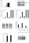
Murine Eμ-Myc lines are sensitive to TGF-β-induced apoptosis. A, murine Eμ-Myc cell lines were analyzed by annexin V staining and flow cytometry for their response to TGF-β treatment (24 h). B, Western blot analysis of lysates from Eμ-Myc cell lines showing cleavage of PARP following 12 h of treatment with TGF-β. C and D, murine Eμ-Myc line 4 (C) and line 8 (D) were treated for 24 h with TGF-β or etoposide (5 μg/ml) in the presence or absence of a TGF-β RI (ALK5) inhibitor, SB-431542 (SBi, 10 μm). Apoptosis induction was determined by annexin V staining and flow cytometry and is expressed as the mean (± S.D.) percentage induction above background levels. E and F, Eμ-Myc cell lines stably transfected with empty vector (pMIH) or Bcl-2 expressing vector (pMIH-Bcl-2) were analyzed by Western blot for Bcl-2 expression (E) and by annexin V staining (F) for susceptibility to TGF-β induced apoptosis following 24 h of treatment. G, wild type Eμ-Myc cell lines were left untreated or treated with TGF-β for 2 h and analyzed by qRT-PCR for Puma mRNA expression levels. The results are expressed as the mean fold mRNA level relative to the untreated control. H, Western blot analysis of PUMA expression in untreated (Con) wild type Eμ-Myc lines or lines treated for 6 h with TGF-β. A blot for tubulin is included as a loading control. DMSO, dimethyl sulfoxide.
FIGURE 7.
FIGURE 7.
Eμ-Myc cell lines derived from Puma−/− mice exhibit a delayed apoptotic response to TGF-β. A, kinetics of TGF-β-mediated Puma RNA induction in wild type Eμ-Myc lymphoma lines as determined by qPCR. B, early kinetics of apoptosis induction in wild type Eμ-Myc lymphoma lines as measured by annexin V staining. C, Western blot analysis of TGF-β signaling (pSmad2) and PUMA expression in WT and Puma null (Puma−/−) Eμ-Myc lymphoma lines in uninduced or induced conditions (6 h of treatment with TGF-β). A blot for total Smad2/3 is included as a loading control. D and E, apoptosis induction above background in lymphoma lines derived from WT (n = 4) and homozygous Puma null (Puma−/−) (n = 4) mice following 6 h (D) and 24 h (E) of treatment with TGF-β. Apoptosis induction above background was determined by annexin V staining and flow cytometry. The mean apoptosis values are shown as bars, and the p values were determined by Mann-Whitney statistical analysis.

Similar articles

Cited by

References

    1. Li M. O., Wan Y. Y., Sanjabi S., Robertson A. K., Flavell R. A. (2006) Transforming growth factor-β regulation of immune responses. Annu. Rev. Immunol. 24, 99–146 - PubMed
    1. Inman G. J., Allday M. J. (2000) Apoptosis induced by TGF-β1 in Burkitt's lymphoma cells is caspase 8 dependent but is death receptor independent. J. Immunol. 165, 2500–2510 - PubMed
    1. Spender L. C., O'Brien D. I., Simpson D., Dutt D., Gregory C. D., Allday M. J., Clark L. J., Inman G. J. (2009) TGF-β induces apoptosis in human B cells by transcriptional regulation of BIK and BCL-XL. Cell Death Differ. 16, 593–602 - PMC - PubMed
    1. Shi Y., Massagué J. (2003) Mechanisms of TGF-β signaling from cell membrane to the nucleus. Cell 113, 685–700 - PubMed
    1. Derynck R., Zhang Y. E. (2003) Smad-dependent and Smad-independent pathways in TGF-β family signalling. Nature 425, 577–584 - PubMed

Publication types

Substances