Frizzled and LRP5/6 receptors for Wnt/β-catenin signaling
- PMID: 23209147
- PMCID: PMC3504444
- DOI: 10.1101/cshperspect.a007880
Frizzled and LRP5/6 receptors for Wnt/β-catenin signaling
Abstract
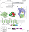
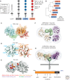
Frizzled and LRP5/6 are Wnt receptors that upon activation lead to stabilization of cytoplasmic β-catenin. In this study, we review the current knowledge of these two families of receptors, including their structures and interactions with Wnt proteins, and signaling mechanisms from receptor activation to the engagement of intracellular partners Dishevelled and Axin, and finally to the inhibition of β-catenin phosphorylation and ensuing β-catenin stabilization.
Figures


Similar articles
-
Oligomerization of Frizzled and LRP5/6 protein initiates intracellular signaling for the canonical WNT/β-catenin pathway.J Biol Chem. 2018 Dec 21;293(51):19710-19724. doi: 10.1074/jbc.RA118.004434. Epub 2018 Oct 25. J Biol Chem. 2018. PMID: 30361437 Free PMC article.
-
Structure and function of Norrin in assembly and activation of a Frizzled 4-Lrp5/6 complex.Genes Dev. 2013 Nov 1;27(21):2305-19. doi: 10.1101/gad.228544.113. Genes Dev. 2013. PMID: 24186977 Free PMC article.
-
β-Catenin-dependent pathway activation by both promiscuous "canonical" WNT3a-, and specific "noncanonical" WNT4- and WNT5a-FZD receptor combinations with strong differences in LRP5 and LRP6 dependency.Cell Signal. 2014 Feb;26(2):260-7. doi: 10.1016/j.cellsig.2013.11.021. Epub 2013 Nov 21. Cell Signal. 2014. PMID: 24269653
-
LRP5 and LRP6 in development and disease.Trends Endocrinol Metab. 2013 Jan;24(1):31-9. doi: 10.1016/j.tem.2012.10.003. Trends Endocrinol Metab. 2013. PMID: 23245947 Free PMC article. Review.
-
Dysregulation of Wnt/β-catenin signaling by protein kinases in hepatocellular carcinoma and its therapeutic application.Cancer Sci. 2021 May;112(5):1695-1706. doi: 10.1111/cas.14861. Epub 2021 Apr 6. Cancer Sci. 2021. PMID: 33605517 Free PMC article. Review.
Cited by
-
Crosstalk between the Hippo Pathway and the Wnt Pathway in Huntington's Disease and Other Neurodegenerative Disorders.Cells. 2022 Nov 16;11(22):3631. doi: 10.3390/cells11223631. Cells. 2022. PMID: 36429058 Free PMC article. Review.
-
Wnt/β-catenin signaling components and mechanisms in bone formation, homeostasis, and disease.Bone Res. 2024 Jul 10;12(1):39. doi: 10.1038/s41413-024-00342-8. Bone Res. 2024. PMID: 38987555 Free PMC article. Review.
-
2,4,5-Trimethoxyldalbergiquinol promotes osteoblastic differentiation and mineralization via the BMP and Wnt/β-catenin pathway.Cell Death Dis. 2015 Jul 16;6(7):e1819. doi: 10.1038/cddis.2015.185. Cell Death Dis. 2015. PMID: 26181200 Free PMC article.
-
AXIN2 germline testing in a French cohort validates pathogenic variants as a rare cause of predisposition to colorectal polyposis and cancer.Genes Chromosomes Cancer. 2023 Apr;62(4):210-222. doi: 10.1002/gcc.23112. Epub 2022 Dec 21. Genes Chromosomes Cancer. 2023. PMID: 36502525 Free PMC article.
-
Identification of therapeutic targets and prognostic biomarkers among frizzled family genes in glioma.Front Mol Biosci. 2023 Jan 9;9:1054614. doi: 10.3389/fmolb.2022.1054614. eCollection 2022. Front Mol Biosci. 2023. PMID: 36699699 Free PMC article.
References
-
- Adamska M, Larroux C, Adamski M, Green K, Lovas E, Koop D, Richards GS, Zwafink C, Degnan BM 2010. Structure and expression of conserved Wnt pathway components in the demosponge Amphimedon queenslandica. Evol Dev 12: 494–518 - PubMed
-
- Ahumada A, Slusarski DC, Liu X, Moon RT, Malbon CC, Wang HY 2002. Signaling of rat Frizzled-2 through phosphodiesterase and cyclic GMP. Science 298: 2006–2010 - PubMed
Publication types
MeSH terms
Substances
Grants and funding
LinkOut - more resources
Full Text Sources
Other Literature Sources
Molecular Biology Databases
