

The viral interferon regulatory factors of kaposi's sarcoma-associated herpesvirus differ in their inhibition of interferon activation mediated by toll-like receptor 3
- PMID: 23115281
- PMCID: PMC3554052
- DOI: 10.1128/JVI.01851-12
The viral interferon regulatory factors of kaposi's sarcoma-associated herpesvirus differ in their inhibition of interferon activation mediated by toll-like receptor 3
Abstract
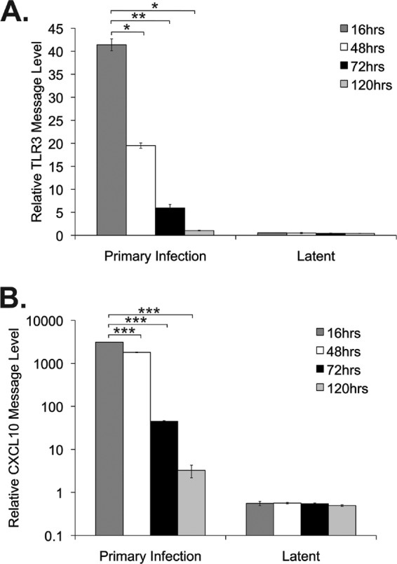
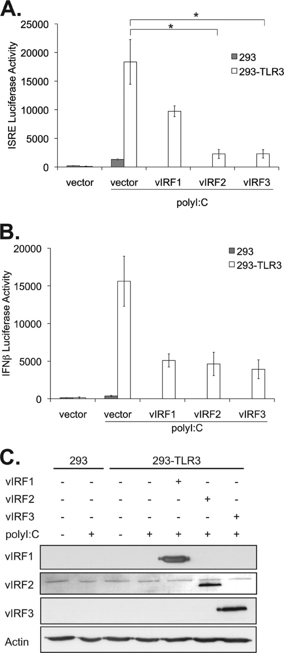
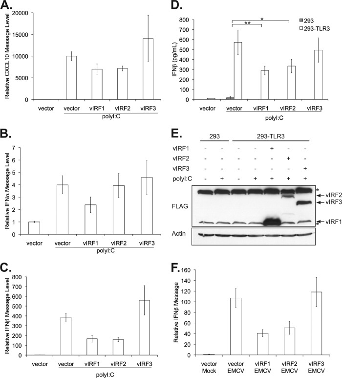
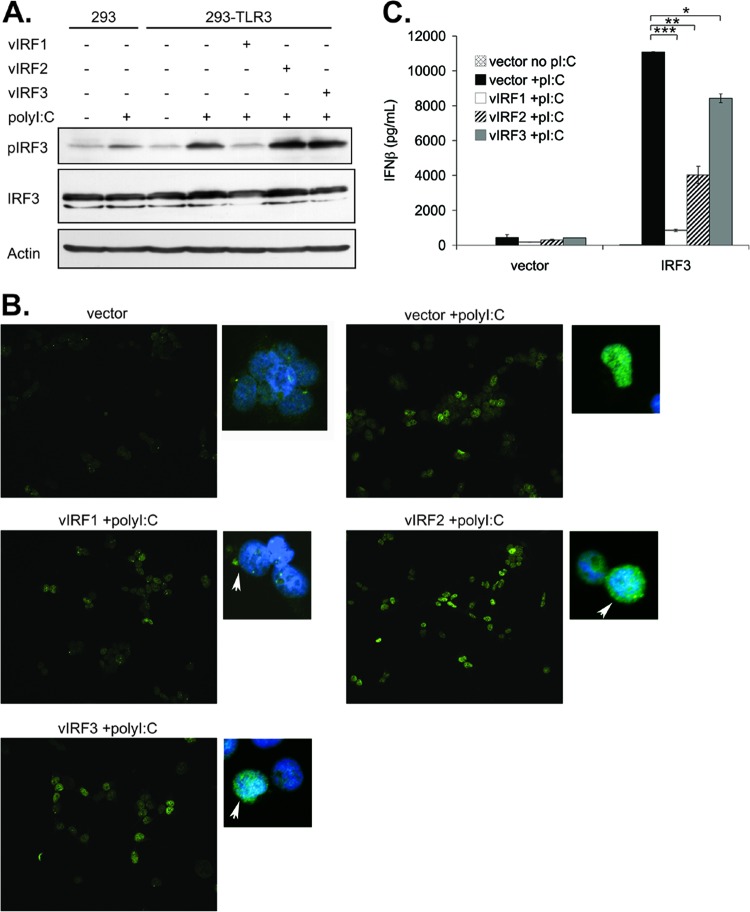
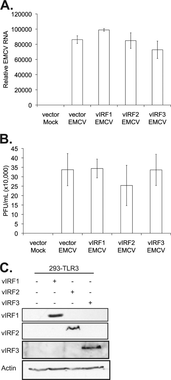
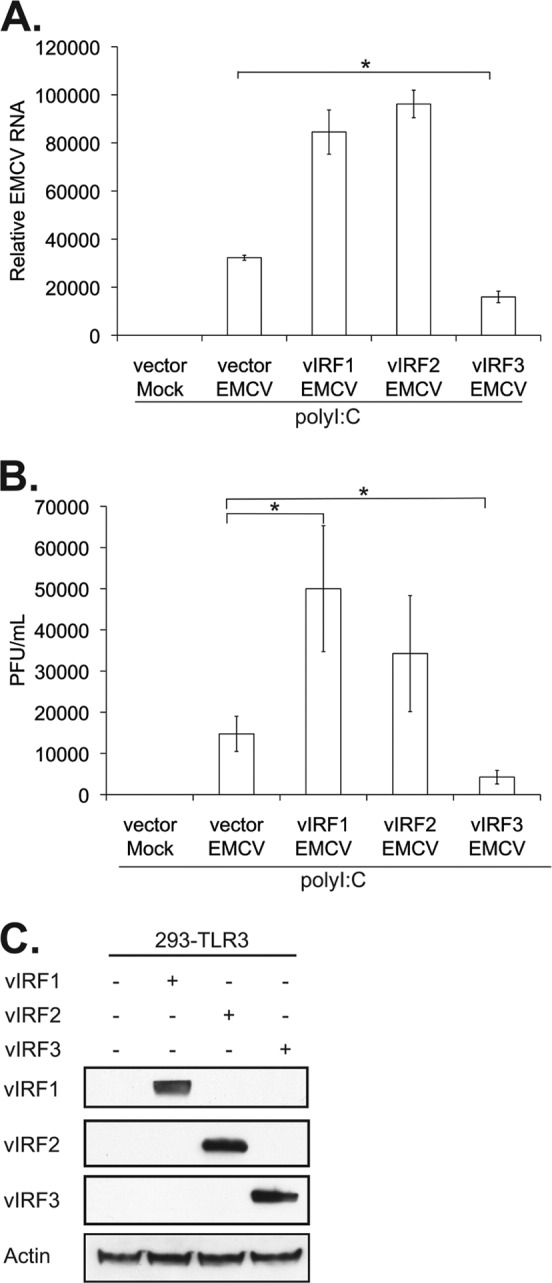
Kaposi's sarcoma-associated herpesvirus (KSHV) infection is correlated with three human malignancies and can establish lifelong latent infection in multiple cell types within its human host. In order to establish and maintain infection, KSHV utilizes multiple mechanisms to evade the host immune response. One such mechanism is the expression of a family of genes with homology to cellular interferon (IFN) regulatory factors (IRFs), known as viral IRFs (vIRFs). We demonstrate here that KSHV vIRF1, -2, and -3 have a differential ability to block type I interferon signaling mediated by Toll-like receptor 3 (TLR3), a receptor we have previously shown to be activated upon KSHV infection. vIRF1, -2, and -3 inhibited TLR3-driven activation of IFN transcription reporters. However, only vIRF1 and vIRF2 inhibited increases in both IFN-β message and protein levels following TLR3 activation. The expression of vIRF1 and vIRF2 also allowed for increased replication of a virus known to activate TLR3 signaling. Furthermore, vIRF1 and vIRF2 may block TLR3-mediated signaling via different mechanisms. Altogether, this report indicates that vIRFs are able to block IFN mediated by TLRs but that each vIRF has a unique function and mechanism for blocking antiviral IFN responses.
Figures
Similar articles
-
Kaposi's Sarcoma-Associated Herpesvirus Viral Interferon Regulatory Factor 1 Interacts with a Member of the Interferon-Stimulated Gene 15 Pathway.J Virol. 2015 Nov;89(22):11572-83. doi: 10.1128/JVI.01482-15. Epub 2015 Sep 9. J Virol. 2015. PMID: 26355087 Free PMC article.
-
Genome-Wide Mapping of the Binding Sites and Structural Analysis of Kaposi's Sarcoma-Associated Herpesvirus Viral Interferon Regulatory Factor 2 Reveal that It Is a DNA-Binding Transcription Factor.J Virol. 2015 Nov 4;90(3):1158-68. doi: 10.1128/JVI.01392-15. Print 2016 Feb 1. J Virol. 2015. PMID: 26537687 Free PMC article.
-
Kaposi's sarcoma-associated herpesvirus vIRF2 protein utilizes an IFN-dependent pathway to regulate viral early gene expression.PLoS Pathog. 2019 May 6;15(5):e1007743. doi: 10.1371/journal.ppat.1007743. eCollection 2019 May. PLoS Pathog. 2019. PMID: 31059555 Free PMC article.
-
Viral interferon regulatory factors.J Interferon Cytokine Res. 2009 Sep;29(9):621-7. doi: 10.1089/jir.2009.0067. J Interferon Cytokine Res. 2009. PMID: 19715458 Free PMC article. Review.
-
Distinct roles of Kaposi's sarcoma-associated herpesvirus-encoded viral interferon regulatory factors in inflammatory response and cancer.J Virol. 2013 Sep;87(17):9398-410. doi: 10.1128/JVI.03315-12. Epub 2013 Jun 19. J Virol. 2013. PMID: 23785197 Free PMC article. Review.
Cited by
-
Activation and counteraction of antiviral innate immunity by KSHV: an Update.Sci Bull (Beijing). 2018 Sep 30;63(18):1223-1234. doi: 10.1016/j.scib.2018.07.009. Epub 2018 Jul 20. Sci Bull (Beijing). 2018. PMID: 30906617 Free PMC article.
-
Viral Inhibition of PRR-Mediated Innate Immune Response: Learning from KSHV Evasion Strategies.Mol Cells. 2016 Nov 30;39(11):777-782. doi: 10.14348/molcells.2016.0232. Epub 2016 Nov 18. Mol Cells. 2016. PMID: 27871174 Free PMC article. Review.
-
Toll-like receptor 3 expression and function in childhood idiopathic nephrotic syndrome.Clin Exp Immunol. 2015 Dec;182(3):332-45. doi: 10.1111/cei.12659. Epub 2015 Sep 30. Clin Exp Immunol. 2015. PMID: 26123900 Free PMC article. Clinical Trial.
-
KSHV-encoded viral interferon regulatory factor 4 (vIRF4) interacts with IRF7 and inhibits interferon alpha production.Biochem Biophys Res Commun. 2017 May 6;486(3):700-705. doi: 10.1016/j.bbrc.2017.03.101. Epub 2017 Mar 22. Biochem Biophys Res Commun. 2017. PMID: 28342865 Free PMC article.
-
KSHV strategies for host dsDNA sensing machinery.Virol Sin. 2016 Dec;31(6):466-471. doi: 10.1007/s12250-016-3877-3. Epub 2016 Dec 5. Virol Sin. 2016. PMID: 27933565 Free PMC article. Review.
References
-
- Chang Y, Cesarman E, Pessin MS, Lee F, Culpepper J, Knowles DM, Moore PS. 1994. Identification of herpesvirus-like DNA sequences in AIDS-associated Kaposi's sarcoma. Science 266:1865–1869 - PubMed
-
- Schulz TF. 1998. Kaposi's sarcoma-associated herpesvirus (human herpesvirus-8). J. Gen. Virol. 79:1573–1591 - PubMed
-
- Alsharifi M, Mullbacher A, Regner M. 2008. Interferon type I responses in primary and secondary infections. Immunol. Cell Biol. 86:239–245 - PubMed
-
- Kawai T, Akira S. 2010. The role of pattern-recognition receptors in innate immunity: update on Toll-like receptors. Nat. Immunol. 11:373–384 - PubMed
Publication types
MeSH terms
Substances
Grants and funding
LinkOut - more resources
Full Text Sources
